4885
Blood-brain barrier permeability in response to caffeine challenge1Department of Radiology, Johns Hopkins University School of Medicine, Baltimore, MD, United States, 2Department of Diagnostic Radiology and Nuclear Medicine, University of Maryland School of Medicine, Baltimore, MD, United States, 3Department of Neurology, Johns Hopkins University School of Medicine, Baltimore, MD, United States
Synopsis
Caffeine is known to alter brain physiology by acting as an adenosine antagonist, but its effect on the BBB permeability to water is not fully elucidated. In this study, we utilized water-extraction-with-phase-contrast-arterial-spin-tagging (WEPCAST) MRI to investigate the change of BBB permeability after caffeine challenge in young healthy adults. Results showed that after caffeine ingestion, water extraction across BBB increased as global cerebral blood flow decreased, while the BBB permeability to water remained unchanged, indicating a relative stable BBB function in response to caffeine challenge, which can be important for normal brain function.
INTRODUCTION
As the most widely used stimulant drug, caffeine is known to have a complicated effect on neurotransmitters, especially its role as an antagonist of adenosine, an inhibitory neurotransmitter1. By reacting with the adenosine receptors, caffeine not only increases neural activity but also constricts cerebral vessels, leading to a profound change in brain physiology, including a decrease in cerebral blood flow (CBF) and venous oxygenation (Yv) but no change in cerebral metabolic rate of oxygen (CMRO2)2. It has also been reported that caffeine has a protective function on the blood-brain barrier (BBB) in Alzheimer’s disease and Parkinson’s disease by blockade of adenosine receptors, inhibition of cAMP phosphodiesterase activity and mobilization of intracellular calcium release3. However, prior studies mainly focused on the chronic effect of caffeine uptake in diseases where BBB was already disrupted, and notably were only performed in animal models due to the invasive techniques, Evans blue, used to measure BBB permeability4,5. Water-extraction-with-phase-contrast-arterial-spin-tagging (WEPCAST) MRI is a recently developed technique to non-invasively measure BBB permeability to water, and is suitable for humans6,7. Thus, in this study, we utilized WEPCAST MRI to investigate the short-term effect of caffeine challenge on BBB in healthy young adults.METHODS
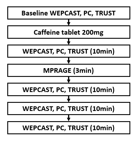
WEPCAST MRI selectively measures ASL signal in the main draining veins of the brain, e.g. superior sagittal sinus (SSS), using a phase-contrast-encoded acquisition, to estimate water extraction fraction (E) and, when combined with CBF (f) measurement, can provide an estimation of BBB permeability in terms of permeability-surface-area product (PS): $$$PS=-ln(1-E)·f$$$ 6,7.Ten young, healthy volunteers were recruited (age 29.1±9.3yrs, 6F/4M). All subjects were studied on a 3T Philips system. The experimental procedure is shown in Figure 1. Each subject was instructed to continue normal caffeine consumption but avoid any caffeine uptake for 4 hours before the study. WEPCAST was performed with a labeling duration of 4000ms and a post-labeling delay of 3000ms. Phase-contrast (PC) MRI was also acquired to measure global CBF. Additionally, T2-Relaxation-Under-Spin-Tagging (TRUST) MRI was performed to estimate Yv. A baseline scan was first performed. Then the subject was taken out of the scanner, sat up on the scanner, ingested a 200mg caffeine tablet and was quickly put back into the scanner. Four sets of WEPCAST, PC and TRUST MRI using the same parameters as baseline were performed immediately after the caffeine ingestion, with each set took around 10 minutes to acquire. A T1-MPRAGE was also acquired for brain volume quantification.
Detailed processing of WEPCAST, PC and TRUST MRI are described elsewhere6-8. For statistical analysis, the physiological parameters (i.e. E, PS, CBF and Yv) measured under baseline were compared with those at each time point after caffeine ingestion using paired t-test.
RESULTS
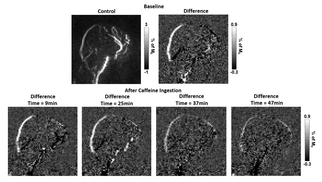
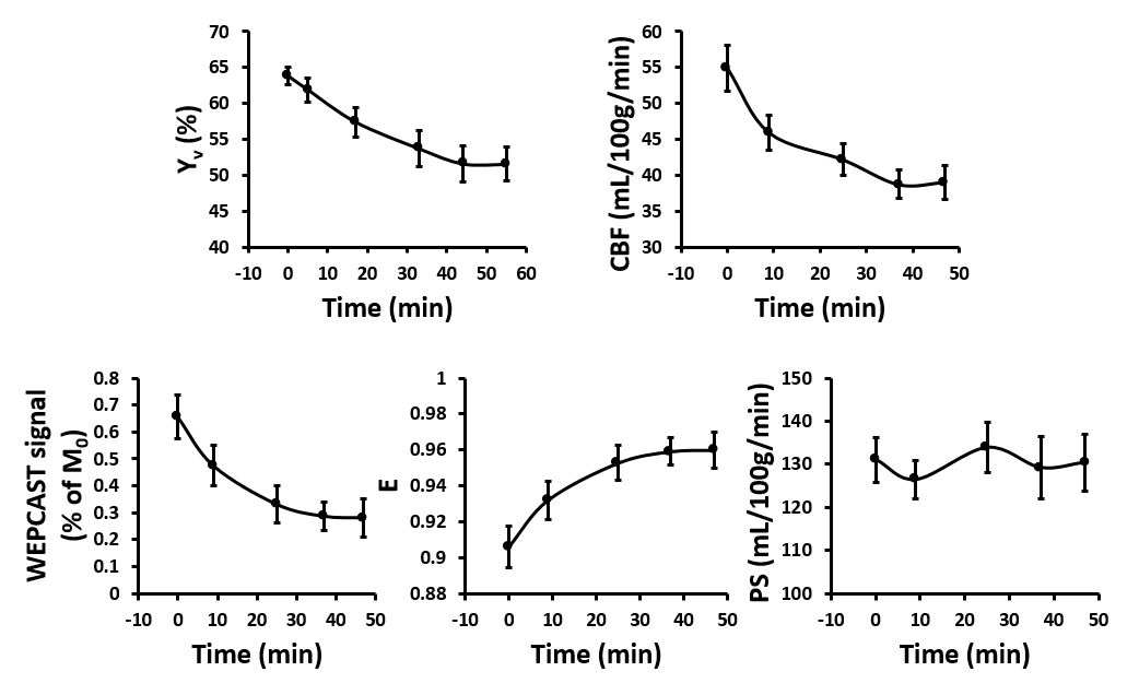
Figure 2 shows representative WEPCAST images before and after caffeine ingestion. At baseline, a prominent signal can be seen at the SSS, representing the labeled water spins that were not extracted by the tissue and drained directly to the venous system. After caffeine uptake, WEPCAST signal decreased significantly, indicating that a larger fraction of water was extracted by the tissue. Peak signal along the SSS was then used to calculate the water extraction fraction.Time courses of the BBB permeability and brain physiological parameters are shown in Figure 3. It can be seen that after caffeine ingestion, venous oxygenation and global CBF gradually decreased while water extraction fraction increased. At around 35 minutes after caffeine uptake, all physiological parameters reached a relatively steady state, with Yv decreased by 19%, CBF decreased by 29%, WEPCAST signal decreased by 56%, and E increased by 6%. Paired t-test of Yv, CBF and E for all time points after caffeine challenge showed a significant difference compared with baseline. On the other hand, no significant changes in the PS values were presented, suggesting an unaltered BBB permeability to water after caffeine ingestion.
DISCUSSION
The impact of caffeine on BBB permeability can be a combination of the vaso-constriction effect and neurotransmitter effect. Our results showed that BBB permeability remained unchanged in response to the caffeine challenge, which could be significant in maintaining a stable microenvironment for normal brain function. Although studies have shown a decrease in BBB permeability in response to caffeine, most studies focused on BBB permeability to large molecules, such as Evans blue and dextran, and were conducted on conditions where BBB was already leaky4,5. One study by Wengler et al. reported a reduction in BBB permeability to water after caffeine ingestion9. However, water exchange across BBB was controlled by both endothelium function and aquaporin-4 on astrocytic endfeet. Current methods on BBB permeability to water can be reflective of different aspects of the water exchange process9-11, and WEPCAST technique mainly measures the endothelium function, in that it cannot differentiate the spin location after it goes across the endothelium, which may be different than Wengler’s method.CONCLUSION
In conclusion, we investigated the effect of caffeine on BBB permeability to water and found that after caffeine ingestion, water extraction across BBB increased while the BBB permeability remained unchanged.Acknowledgements
No acknowledgement found.References
1. Fredholm BB. Adenosine, Adenosine Receptors and the Actions of Caffeine *. Pharmacology & Toxicology 1995;76:93-101.
2. Xu F, Liu P, Pekar JJ, Lu H. Does acute caffeine ingestion alter brain metabolism in young adults? NeuroImage 2015;110:39-47.
3. Chen X, Ghribi O, Geiger JD. Caffeine Protects Against Disruptions of the Blood-Brain Barrier in Animal Models of Alzheimer's and Parkinson's Diseases. Journal of Alzheimer's Disease 2010;20:S127-S141.
4. Chen X, Gawryluk JW, Wagener JF, Ghribi O, Geiger JD. Caffeine blocks disruption of blood brain barrier in a rabbit model of Alzheimer's disease. Journal of Neuroinflammation 2008;5:12.
5. Chen X, Lan X, Roche I, Liu R, Geiger JD. Caffeine protects against MPTP-induced blood-brain barrier dysfunction in mouse striatum. J Neurochem 2008;107:1147-1157.
6. Lin Z, Jiang D, Liu D, Li Y, Uh J, Hou X, Pillai JJ, Qin Q, Ge Y, Lu H. Non-contrast assessment of blood-brain barrier permeability to water: shorter acquisition, test-retest reproducibility, and comparison with contrast-based method. Magn Reson Med 2021;86:143-156.
7. Lin Z, Li Y, Su P, Mao D, Wei Z, Pillai JJ, Moghekar A, van Osch M, Ge Y, Lu H. Non-contrast MR imaging of blood-brain barrier permeability to water. Magn Reson Med 2018;80:1507-1520.
8. Peng SL, Dumas JA, Park DC, Liu P, Filbey FM, McAdams CJ, Pinkham AE, Adinoff B, Zhang R, Lu H. Age-related increase of resting metabolic rate in the human brain. Neuroimage 2014;98:176-183. 9. Wengler K, Bangiyev L, Canli T, Duong TQ, Schweitzer ME, He X. 3D MRI of whole-brain water permeability with intrinsic diffusivity encoding of arterial labeled spin (IDEALS). Neuroimage 2019;189:401-414.
10. Ohene Y, Harrison IF, Nahavandi P, Ismail O, Bird EV, Ottersen OP, Nagelhus EA, Thomas DL, Lythgoe MF, Wells JA. Non-invasive MRI of brain clearance pathways using multiple echo time arterial spin labelling: an aquaporin-4 study. Neuroimage 2019;188:515-523.
11. Shao X, Ma SJ, Casey M, D’Orazio L, Ringman JM, Wang DJJ. Mapping water exchange across the blood-brain barrier using 3D diffusion-prepared arterial spin labeled perfusion MRI. Magn Reson Med 2019;81:3065-3079.
Figures