4856
Increased hippocampal cerebrospinal fluid fraction is associated with episodic verbal learning and memory deficits in multiple sclerosis1Weill Cornell Medicine, New York, NY, United States
Synopsis
We applied FAST-T2 multi-component T2 relaxometry to 145 MS patients and found that the mean hippocampal CSF fraction, a measure of glymphatic clearance dysfunction in the hippocampus, was negatively associated with verbal learning and memory performance measured by the California Verbal Learning Test after adjusting for age, sex, MS phenotype, disease duration, and log total lesion volume.
INTRODUCTION
The glial-lymphatic system facilitates waste removal from the brain parenchyma via perivascular cerebrospinal fluid (CSF) water channels with important implications for pathogenic mechanisms of many neurological disorders (1,2). Impaired parenchymal fluid clearance has been postulated to play a role in neuroinflammation and autoimmune response underlying multiple sclerosis (MS), supported by recent data demonstrating reduced CSF circulation on an animal model of MS (3). Parenchymal CSF water fraction (CSFF) can be mapped by separating the signal of the highly mobile CSF water that occupies the perivascular space and has very long T2 (~2 sec) from the signal of restricted water in the brain tissue that has much shorter T2, including myelin water (T2<20 msec) and intra/extracellular water (T2~50-80 msec). The increase in parenchymal CSFF is a potential indicator of glymphatic clearance dysfunction. CSFF has been shown to be associated with age in normal aging and MS (4,5). The objective of this MS patient study was to apply the efficient Fast Acquisition with Spiral Trajectory and adiabatic T2prep (FAST-T2) sequence (6) to test the hypothesis that CSFF measured in the hippocampus, which is central to episodic memory formation and learning, is associated with verbal learning and memory disability measured by the California Verbal Learning Test version II (CVLT).METHODS
MS cohort. A total of 145 MS patients was included in the study (mean age 43.5±10.4 years, range 22-65 years, 106 women (73.1%), 39 men (26.9%), 140 RRMS, 5 SPMS, mean disease duration 11.2±7.7 years). Patients with active inflammatory disease as evidenced by new Gd-enhancing lesions indicating blood-brain barrier breakdown were excluded. CVLT was administered as part of the Brief International Cognitive Assessment for Multiple Sclerosis (BICAMS) test battery for cognitive evaluation (7). CVLT consisted of five learning trials in which test subjects were asked to recall a list of 16 words, and a total score of correctly remembered words was recorded for each subject. A lower score indicates increased disability.MRI acquisition and data analysis. The 3T brain MRI protocol included a 4 min FAST-T2 sequence with geometric echo spacing for multi-component T2 relaxometry (6) in addition to the conventional T1W (without and with Gd contrast), T2W and T2FLAIR sequences. A spatially regularized three-pool non-linear least squares algorithm using the L-BFGS iterative solver was used to extract myelin water fraction (MWF), intra/extracellular water fraction (IEWF), and CSFF maps from the six-echo T2 decay data. The lower and upper T2 bounds for each of the three water pools (in msec) were set to [5 20], [20 200], and [200 2000], respectively. FreeSurfer’s recon-all command was applied to the T1W structural image to obtain brain segmentation including the hippocampus (8), which was then aligned to CSFF map using FMRIB's Linear Image Registration Tool (9). We used a linear regression model and regressed CLVT total score on mean hippocampal CSFF, adjusting for age, sex, MS phenotype (RRMS, SPMS), disease duration, and log total T2FLAIR lesion volume.
RESULTS
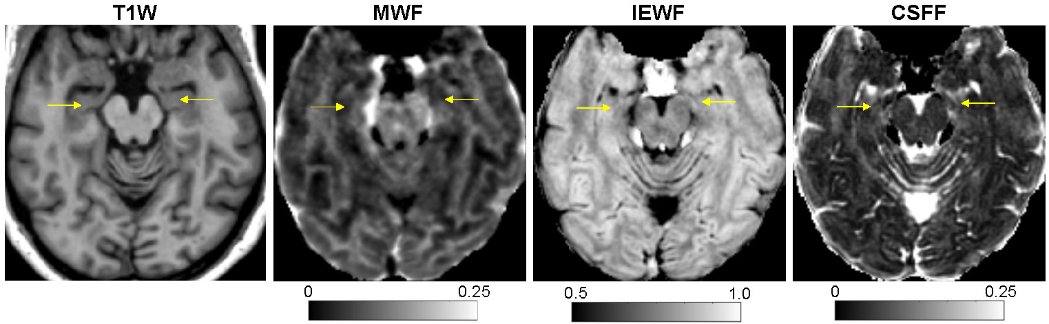
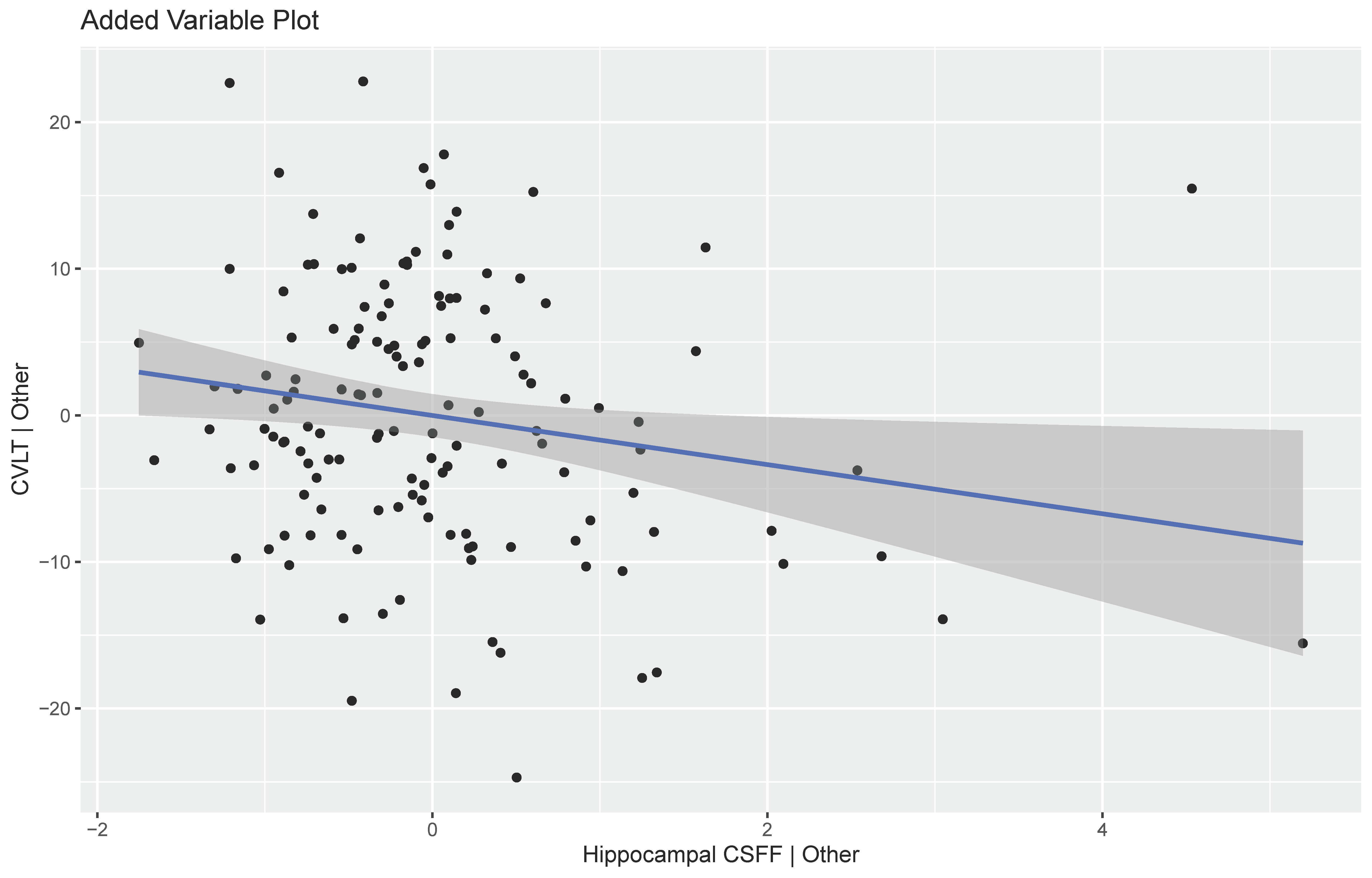
Figure 1 shows an example of the three water fraction maps derived from FAST-T2 which can be interpreted as the relative contribution of water residing within the myelin sheath (myelin water), water residing inside and between cells (intra/extracellular water), as well as free water occupying the brain ventricles and perivascular space. In the linear model with CVLT score as outcome, hippocampal CSFF was found to be statistically significant (beta = -1.68, p-value = 0.027). Besides hippocampal CSFF, only sex was found to be statistically significant (beta = -4.42, p-value = 0.012). Figure 2 shows the added variable plot for the regression, which shows the relationship between the CLVT score and the hippocampal CSFF, adjusting for all other variables.DISCUSSION
Our results provided the first in vivo evidence in MS patients that glymphatic clearance impairment in the hippocampus, as measured by the increased regional CSFF, is associated with episodic learning and memory deficits measured by CVLT total score. FAST-T2 sequence has the potential to improve disease characterization and predicting clinical outcome in MS. Histopathological validation is needed to establish the biological underpinnings of parenchymal CSFF in the MS brain.Acknowledgements
No acknowledgement found.References
1. Rasmussen MK, Mestre H, Nedergaard M. The glymphatic pathway in neurological disorders. Lancet Neurol 2018;17:1016-24.
2. Wardlaw JM, Benveniste H, Nedergaard M, Zlokovic BV, Mestre H, Lee H, Doubal FN, Brown R, Ramirez J, MacIntosh BJ, Tannenbaum A, Ballerini L, Rungta RL, Boido D, Sweeney M, Montagne A, Charpak S, Joutel A, Smith KJ, Black SE; colleagues from the Fondation Leducq Transatlantic Network of Excellence on the Role of the Perivascular Space in Cerebral Small Vessel Disease. Perivascular spaces in the brain: anatomy, physiology and pathology. Nat Rev Neurol 2020;16:137-53.
3. Fournier AP, Gauberti M, Quenault A, Vivien D, Macrez R, Docagne F. Reduced spinal cord parenchymal cerebrospinal fluid circulation in experimental autoimmune encephalomyelitis. J Cereb Blood Flow Metab 2019;39:1258-65.
4. Nguyen TD, Zhou L, Sweeney EM, Wang XH, Gauthier SA, Wang Y, Kuceyeski A, Li Y. Cerebrospinal fluid water fraction increases with age in normal aging. ISMRM 2021;2367.
5. Zhou L, Li Y, Wang XH, Sweeney EM, Zhang H, Tanzi EB, Prince J, Su-Ortiz VA, Gauthier SA, Nguyen TD. Increasing age is independently associated with higher free water in non-active MS brain - A multi-compartment analysis using FAST-T2. ISMRM 2021;2825.
6. Nguyen TD, Deh K, Monohan E, Pandya S, Spincemaille P, Raj A, Wang Y, Gauthier SA. Feasibility and reproducibility of whole brain myelin water mapping in 4 minutes using fast acquisition with spiral trajectory and adiabatic T2prep (FAST-T2) at 3T. Magn Reson Med 2016;76:456-65.
7. Langdon DW, Amato MP, Boringa J, Brochet B, Foley F, Fredrikson S, Hämäläinen P, Hartung HP, Krupp L, Penner IK, Reder AT, Benedict RH. Recommendations for a Brief International Cognitive Assessment for Multiple Sclerosis (BICAMS). Mult Scler 2012;18:891-8.
8. Fischl B. FreeSurfer. Neuroimage 2012;62:774-81.
9. Jenkinson M, Bannister P, Brady M, Smith S. Improved optimization for the robust and accurate linear registration and motion correction of brain images. Neuroimage 2002;17(2):825-41.
Figures