4846
Addressing ultra highfield MRI challenges in ex-vivo
1University of Pittsburgh, pittsburgh, PA, United States, 2Bioengineering, University of Pittsburgh, pittsburgh, PA, United States, 3UPMC Presbyterian Hospital, Pittsburgh, PA, United States
Synopsis
Introduction
Ex-vivo ultra-high field brain MRI can play a crucial role in neuroimaging and neuropathology: it can archive brain specimens for future reconstructions before permanent sectioning [1], and it can help pathologists in identifying regions of interest while addressing sampling limitations [2]. 7T ex-vivo brain images, for instance, can represent an intermediate transform that is used in the alignment of in-vivo to histology. However, ex-vivo at ultra-high fields has several challenges. A low grey to white matter contrast in T1 weighted (T1w) sequences is usually present due to the proportional relationship of T1 with temperature as a result most groups have avoided using T1w sequences in ex-vivo [3]. field inhomogeneities are also present requiring an ex-vivo specific RF coil [4], which can be expensive and requires expertise that few research groups have. To circumvent field inhomogeneities. MRI scans have been performed on pre-cut brain slices, which can negatively impact successful registration between premortem and postmortem MRI [5]. In our previous work [6], we developed a comprehensive workflow that can perform ex-vivo volumetric brain imaging and allows the registration of premortem brain MRI to postmortem brain MRI and align it with histology[12]. In this current work, we show how our ex-vivo workflow addresses field inhomogeneities without building a dedicated 7T ex-vivo coil and provide an ex-vivo adapted MP2RAGE sequence with a good grey to white matter contrast.Methods
We acquired 3 postmortem left hemisphere brains from the University of Pittsburgh Alzheimer's Disease Research Center (ADRC) brain’s bank. We have embedded the brain in agarose in a brain conforming container that minimizes the amount of the surrounding embedding media [6]. A 30% per volume of water sugar was added to the agarose embedding media [7] which reduced the relative permittivity of the media from ε = 78 to ε= 68 at room temperature. We have placed the container in our 16Tx/32Rx channels TicTacToe (TTT) coil [8] and collected 7T MRI (Magnetom, Siemens , Germany) sequences: MP2RAGE sequences were acquired with TR = 6000 ms, TE = 4.1 ms, TI1 = 800 ms, α1 = 6°, TI2 = 2500 ms, α2 = 6°, Matrix = 228x514, Slice thickness 0.37 mm. Flip Angle (FA) Maps were acquired with a Ref amplitude 1 H 200V, TR = 2000 ms, TE = 1.08 ms, FOV =250 mm, Matrix = 44x64, Slice thickness 3.2 mm. The same shimming (amplitudes and phases) of the in-vivo setup was used on the 3 ex-vivo brains. To restrict the coefficient of variance (CV) computation of the (FA-map) to the brain regions, we have used ITK-snap(V3.8) to create a brain mask from the MP2RAGE and we resampled the mask to include to the FA resolution. We manually adjusted the automatic brain masks to remove areas where air bubbles were causing erroneous measurements.Results
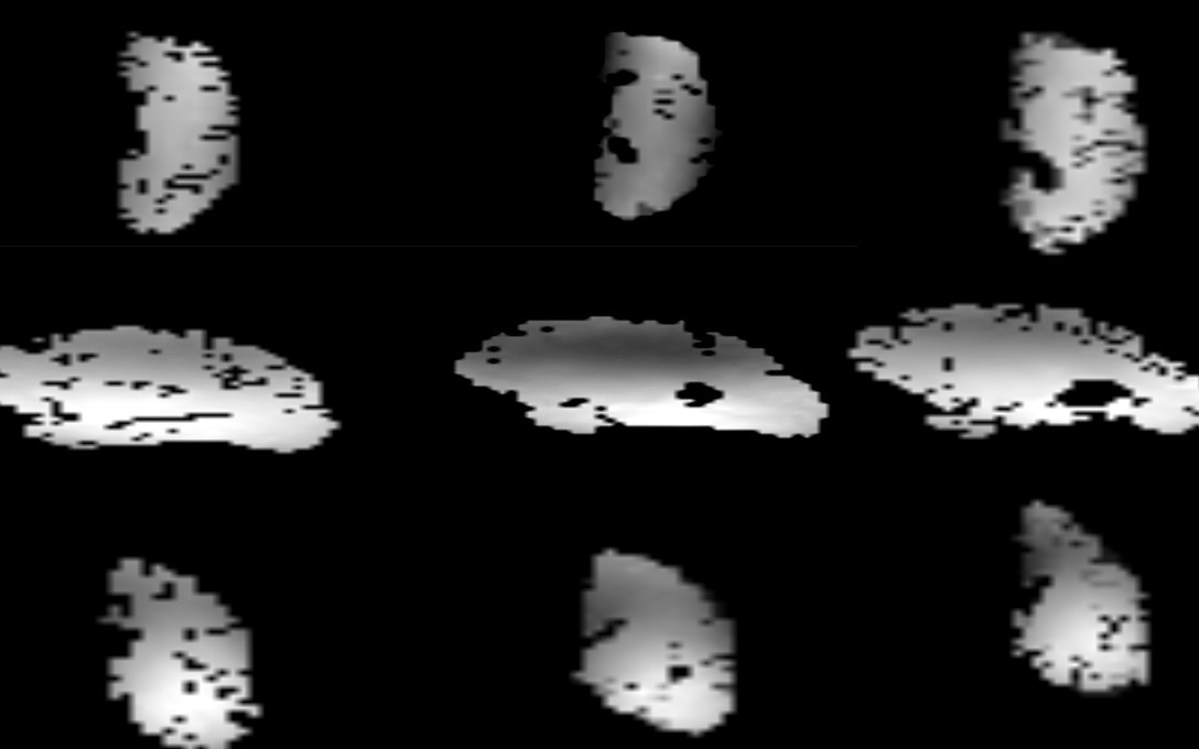
Figure 1 shows the (FA) Maps 200 v of 1 ms square pulse from the three different ex-vivo brains of the mask that has been applied. The dark spots in the brain are the location of air bubbles that were excluded from the FA map. The 3 brains are almost in identical locations in the Superior Inferior and Right Left (direction). The CV of FA in the masked brain regions for the three subjects are 0.25, 0.29, and 0.31 with mean FA μ1= 230 °, μ2= 254 °, μ3= 234 °. In Figure 2, we show an MP2RAGE with a high grey matter to white matter contrast, also the high contrast is able to depict the formalin boundary.Discussion and Conclusion
The TTT coil is highly insensitive to changes in head sizes in-vivo; therefore, subject-dependent shimming is unnecessary and the variance in the CV between subjects is typically minimal [8]. In, this ex-vivo setup we have used the same in-vivo parameters for the ex-vivo brains and were able to show that the variance in the CV for the TTT is relatively constrained to 0.06 difference between 3 different ex-vivo brains. While the CV value of the field is higher than our in-vivo counterparts (~ 0.17-0.2 in-vivo [11] versus ~0.25-0.31 ex-vivo), they are still comparable to the performance of a TEM coil design in-vivo [8]. We think the difference in the CV between ex-vivo and in-vivo is due to the high dielectric effect of the embedding media ( ε= 68 ) and lack of RF shimming specific to the post mortem scans. MP2RAGE sequences minimize the effect of the heterogeneity and susceptibility effects [9]. We have adapted an in-vivo MP2RAGE sequence to be used ex-vivo and to the best of our knowledge, it is the first time an MP2RAGE was applied in ex-vivo human brains at 7T. T1 values are temperature-dependent therefore MPRAGE sequences are not easily optimized for ex-vivo settings at room temperature. We collected an MP2RAGE (Figure 2) that shows a high contrast of grey to white matter. There are several limitations to this work; fixation, and tissue degeneration varies between ex-vivo brains and therefore image quality might vary. In our future work, we are planning to design and implement a cradle that fixes the container to a specific position in the coils, and to implement a vacuum system to remove air bubbles.Acknowledgements
This work was supported by the National Institutes of Health under award numbers R01MH111265, R01AG063525, T32MH119168, and U19AG068054 and CRC.References
[1] Yacoub, E., Shmuel, A., Pfeuffer, J., Van De Moortele, P. F., Adriany, G., Andersen, P., Thomas Vaughan, J., Merkle, H., Ugurbil, K., & Hu, X. (2001). Imaging brain function in humans at 7 Tesla. Magnetic Resonance in Medicine, 45(4), 588–594. https://doi.org/10.1002/mrm.1080
[2] Guasp-Verdaguer, M., Grau-Rivera, O., Prats-Galino, A., Bargalló, N., Sánchez-Valle, R., Gelpi, E., & Soria, G. (2017). Clinical Neuropathology image 4-2017: High-resolution 7 Tesla MRI of postmortem brain specimens: improving neuroimaging-neuropathology correlations. Clinical Neuropathology, 36(07), 162–163. https://doi.org/10.5414/NP301049
[3] Birkl, C., Langkammer, C., Haybaeck, J., Ernst, C., Stollberger, R., Fazekas, F., & Ropele, S. (2014). Temperature-induced changes of magnetic resonance relaxation times in the human brain: A postmortem study. Magnetic Resonance in Medicine, 71(4), 1575–1580. https://doi.org/10.1002/mrm.24799
[4] Edlow, B. L., Mareyam, A., Horn, A., Polimeni, J. R., Witzel, T., Tisdall, M. D., Augustinack, J. C., Stockmann, J. P., Diamond, B. R., Stevens, A., Tirrell, L. S., Folkerth, R. D., Wald, L. L., Fischl, B., & van der Kouwe, A. (2019). 7 Tesla MRI of the ex vivo human brain at 100 micron resolution. Scientific Data, 6(1), 244. https://doi.org/10.1038/s41597-019-0254-8
[5] Dusek, P., Madai, V. I., Huelnhagen, T., Bahn, E., Matej, R., Sobesky, J., Niendorf, T., Acosta‐Cabronero, J., & Wuerfel, J. (2019). The choice of embedding media affects image quality, tissue R 2 * , and susceptibility behaviors in post‐mortem brain MR microscopy at 7.0T. Magnetic Resonance in Medicine, 81(4), 2688–2701. https://doi.org/10.1002/mrm.27595
[6] Nadim Farhat*, Julia Kofler* Jacob Berardinelli, Mark Stauffer, Tales Santini, Neilesh Vinjamuri, Andrea Sajewski , Salem Alkhateeb, Tiago Martins, Noah Schweitzer , Milos Ikonomovic, Howard J. Aizenstein, and Tamer S Ibrahim1 , Reusable 3D printed enclosure with integrated cutting guides for the alignment of ex-vivo MRI with ex-vivo gross brain photographs. ISMRM Annual Meeting & Exhibition, 15-20 May 2021
[7] Duan, Q., Duyn, J. H., Gudino, N., de Zwart, J. A., van Gelderen, P., Sodickson, D. K., & Brown, R. (2014). Characterization of a dielectric phantom for high-field magnetic resonance imaging applications. Medical Physics, 41(10), 102303. https://doi.org/10.1118/1.4895823
[8] Krishnamurthy, N., Santini, T., Wood, S., Kim, J., Zhao, T., Aizenstein, H. J., & Ibrahim, T. S. (2019). Computational and experimental evaluation of the Tic-Tac-Toe RF coil for 7 Tesla MRI. PloS one, 14(1), e0209663. https://doi.org/10.1371/journal.pone.0209663
[9] Marques, J. P., Kober, T., Krueger, G., van der Zwaag, W., Van de Moortele, P.-F., & Gruetter, R. (2010). MP2RAGE, a self bias-field corrected sequence for improved segmentation and T1-mapping at high field. NeuroImage, 49(2), 1271–1281. https://doi.org/10.1016/j.neuroimage.2009.10.0
[10] Yong-Hing, C. J., Obenaus, A., Stryker, R., Tong, K., & Sarty, G. E. (2005). Magnetic resonance imaging and mathematical modeling of progressive formalin fixation of the human brain. Magnetic Resonance in Medicine, 54(2), 324–332. https://doi.org/10.1002/mrm.20578
[11] Tales Santini, Sossena Wood, Narayanan Krishnamurthy, Tiago Martins, Howard J. Aizenstein, Tamer S. Ibrahim. Improved 7 Tesla Transmit Field Homogeneity with Reduced Electromagnetic Power Deposition Using Coupled Tic Tac Toe Antennas. Scientific Reports, 2021.
[12] A novel method for tracking the progression of WMHs through the alignment of premortem in-vivo to postmortem ex-vivo MRI and histopathology. 2021 Alzheimer's Association International Conference, 2021
Figures