4845
Improved Structural Neuro-MRI with Triple TSE-based Synthetic MP-RAGE
Lei Zhang1, Ryan McNaughton2, Hernan Jara2, David N. Kennedy3, Carmen A. Marable1, Ning Hua4, Karl C. K. Kuban4, T. Michael O'Shea1, and Rebecca C. Fry1
1University of North Carolina at Chapel Hill, Chapel Hill, NC, United States, 2Boston University, Boston, MA, United States, 3University of Massachusetts Chan Medical School, Worcester, MA, United States, 4Boston University Medical Center, Boston, MA, United States
1University of North Carolina at Chapel Hill, Chapel Hill, NC, United States, 2Boston University, Boston, MA, United States, 3University of Massachusetts Chan Medical School, Worcester, MA, United States, 4Boston University Medical Center, Boston, MA, United States
Synopsis
Purpose: To develop and evaluate a
metal-artifact robust triple turbo-spin-echo (TSE)-based synthetic MP-RAGE method. Methods: T1, T2 and PD maps generated
from triple TSE, two MP-RAGE image sets simulated by the Bloch equation, and FreeSurfer
brain segmentation were performed on 44 participants at age of fifteen. Results: Reduced artifact and
homogenous synthetic MP-RAGE images were produced in orthodontic brace-wearing
subjects. Comparable image contrast and FreeSurfer brain segmentation results were
observed between synthetic and acquired MP-RAGE in normal subjects. Conclusion: Triple TSE based synthetic
MP-RAGE may enhance conventional MP-RAGE in the neuroimaging studies of orthodontic
brace-wearing subjects with reduced metal artifacts.
Introduction
Adolescent brain MRI and image analysis are important for the study of the development, health and disease of human brain. Magnetization-prepared rapid gradient echo (MP-RAGE) is a popular sequence providing distinct brain image quality. Most brain image analysis tools, such as FreeSurfer, use MP-RAGE as a standard source image, and have been implemented in large national and international cohort brain studies of adolescents.1 Presence of orthodontic metal braces in adolescents, however, may detrimentally affect neuroimaging studies by imposing susceptibility artifact on MP-RAGE images. Triple turbo spin echo (TSE) sequences with T1, T2 and proton density (PD) weighting have been recently used in our multiparametric quantitative MRI (MP-qMRI) imaging protocol for the Extremely Low Gestational Age Newborn – Environmental Influences on Child Health Outcomes (ELGAN-ECHO) study. We hypothesize that through the use of the triple TSE based relaxometry parameters and Bloch equation simulation, we could synthesize MP-RAGE images with reduced metal artifacts. The purpose of this study was: 1) to compare the image quality between triple TSE based synthetic MP-RAGE and acquired MP-RAGE in orthodontic metal-brace wearing adolescents; and 2) to compare the image quality and FreeSurfer brain segmentation results between the synthetic and acquired MP-RAGE in non-brace wearing adolescents.Materials and Methods
The study was approved by the Institutional Review Board (IRB) of the University of North Carolina at Chapel Hill (UNC-CH), a participating institution of the ELGAN-ECHO study.2 A cohort of all 44 participants – 18 females and 26 males, mean age 15.2 ± 0.5 who underwent both triple TSE and MP-RAGE brain imaging at two sites (UNC-CH and Boston Children’s Hospital) was studied retrospectively. Among them, five wore orthodontic braces and 39 did not, where 30 of the 39 passed quality assurance and were evaluated quantitatively. All scans were performed on Siemens Prisma 3T MRI. As shown in Figure 1, the triple TSE acquisition consists of concatenated long repetition time (TR) dual echo turbo spin echo (DE-TSE) sequence for PD- and T2-weighting and short TR single echo TSE (SE-TSE) sequence for T1-weighting implemented with identical scan geometry and receiver settings. Typical imaging parameters: voxel = 0.5 x 0.5 x 2 mm3, TE1,2eff = 12 ms, 102 ms, TRlong = 10 s, TRshort = 0.5 s. Standard MP-RAGE sequence was used with parameters: TR = 2400 ms, TE = 2.15 ms. TI = 1000 ms, Flip angle = 8°, BW = 220 Hz/Px, FoV = 256 x 256 mm2, sagittal 160 slices, voxel = 1 x 1 x 1 mm3, and GRAPPA PE acceleration factor of 2. Calculation of T1, T2, PD and synthetic MP-RAGE generation were performed by in-house developed program in Matlab 2018b (Mathworks, Natick, MA). T1, T2, PD were calculated following Equations 1-3:$$ T_2{}={}\mid\frac{TE_{2eff}-TE_{1eff}}{ln(DA_1/DA_2)}\mid \:\:\:\:\:(1) $$
$$T_1{}={}\mid\frac{TR_{short}}{ln(1-DA_3/DA_1)}\mid \:\:(2) $$
$$PD{}={}DA_1 \cdot exp(\frac{TE_{1eff}}{T_2 }) \:\:\:\:\:\:\:(3)$$
where, DA1,2,3 are the PD-weighted, T2-weighted and T1-weighted images, respectively. As a proof-of-concept, MP-RAGE synthesis was performed following a published method.3 In our study, the parameters were: TR = 2500 ms; TE = 2.15 ms; TI = 1000 ms;
Results
From the triple TSE, T1, T2 and PD-weighted images were acquired, and T1, T2 and PD maps were generated for all subjects (Figure 2). Two types of synthetic MP-RAGE images were generated, one with PD and T1-weighting in the simulation (Syn-PD1) and one with pure T1-weighting (Syn-PD0). In the orthodontic brace-wearing subjects (Figure 3), acquired MP-RAGE showed susceptibility artifacts near the oral cavity affecting the frontal brain, while Syn-PD1 and Syn-PD0 showed decreased metal artifacts with an improved image homogeneity, where Syn-PD0 was more homogenous than Syn-PD1. In the non-brace wearing subjects, acquired MP-RAGE showed no metal-induced susceptibility artifacts. Syn-PD1 and Syn-PD0 showed comparable or superior image contrast to acquired MP-RAGE (Figure 4). FreeSurfer brain segmentation was performed successfully on the acquired and two synthetic MP-RAGE (Figure 5) on all subjects. Cerebral cortex volume showed r = 0.92 and 1.01, R2 = 0.60 and 0.76 (absolute and normalized); cerebral white matter volume showed r = 0.87 and 0.96, R2 = 0.75 and 0.75 (absolute and normalized); and subcortical gray matter volume showed r = 0.93 and 1.03, R2 = 0.90 and 0.37 (absolute and normalized) between Syn-PD0 and acquired MP-RAGE.Discussion and Conclusions
A triple turbo spin echo sequence-based MP-RAGE synthesis method has been developed and evaluated. In adolescent subjects wearing orthodontic braces, it has shown to buffer against metal artifacts and achieved improved image homogeneity. In the non-brace wearing subjects, it has shown comparable or higher image contrast than acquired MP-RAGE. FreeSurfer brain segmentation showed comparable structural volumes on synthetic and acquired MP-RAGE. Further studies are needed to evaluate the similarities and differences between triple TSE-based synthetic MP-RAGE and acquired MP-RAGE.Acknowledgements
This work was supported in part by the National Institutes of Health Office of the Director (5UH3OD023348-06), National Institute of Neurological Disorders and Stroke (5U01NS040069-05 and 2R01NS040069-09), and the National Institute of Child Health and Human Development (5P30HD018655-28).References
1. Hagler DJ, Jr., Hatton S, Cornejo MD, et al. Image processing and analysis methods for the Adolescent Brain Cognitive Development Study. Neuroimage 2019;202:116091.2. O'Shea TM, Allred EN, Dammann O, et al. The ELGAN study of the brain and related disorders in extremely low gestational age newborns. Early Hum Dev 2009;85:719-25.
3. Noth U, Hattingen E, Bahr O, Tichy J, Deichmann R. Improved visibility of brain tumors in synthetic MP-RAGE anatomies with pure T1 weighting. NMR Biomed 2015;28:818-30.
Figures
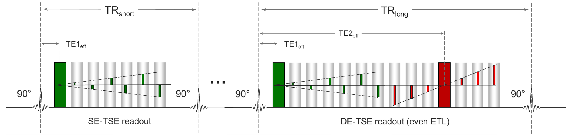
Figure 1. Triple TSE pulse sequence in
ELGAN-ECHO study, consisting of SE-TSE and DE-TSE acquisitions.
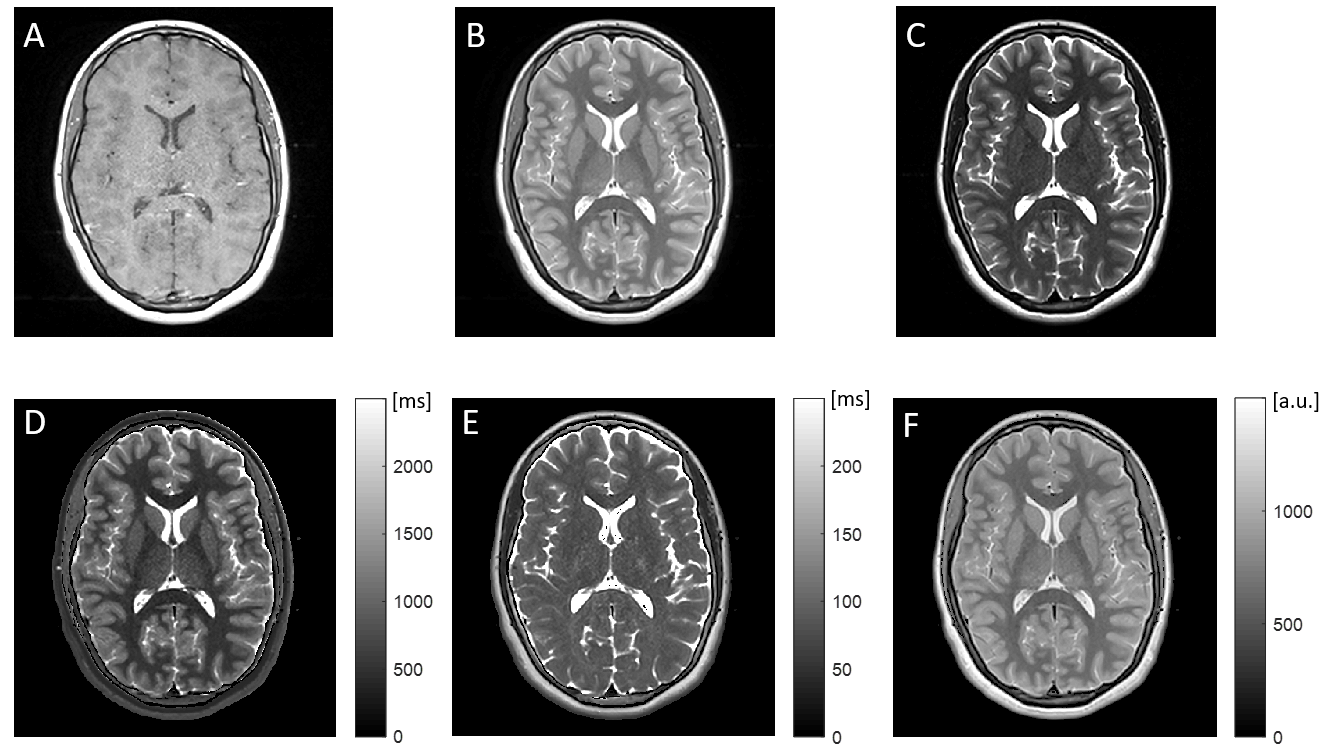
Figure 2. Triple TSE images were acquired (A)
SE-TSE T1-weighted, (B) DE-TSE PD-weighted, and (C) DE-TSE T2-weighted. Three
quantitative maps were calculated: (D) T1, (E) T2 and (F) PD, for the
generation of synthetic MP-RAGE.
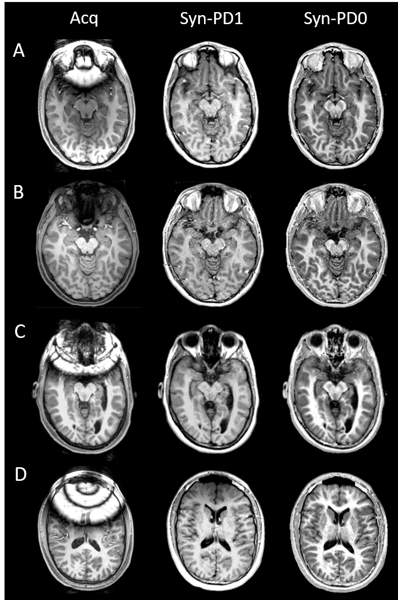
Figure 3. Brace-wearing subjects acquired
MP-RAGE and synthetic MP-RAGE (Syn-PD1, Syn-PD0) images. Acquired MP-RAGE
showed different degrees of metal artifact (left). Syn-PD1 (middle) and Syn-PD0
(right) showed near homogenous brain images without substantial metal artifact.
(A-D) four representative subjects. Syn-PD1: synthetic MP-RAGE with T1 and PD-weighting;
Syn-PD0: synthetic MP-RAGE with pure T1-weighting.
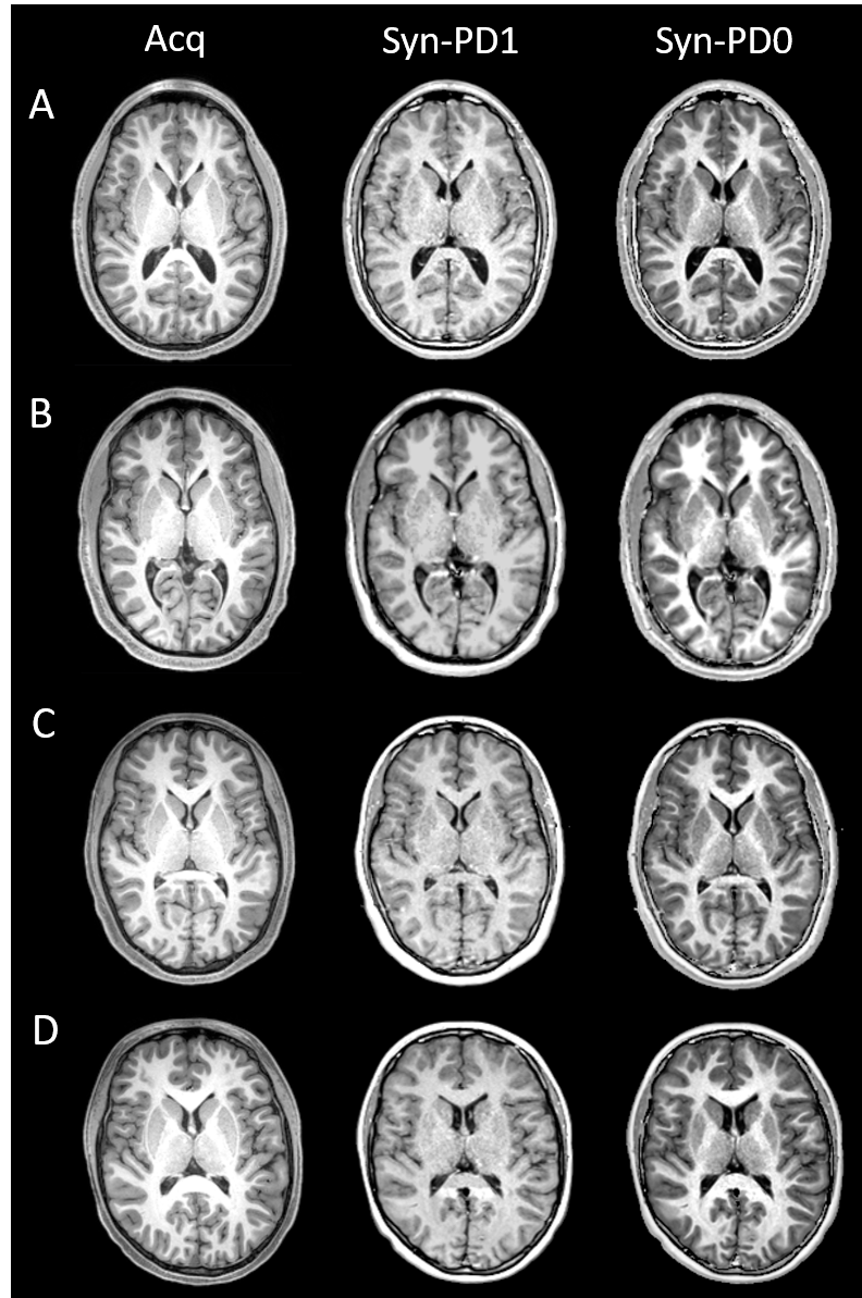
Figure 4. Non-brace-wearing subjects acquired
MP-RAGE and synthetic MP-RAGE (Syn-PD1, Syn-PD0) images. Acquired MP-RAGE
showed no metal artifact (left). Syn-PD1 (middle) and Syn-PD0 (right) showed
comparable and improved image contrast, respectively. (A-D) four representative
subjects. Syn-PD1: synthetic MP-RAGE with T1 and PD-weighting; Syn-PD0: synthetic
MP-RAGE with pure T1-weighting.
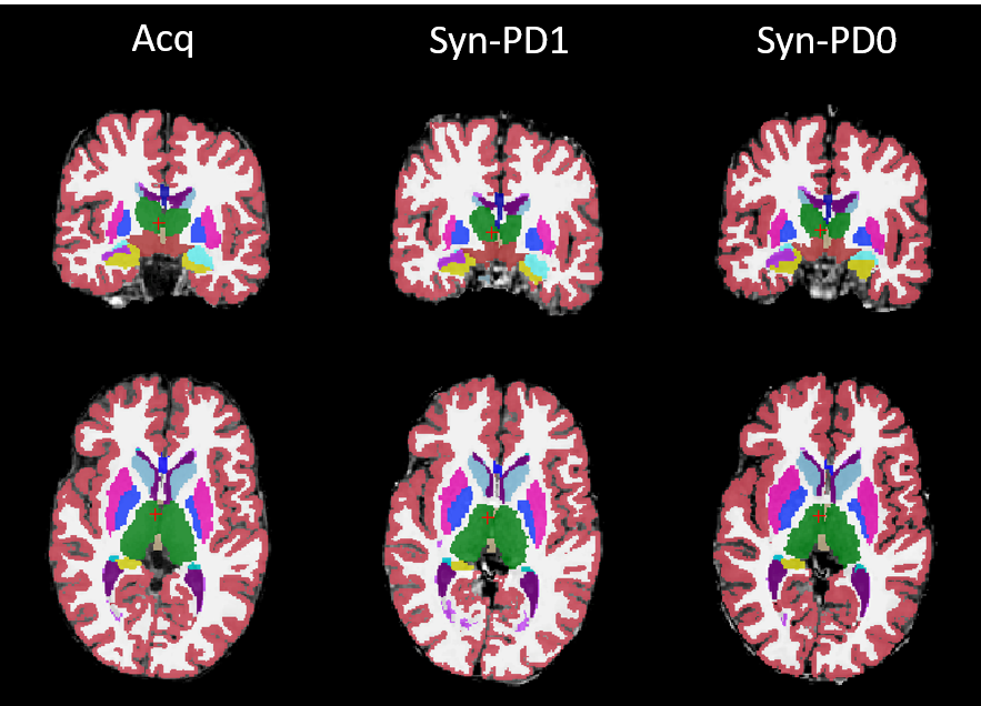
Figure 5. Brain segmentation by FreeSurfer
on non-brace wearing subject acquired MP-RAGE and synthetic MP-RAGE (Syn-PD1, Syn-PD0)
images. Brain segmentations were qualitatively and quantitatively similar when compared
between the three types of MP-RAGE images. Syn-PD1: synthetic MP-RAGE with T1
and PD-weighting; Syn-PD0: synthetic MP-RAGE with pure T1-weighting.
DOI: https://doi.org/10.58530/2022/4845