4841
Rapid, high-spatial resolution in vivo diffusion MRI with joint subsampling and reconstruction in k-,q- and RF-space1Athinoula A. Martinos Center for Biomedical Imaging, Massachusetts General Hospital, Harvard Medical School, Charlestown, MA, United States, 2Harvard-MIT Division of Health Sciences and Technology, Cambridge, MA, United States
Synopsis
In this work, we accelerate the high-SNR, high-spatial resolution in vivo diffusion MRI technique gSlider by applying subsampling jointly in the k,-q, and RF-encoding space. Good reconstruction quality is achieved if complementary information from the multi-dimensional space k-,q-, and RF-space is combined in a complementary fashion and the reconstruction problem is solved jointly.
Introduction
Pursing in vivo high-resolution diffusion MRI with high SNR, minimal distortion artifacts, and in efficient scan time, is of great importance for many neuroscientific and clinical applications. In this work, we accelerate the high-resolution diffusion MRI technique, gSlider1, by applying complementary data undersampling along the k-,q, and RF-encoding space, simultaneously. We first demonstrate a two-fold acceleration in a real scenario with the recently proposed blip-up blip-down gSlider method (BUDA-gSlider2), accomplishing distortion-free, whole brain 1 mm isotropic diffusion MRI (b =1500 s/mm2) with 64 diffusion directions in 17 min. We then investigate the potential to achieve higher acceleration factors in simulations with high image fidelity by varying the k-space sampling scheme for each RF-encoding profile in an interleaved fashion, in combination with undersampling along q-space as well.Methods
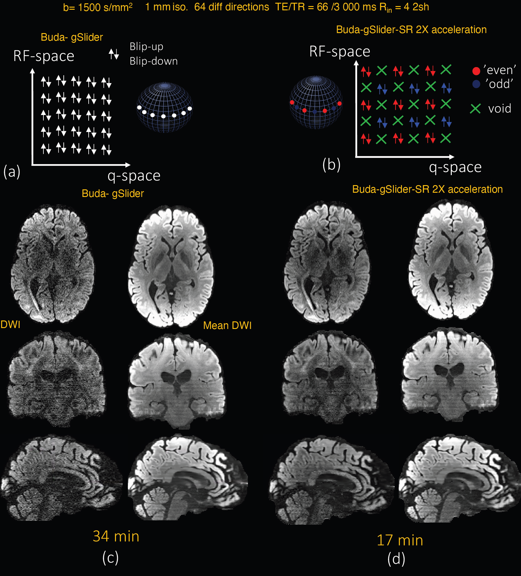
Accelerated BUDA-gSlider with complementary undersampling in the RF-q spaceWe propose to improve on the recently proposed distortion-free BUDA-gSlider by shortening the acquisition by half via exploiting redundancies along RF and q-space together3. Using the same sparsity basis for the diffusions signal used in3, a spherical ridgelets basis, and similar complimentary, non-orthogonal, undersampling pattern in RF-q space, we reconstruct high-fidelity, high-SNR, 1 mm isotropic DWI volumes of an in vivo whole human brain with a total time of 17 min (64 directions).
BUDA g-slider acquisition:
Whole-brain 1-mm isotropic resolution data were acquired with a Siemens 32-channel head coil on a 3T Siemens Prisma scanner. FOV: 224 x 224 x 130 mm3 (26 slabs, 5-mm slab thickness), MB x Rin x gSlider = 1 x 4 x 5 per shot, PF = 6/8, two BUDA shots (A>>P, P>>A), b = 1500 s/mm2, 64 directions + 4 b0s, and TR/TE =3000/66 ms. To create reference volumes, RF-encoded images are first reconstructed from multi-shot k-space data using Buda. Then a high-resolution image is obtained with gSlider. We compare this reference to a high-resolution image obtained with undersampled Rf-q space. To that end, we subsampled retrospectively the reconstructed RF-encoded images along the q-space as shown in Fig1b. We then applied gSlider-SR to estimate high-resolution diffusion MR image.
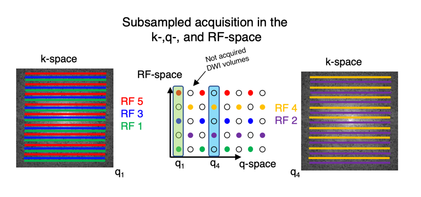
Complementary k-space sampling along the RF-encoding dimension
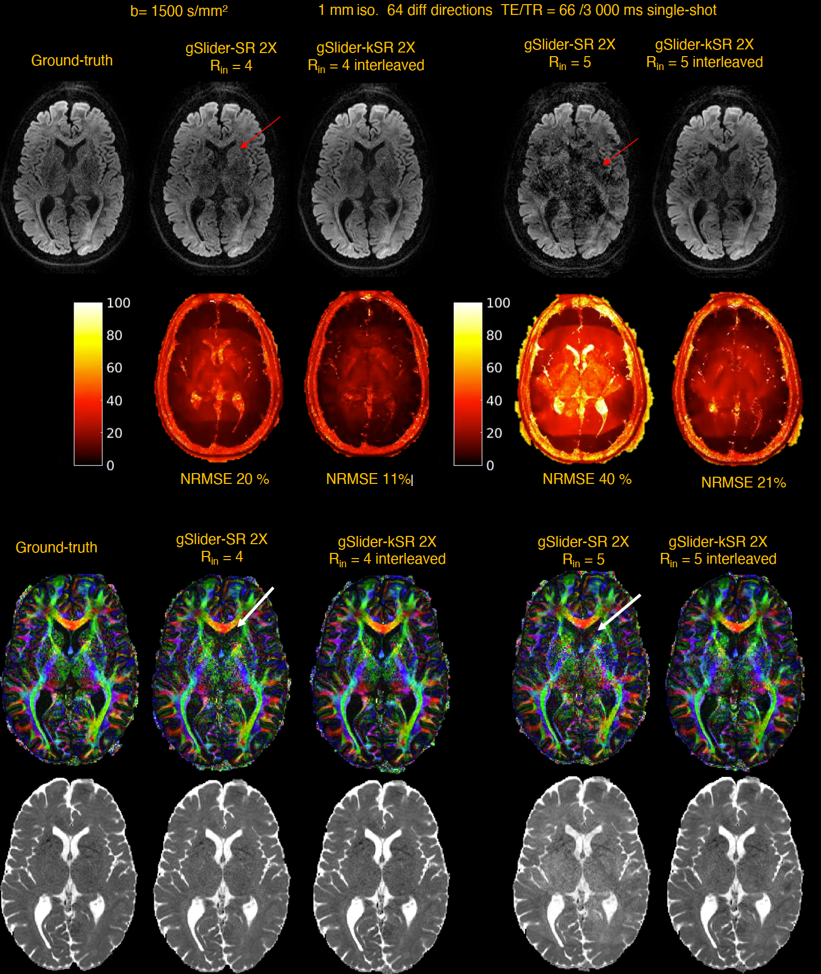
We also investigated whether the total acceleration of single-shot gSlider can be increased if the k-space sampling scheme varies along the RF-encoding dimension in an interleaved fashion. In combination with the already subsampled RF-q space, this approach constitutes a unified sampling scheme in the k,-q-, and RF-encoding space, see Fig.3. Realistic simulations were performed with the in vivo BUDA-reconstructed gSlider data. This ground-truth was propagated through a forward model that includes the gSlider-encoding matrix, coil sensitivities, 2D Fourier transform, k-space sampling mask that varies per each RF-encoding, and additive white Gaussian noise. Acceleration factors of Rin = 4,5 were simulated with no PF and interleaved encoding as shown in Fig3. From the k-q-RF-space, a high-resolution image was estimated in a compressed sensing framework with spherical ridgelets as sparse basis. The method, termed gSlider-KSR, was compared against gSlider-SR, i.e., k-space data are acquired with identical pattern per RF shot (same Rin), thick-slices are reconstructed with SENSE, and the high-resolution image is obtained with gSlider-SR.
Results
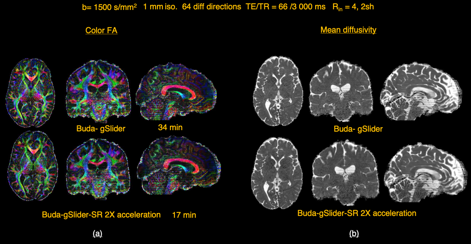
Reconstructed images with BUDA-gSlider (full q-RF space) and BUDA-gSlider-SR (2x speed-up) are shown in Fig.1c and 1d respectively. Despite lacking half of the data, buda-gSlider-SR is able to reconstruct high fidelity, high SNR, DWI images in only 17 min. Note as well the improvement in SNR due to the regularization of spherical ridgelets. Color-coded FA and MD show no appreciable difference (Fig.2), suggesting the acquisition time can be reduced to half without compromising dMRI signal quality.Results from the experiments with synergistic sampling in the k-,q-, and RF-space are shown in Fig.4. For illustration, only a high-resolution axial slice is shown for each method. As observed in the DWI images and FA maps, and confirmed by the RMSE, superior reconstruction quality can be obtained when interleaved k-space sampling is used especially for very high in-plane acceleration (Rin=5)
Discussion and Conclusions
We have shown that is possible to achieve 64-directions, distortion-free high-resolution diffusion MRI (1 mm) in acquisition times less than 20 minutes with BUDA-gSlider-SR, where the RF and q-space is jointly undersampled. Aiming at further mitigating EPI distortions and reducing echo times, we have investigated whether very high in-plane accelerations (Rin = 5) can be achieved in a single in-plane shot gSlider acquisition. Realistic simulations results suggest that reliable DWI images can be reconstructed if the k, q, and RF-encoding space is undersampled in a complementary way, where the set of RF-encoding profiles varies along the q-space direction, and the k-space sampling do so as well along the RF-encoding direction. Interleaving k-space EPI readout for different RF-encoding provides an efficient way to achieve higher acceleration factor with mitigated reconstruction artifacts.Acknowledgements
This work was supported by the NIH (grants P41-EB030006, U01-EB026996, R01-EB021265, K23-NS096056,R01 EB028797, R03 EB031175, U01 EB025162, P41 EB030006)., and the NVidia Corporation for computing support.References
1Setsompop K, Fan Q, Stockmann J, Bilgic B, Huang S, Cauley SF, Nummenmaa A, Wang F, Rathi Y, Witzel T, Wald LL. High-resolution in vivo diffusion imaging of the human brain with generalized slice dithered enhanced resolution: Simultaneous multislice (gSlider-SMS). Magn Reson Med. 2018 Jan;79(1):141-151. doi: 10.1002/mrm.26653. Epub 2017 Mar 5. PMID: 28261904; PMCID: PMC5585027.
2Liao C, Bilgic B, Tian Q, Stockmann JP, Cao X, Fan Q, Iyer SS, Wang F, Ngamsombat C, Lo WC, Manhard MK, Huang SY, Wald LL, Setsompop K. Distortion-free, high-isotropic-resolution diffusion MRI with gSlider BUDA-EPI and multicoil dynamic B0 shimming. Magn Reson Med. 2021 Aug;86(2):791-803. doi: 10.1002/mrm.28748. Epub 2021 Mar 10. PMID: 33748985; PMCID: PMC8121182.
3Ramos-Llordén G, Ning L, Liao C, Mukhometzianov R, Michailovich O, Setsompop K, Rathi Y. High-fidelity, accelerated whole-brain submillimeter in vivo diffusion MRI using gSlider-spherical ridgelets (gSlider-SR). Magn Reson Med. 2020 Oct;84(4):1781-1795. doi: 10.1002/mrm.28232. Epub 2020 Mar 3. PMID: 32125020.
Figures