4832
Physical Activity during Pregnancy is Associated with Newborn’s Brain Cortical Development1Radiology, University of Arkansas for Medical Sciences, Little Rock, AR, United States, 2Pediatrics, University of Arkansas for Medical Sciences, Little Rock, AR, United States, 3Arkansas Children’s Nutrition Center, Little Rock, AR, United States, 4Arkansas Children's Research Institute, Little Rock, AR, United States
Synopsis
This study examined relationships between mother’s physical activity during pregnancy and newborn’s brain cortical development. Healthy pregnant women were recruited and their physical activity level were monitored throughout pregnancy. Their newborns underwent a brain MRI examination at 2 weeks of postnatal age. Brain structural images were post-processed and mean cortical thickness for different brain regions was calculated. We found that at both 1st and 2nd trimester of pregnancy, there were significant positive correlations between mother’s physical activity level and newborn’s brain cortical thickness in multiple regions, suggesting positive impact of mother’s physical activity during pregnancy on offspring brain cortical development.
INTRODUCTION
Physical activity is an essential element of a healthy lifestyle, and the American College of Obstetricians and Gynecologists encourages pregnant women without obstetric/medical complications/contraindications to continue or to initiate safe physical activities. In this study, we aimed to investigate whether there is a relationship between mother’s physical activity at different time points during pregnancy and newborn’s brain cortical development.METHODS
Healthy pregnant women without medical complications were recruited. Inclusion criteria: second parity, singleton pregnancy; ≥21 years of age; conceived without assisted fertility treatments. Exclusion criteria: pre-existing medical conditions such as diabetes mellitus, seizure disorder, and serious psychiatric disorders; drug or alcohol abuse; sexually transmitted diseases; medical complications developed during pregnancy such as gestational diabetes and pre-eclampsia. In addition, neonates born pre-term (<37 weeks of gestation) or with medical conditions or medications known to influence growth and development, or those unable to complete a brain MRI examination during natural sleep were also excluded. All women had their physical activity measured at <10 weeks, ~12 weeks, ~18 weeks, ~24 weeks, ~30 weeks, and ~36 weeks of pregnancy, respectively, by wearing an accelerometer (Actical, Philips Respironics, Bend, OR) at the ankle for 3-7 days at each time point. Physical activity parameters including daily step amounts, total activity counts, and average daily time spent in sedentary or light/moderate/vigorous activity modes were calculated. At ~2 weeks of postnatal age, the newborns underwent an MRI examination of the brain on a Philips Acheiva 1.5T scanner during natural sleep without sedation. A neonatal brain MRI protocol was used which included a 3D-T1 scan covering the whole brain with a resolution of 1mm x 1mm x 1mm. In total, 44 pregnant women and their newborns (23 boys and 21 girls) completed the study. The T1-weighted images were post-processed by the Infant Brain Extraction and Analysis Toolbox software (iBEAT V2.0, developed by the Developing Brain Computing lab and Baby Brain Mapping lab at the University of North Carolina at Chapel Hill). Specifically, steps including inhomogeneity correction, skull stripping, tissue segmentation to gray matter, white matter and cerebrospinal fluid, left/right hemisphere separation, topology correction, cortical surfaces (inner and outer) reconstruction and cortical parcellation were performed. The UNC neonatal cortical surface atlas was used for parcellation the brain MRI into 34 regions per hemisphere, and the mean cortical thickness (defined as the closest distance from the white surface to the pial surface at each surface’s vertex) of each cortical region was calculated. Spearman’s rank partial correlation tests were used to evaluate potential relationships between mother’s physical activity level at different time points during pregnancy and neonatal brain cortical thickness in all brain regions. Sex and brain cortical volume were included as covariates to account for sex differences and variations in individual brain size that may be associated with factors such as birth weight/length, head circumference, gestational age at birth and postnatal age at MRI. False discovery rate (FDR) correction was used for multiple-comparison correction associated with the 68 brain cortical regions (left and right hemisphere combined), and correlations with FDR-corrected P values ≤.05 were regarded as significant. All statistics analyses were done in Matlab software (Version R2018b, The MathWorks, Inc., Natick, MA).RESULTS
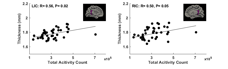
Significant correlations (FDR-corrected P ≤ 0.05) between mother’s physical activity at <10 weeks of pregnancy and newborn’s cortical thickness in 4 brain regions were identified (Figure 1). Specifically, mother’s average daily total activity count positively correlated with newborn’s cortical thickness in the left caudal middle frontal gyrus (R = 0.48, P = 0.04), right medial orbital frontal gyrus (R = 0.48, P = 0.04), and right transverse temporal gyrus (R = 0.48, P = 0.04); mother’s average daily time spent in moderate activity mode positively correlated with newborn’s cortical thickness in the right transverse temporal gyrus (R = 0.53, P = 0.03). Furthermore, significant correlations between mother’s physical activity at ~24 weeks of pregnancy and newborn’s cortical thickness in 2 brain regions were identified (Figure 2). Specifically, mother’s average daily total activity count positively correlated with newborn’s cortical thickness in the left isthmus cingulate gyrus (R = 0.56, P = 0.02) and right isthmus cingulate gyrus (R = 0.50, P = 0.05).CONCLUSIONS
Our results show that higher maternal physical activity level during uncomplicated pregnancy is associated with greater neonatal brain cortical thickness, and indicate that physical activity during pregnancy may be beneficial for offspring brain development.Acknowledgements
This project was supported by NIH 1R01HD099099 and USDA-ARS 6026-51000-012-06S.References
No reference found.Figures