4818
Low-field Magnetic Resonance Imaging with MaRCoS1Oxford Ionics, Oxford, United Kingdom, 2University of Applied Sciences and Arts Dortmund, Dortmund, Germany, 3Department of Radiology, Leiden University Medical Centre, Leiden, Netherlands, 4MRILab, Institute for Molecular Imaging and Instrumentation (i3M), Spanish National Research Council (CSIC), Valencia, Spain, 5MRILab, Institute for Molecular Imaging and Instrumentation (i3M), Universitat Politècnica de València (UPV), Valencia, Spain, 6Tesoro Imaging S.L., Valencia, Spain, 7Department of Electronic Engineering, Universidad de Valencia, Valencia, Spain, 8Massachusetts General Hospital, A. A. Martinos Center for Biomedical Imaging, Charlestown, MA, United States, 9Physikalisch-Technische Bundesanstalt (PTB), Braunschweig and Berlin, Germany, 10Department of Biomagnetical Resonance, Otto-von-Guericke University Magdeburg, Forschungscampus STIMULATE, Magdeburg, Germany, 11Q Bio Inc., San Carlos, CA, United States
Synopsis
In recent years, low-field scanners have proven their potential for clinical, preclinical, research and teaching applications. However, a critical milestone is yet to be achieved on the path to affordable, high-performance MRI systems: the availability of an inexpensive, versatile electronic control system (MR console), competitive with commercial spectrometers, but without dramatically increasing the overall scanner cost. In this work we show the status of MaRCoS (MAgnetic Resonance COntrol System), an open-source MR console and software package with powerful features, that offers higher versatility than many commercial spectrometers, and show its use in several MR setups, including in vivo acquisitions.
Introduction
The interest in low-cost MRI is presently reigniting, stimulated by the generalized lack of MRI accessibility in emergency rooms, points of care, medicalized vehicles or developing regions [1-3]. In recent years, low-field scanners have proven their potential for clinical, preclinical, research and teaching applications [4-9]. However, a critical milestone is yet to be achieved on the path to affordable, high-performance MRI systems: the availability of an inexpensive, versatile electronic control system (MR console), competitive with commercial spectrometers, but without dramatically increasing the overall scanner cost.Based on a Red Pitaya SDRLab ([10], $600), OCRA (Open-source Console for Real-time Acquisition, [11]) was a first decided step in this direction. In this work we show the status of MaRCoS (MAgnetic Resonance COntrol System, [12-14]), an open-source MR console and software package with powerful features, that offers higher versatility than many commercial spectrometers, and show its use in several MR setups.
Methods
Figure 1 shows the MaRCoS stack, an evolution of OCRA, upgraded to improve its features. The SDRLab FPGA (Field Programmable Gate Array) runs the flocra firmware. Compared to OCRA, flocra uses a 'FLow-based' structure with two RF transmit and two receive channels with independent frequencies, phases, amplitudes, downmixing parameters and sampling data rates. It also provides four gradient outputs and six TTL I/Os, with a timing resolution of 8.13 ns. The shortest time between RF, TTL or receiver events is 33 ns, and ~1 us for gradients. Sequence length, timing and complexity have fewer restrictions than before, e.g. there are no raster clocks, and sequences with hundreds of coherent TRs, soft pulses, and arbitrary gradient waveforms have been tested.The SDRLab is controlled using Python libraries. Sequences can be written using i) Pulseq [17], a hardware-agnostic pulse sequence prototyping framework, ii) a simple domain-specific language, or iii) sets of time/value Numpy arrays. A hardware simulator and a test suite greatly simplify new feature development.
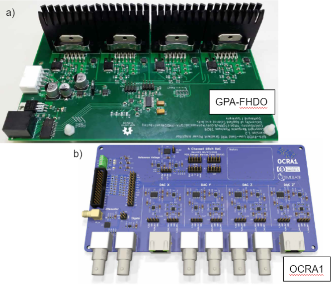
MaRCoS has been tested with two gradient boards: a) the GPA-FHDO board, a power gradient amplifier board that can directly drive gradient coils [15], and b) the OCRA1 board, a DAC board that connects to any gradient power amplifier with analogue inputs [16].
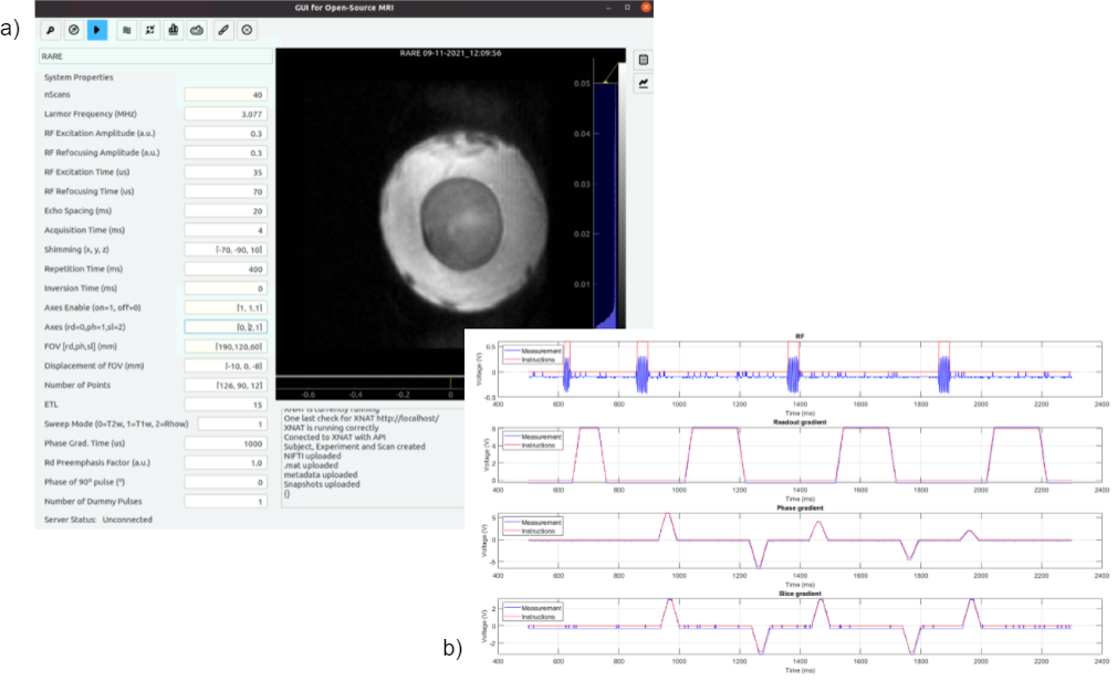
The Graphical User Interface of MaRCoS (GUI, Fig. 3) is programmed in Python using the pyQt5 package. The GUI comes with pre-installed standard sequences (turbo spin echo, gradient echo, etc.), and arbitrary sequences can be easily incorporated. Multiple calibration functions are also readily available, including Larmor frequency search, T1 time determination through inversion recovery, Rabi frequency and pulse length calibrations, etc. To make the most of MaRCoS-powered scanners, the GUI allows remote control and we have developed a batch system for running queued jobs sequentially and automatically, enabling continuous operation. Besides, the GUI is connected to XNAT [18], an open-source imaging platform for importing, archiving, processing and securely distributing imaging.
Results
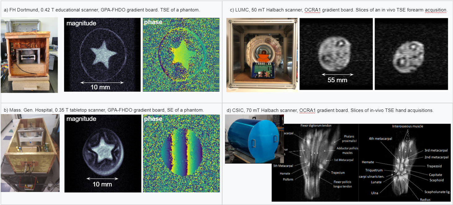
Figure 4 shows images acquired at four sites using MaRCoS, demonstrating the capabilities of the system for diverse and high-quality imaging, with magnets ranging from 0.05 to 0.42 T and fields of view from 10 to > 200 mm.Figure 4a shows the 0.42 T educational scanner at FH Dortmund, with the GPA-FHDO gradient board (Fig. 2a), and the reconstruction of a phantom imaged with a TSE pulse sequence. The echo train length (ETL) is 8, with an echo spacing (ESP) of 8 ms, and a total acquisition time (TACQ) of 80 s for a voxel resolution (RES) of 75 um x 117 um x 1 cm.
Figure 4b shows the 0.35 T tabletop scanner at Massachusetts General Hospital, also with the GPA-FHDO gradient board (Fig. 2a), and the reconstruction of a phantom imaged with a SE pulse sequence. Here, TACQ = 190 s for RES = 156 um x 156 um.
Figure 4c shows the 0.05 T scanner at Leiden University Medical Center, with the OCRA1 gradient board (Fig. 2b), and a slice from an in vivo acquisition of a volunteer's forearm imaged with a TSE pulse sequence.
Figure 4d shows the 0.07 T scanner at the Spanish National Research Council, also with the OCRA1 gradient board (Fig. 2b), and slices from in vivo acquisitions of two volunteers' hands and wrists imaged with TSE pulse sequences. For the middle image, ETL = 15, TR = 400 ms, ESP = 20 ms, TACQ = 600 s and RES = 2 mm x 1.5 mm x 10 mm. For the right image, ETL = 15, TR = 500 ms, ESP = 20 ms, TACQ = 600 s and RES = 1 mm x 1.17 mm x 5mm.
Conclusion
Encouraged by its demonstrated performance, wide applicability to low field MR and accessible documentation, the expanding community of developers continues to further the capabilities of MaRCoS [14]. Amongst others, future developments aim to interconnect multiple RedPitaya boards to increase the number of input and output ports and processing power of the system. MaRCoS already provides much of the functionality of modern MR consoles at a fraction of the cost and facilitates customization as compared to proprietary systems.Acknowledgements
We acknowledge the generous contributions of Suma Anand, Hengjie Liu and Ivan Fomin.
References
[1] Marques, José P., Frank F.J. Simonis, and Andrew G. Webb, ‘Low‐field MRI: An MR Physics Perspective’, Journal of Magnetic Resonance Imaging, 49 (2019), 1528–42
[2] Sarracanie, Mathieu, and Najat Salameh, ‘Low-Field MRI: How Low Can We Go? A Fresh View on an Old Debate’, Frontiers in Physics, 8 (2020), 172
[3] https://www.opensourceimaging.org/
[4] Sarracanie, Mathieu, Cristen D. LaPierre, Najat Salameh, David E. J. Waddington, Thomas Witzel, and Matthew S. Rosen, ‘Low-Cost High-Performance MRI’, Scientific Reports, 5 (2015), 15177
[5] Sheth, Kevin N, Mercy H Mazurek, Matthew M Yuen, Bradley A Cahn, Jill T Shah, Adrienne Ward, and others, ‘Assessment of Brain Injury Using Portable, Low-Field Magnetic Resonance Imaging at the Bedside of Critically Ill Patients’, JAMA Neurology, 78 (2021), 41–47
[6] O’Reilly, Thomas, Wouter M. Teeuwisse, Danny Gans, Kirsten Koolstra, and Andrew G. Webb, ‘In Vivo 3D Brain and Extremity MRI at 50 mT Using a Permanent Magnet Halbach Array’, Magnetic Resonance in Medicine, 2020, mrm.28396
[7] Cooley, Clarissa Z., Patrick C. McDaniel, Jason P. Stockmann, Sai Abitha Srinivas, Stephen Cauley, Monika Sliwiak, and others, ‘A Portable Brain MRI Scanner for Underserved Settings and Point-Of-Care Imaging’, 2020
[8] Algarín, José M, Elena Díaz-Caballero, José Borreguero, Fernando Galve, Daniel Grau-Ruiz, Juan P Rigla, and others, ‘Simultaneous Imaging of Hard and Soft Biological Tissues in a Low-Field Dental MRI Scanner’, Scientific Reports, 10 (2020), 21470
[9] Cooley, Clarissa Zimmerman, Jason P. Stockmann, Thomas Witzel, Cris LaPierre, Azma Mareyam, Feng Jia, and others, ‘Design and Implementation of a Low-Cost, Tabletop MRI Scanner for Education and Research Prototyping’, Journal of Magnetic Resonance, 310 (2020), 106625
[10] https://redpitaya.com/
[11] https://zeugmatographix.org/ocra
[12] ISMRM 2021, Abstract Nr. 0748
[13] ESMRMB 2021, Abstract Nr. S5.P15
[14] https://github.com/vnegnev/marcos_extras/wiki
[15] https://github.com/menkueclab/gpa-fhdo
[16] https://zeugmatographix.org/ocra/2020/11/27/ocra1-spi-controlled-4-channel-18bit-dac-and-rf-attenutator/
[17] Layton, Kelvin J., Stefan Kroboth, Feng Jia, Sebastian Littin, Huijun Yu, Jochen Leupold, and others, ‘Pulseq: A Rapid and Hardware-Independent Pulse Sequence Prototyping Framework’, Magnetic Resonance in Medicine, 77 (2017), 1544–52
[18] https://www.xnat.org/
Figures