4780
Neural style transfer: Applications in cardiac MR image registration1School of Medicine, University of Virginia, Charlottesville, VA, United States, 2Biomedical Engineering, University of Virginia, Charlottesville, VA, United States, 3Department of Medicine and Radiology, Stanford University, Stanford, CA, United States
Synopsis
Contrast-enhanced cardiac magnetic resonance (CMR) stress perfusion imaging shows excellent utility in evaluating coronary artery disease1. Registering perfusion CMR image series is difficult due to the varying image contrast. Neural style transfer is a deep learning method used to transfer the “style” of one domain to another while preserving the content. Two neural style transfer networks were implemented in Python using TensorFlow and PyTorch. Training of each network was done using three, slice matched patient profiles and cine-like perfusion images were generated and registered. This method is compared to a KL-transform based registration approach.
Introduction
Contrast-enhanced cardiac magnetic resonance (CMR) stress perfusion imaging shows excellent diagnostic and prognostic utility in evaluating coronary artery disease1. However, respiratory motion remains a challenge in achieving optimal perfusion quantification. Registration of CMR perfusion data is challenging due to the large contrast variations in the temporal series. Previous registration methods have relied on computationally expensive calculations of eigen-images using principal component analysis (PCA), either in a bulk or iterative fashion2,3. Neural style transfer is a deep learning method used to transfer the “style” of one domain to the target image, while preserving the overall content of the target image. Rather than use a traditional PCA approach to generate eigen-images that have temporally unvarying contrast, which requires large computational time, a neural style transfer network can be trained to apply the more-desirable style of another CMR sequence to that of perfusion series. In our case, the unvarying contrast of cine can be applied to a perfusion image series to obtain flattened contrast while preserving the content of the perfusion series. This method has the potential for widespread application to other CMR problems.Methods
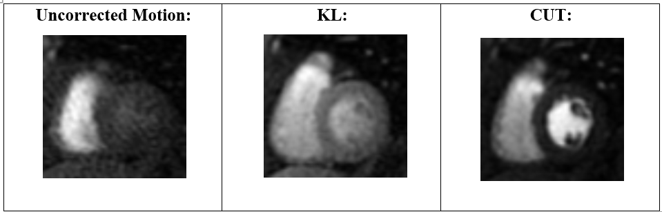
Training of neural style transfer networks:To achieve Cine-like contrast for a perfusion series, two style transfer neural networks were implemented in Python using TensorFlow and PyTorch and compared. These style transfer neural networks are from the Berkeley Artificial Intelligence Research Lab and named Cycle Generative Adversarial Network (CycleGAN) and Contrastive Unpaired Image-to-Image Translation (CUT)4,5. CycleGAN is a traditional generative adversarial network that adds a cycle loss, enforcing reciprocal domain style emulation, while preserving the content of the original image. CUT is a completely different approach than that of CycleGAN. CUT uses an InfoNCE loss to maximize mutual information between corresponding input and output patches, while drawing upon other patches in the image as contrastive negatives. CUT was implemented using PyTorch and CycleGAN was implemented using TensorFlow. Training of each network was done using three, slice matched patient profiles. Total training time was 3.5 hours for CUT and 12 hours for CycleGAN. Based on initial experiments, CUT preserved the content of the perfusion image while applying the style of cine in a higher degree than that of CycleGAN (Figure 1). Because of this, the trained CUT network was chosen as the model to apply to a non-motion corrected image series.
Generation of ‘pseudo-cine’ perfusion series and registration algorithm:
Five non-motion corrected perfusion series from healthy volunteers were used as input series into the trained CUT network. Generation of a perfusion in the style of cine (pseudo-cine) image series took only ~10 seconds for each patient. A mid-temporal frame was selected as a reference frame, and the pseudo-cine image series was registered pairwise to this reference frame using non-rigid registration. The deformation fields generated in this step were then applied pairwise to the original perfusion series for each corresponding temporal frame. This gave the resultant registered perfusion series (Figure 2).
Comparison to iterative KL-transform based registration:
The KL-transform based registration technique was implemented in MATLAB. This registration technique was compared to that of the style transfer registration technique2. The registration quality was rated by a cardiologist blinded to the registration technique (5=excellent, 1=poor).
Results
The CUT registration algorithm shows promising motion correction comparable to that of the KL-transform based approach (Figures 3, 5). Clinical grading of motion correction demonstrated scores of 4.2 and 4.1 for the KL technique and CUT techniques respectively (0.72). Figure 4 shows temporal profiles from the unregistered, KL and CUT techniques. The CUT technique performed well both for abrupt breathing motion (patient 1) and free-breathing sinusoidal motion (patient 2). The CUT technique showed less drift of the registration than the KL-based technique.Discussion and Conclusion
The CUT registration approach shows promising results for motion correction. Registration of perfusion images is inherently difficult due to a dynamic contrast range, but the CUT approach alleviates this by generating a flattened contrast image series using neural style transfer. It has minimal computational cost and only requires training a single neural network for fast generation of pseudo-cine series for any perfusion input. This method has the potential for widespread application to other CMR registration and segmentation problems.Acknowledgements
The primary author would like to thank Nick Tustison for his advice and guidance on using the Advanced Normalization Tools package for non-rigid image registration.References
1. T. Makela et al., "A review of cardiac image registration methods," in IEEE Transactions on Medical Imaging, vol. 21, no. 9, pp. 1011-1021, Sept. 2002, doi: 10.1109/TMI.2002.804441.
2. Xue, H, Brown, LAE, Nielles-Vallespin, S, Plein, S, Kellman, P. Automatic in-line quantitative myocardial perfusion mapping: Processing algorithm and implementation. Magn Reson Med. 2020; 83: 712– 730. https://doi.org/10.1002/mrm.27954
3. C. Scannell, A. Villa, J. Lee, M. Breeuwer, and A. Chiribiri, “Robust Non-Rigid Motion Compensation of Free-Breathing Myocardial Perfusion MRI Data,” IEEE Transactions on Medical Imaging, vol. 38, no. 8, pp. 1812–1820, Aug. 2019.
4. J.-Y. Zhu, T. Park, P. Isola, and A. Efros, “Unpaired Image-to-Image Translation using Cycle-Consistent Adversarial Networks,” arXiv:1703.10593v7(cs), 2017.
5. T. Park, A. Efros, R. Zhang, and J.-Y. Zhu, “Contrastive Learning for Unpaired Iamge-to-Image Translation,” arXiv:2007.15651(s), 2020.
6. Tustison, N.J., Cook, P.A., Holbrook, A.J. et al. The ANTsX ecosystem for quantitative biological and medical imaging. Sci Rep 11, 9068 (2021). https://doi.org/10.1038/s41598-021-87564-6
Figures