4766
Effect of Meditation on Brain Function During an Attention Task Using ASL and BOLD fMRI
Yakun Zhang1, Shichun Chen1, Zongpai Zhang1, Wenna Duan1, Li Zhao2, George Weinschenk1, Wen-Ming Luh3, Adam Anderson4, and Weiying Dai1
1Department of Computer Science, State University of New York at Binghamton, Binghamton, NY, United States, 2College of Biomedical Engineering & Instrument Science, Zhejiang University, Hangzhou, China, 3National Institute on Aging, National Institutes of Health, Baltimore, MD, United States, 4Department of Human Development, Cornell University, Ithaca, NY, United States
1Department of Computer Science, State University of New York at Binghamton, Binghamton, NY, United States, 2College of Biomedical Engineering & Instrument Science, Zhejiang University, Hangzhou, China, 3National Institute on Aging, National Institutes of Health, Baltimore, MD, United States, 4Department of Human Development, Cornell University, Ithaca, NY, United States
Synopsis
The effect of meditation on brain functional activation when engaged in an attention task was evaluated longitudinally using dASL and BOLD fMRI in nine healthy subjects. Functional activation before and after meditation practice was compared and the change of functional activation was correlated with practice time. Using dASL, functional activation in the occipital region was significantly reduced; more practice time was associated with more reduced activation in the mediofrontal, temporal, and precuneus regions. Using BOLD fMRI, no significant activation was found. The findings suggest that dASL has superior performance in detecting task performance and that meditation can improve brain efficiency.
Introduction
Focused attention meditation (FAM) has been shown to impact attention 1-3. Blood oxygenation level dependent (BOLD) fMRI reported altered brain activation during an attention task in experienced meditators or novice meditators after a brief and short-term meditation training 2,4. BOLD fMRI signals contain contributions from both vascular supply and neural oxygenation consumption 5, and physiological noises represent an important confound in BOLD fMRI studies 6. By contrast, dynamic arterial spin labeling (dASL) with markedly suppressed tissue signals can be used to understand the vascular contribution with minimal confounds from physiological noises 7. We used dASL and BOLD fMRI to evaluate longitudinal effects of 2-month meditation on novice meditators when engaged in an attention task.Methods
Nine young healthy college students (age: 19.11 ± 0.60 years old, 4 females) participated in the study. Each subject was scanned twice, before and after a two-month meditation training. During each scanning session, each subject was scanned by dynamic pseudo-continuous arterial spin labeling (PCASL, labeling duration = 2s, postlabeling delay = 1.8s, TR =5s) and BOLD fMRI (TR = 2s) with the visual oddball task for measuring the brain activation. Visual stimuli, ‘X’ and ‘O’ were randomly presented in the center of the screen with a duration of 500ms. The inter-stimulus interval (ISI) was ranging from 3.5s to 4.5s. Subjects were instructed to press the left button for ‘X’ and the right button for ‘O’. During the remaining time (3s to 4s), subjects were asked to focus on the fixation point. Each session consisted of 100 trials in total with 20% of ‘X’ (target) and 80% of ‘O’ (standard).ASL and BOLD images were processed with motion correction and normalization to the standard MNI space using a reference image as intermediate. The individual activation maps were produced using a SPM12 general linear model with the onset times of ‘X’ and ‘O’ and their temporal derivatives as regressors. Images from individual contrasts, ‘X’ and ‘X-O’, were generated for each subject and each condition (baseline or follow-up). The individual contrast images were compared between baseline and follow-up using a paired-t test. For each contrast, the multiple regression model was used to detect the potential association between meditation practice time and the activation changes with gender as a covariate. The statistical maps were thresholded using a voxel-level p value of 0.005. A cluster-level p value of 0.05 was used to correct for multiple comparisons.
Results
Response time to ‘X’ and ‘O’ was significantly shorter (baseline: 387.71 ± 19.61ms, follow-up: 365.84 ± 47.12ms, p = 0.032). Using dASL fMRI, significant reduced activation (voxel-level p value of 0.005) was found after two-month meditation training in the occipital region for contrast ‘X’ (Fig. 1A). Three clusters were found to exhibit significantly negative association between practice time and the changes of activation, mainly in the precuneus (Fig. 1B), superior/middle temporal (Fig. 1C), and superior medial frontal (Fig. 1D) regions for contrast ‘X’. We also found a significantly negative association between practice time and the changes of activation in the temporal and parietal regions for contrast ‘X-O’ (Fig. 1E). Table 1 summarizes the statistics of these significant clusters. Fig. 2 plots the relationship between the changes of average activation in the clusters and meditation practice time. Using BOLD fMRI, no significant activation and association with meditation practice time were found with the same significance threshold. The results of BOLD fMRI were reported in a preliminary format 8 with reduced significance threshold (voxel level significance of 0.01/0.02).Discussion
Following 2-month meditation practice, novice meditators had reduced activation in the occipital region. The reduced activation in the occipital region indicates that less effort is required after meditation training to maintain the visual focus when performing the attention task. We also observed negative association between changes of activation amplitudes and meditation practice time in the precuneus, temporal/insular, and mediofrontal/anterior cingulate cortex regions. These results are in a good agreement with the decreased activation during the Stroop Word-Color Task (SWCT, requiring attention control) in these regions in experienced meditators compared to nonmeditators 2 and novice meditators after a 7-day meditation retreat 4. The findings suggest that meditation improves brain efficiency with less functional activation in attention-related regions to achieve an improved task performance. The observed meditation effects on brain activation during an attention task using dASL are most significant than using BOLD fMRI, indicating more sensitive detection of dASL on vascular contribution of task activation. This study provides further support of short-term meditation as a potentially beneficial method for efficient attention control.Acknowledgements
No acknowledgement found.References
- Goldin P, Ziv M, Jazaieri H, Hahn K, Gross JJ. MBSR vs aerobic exercise in social anxiety: fMRI of emotion regulation of negative self-beliefs. Soc Cogn Affect Neurosci. 2013;8(1):65-72. Epub 2012/05/16. doi: 10.1093/scan/nss054. PubMed PMID: 22586252; PMCID: PMC3541489.
- Kozasa EH, Sato JR, Lacerda SS, Barreiros MA, Radvany J, Russell TA, Sanches LG, Mello LE, Amaro E, Jr. Meditation training increases brain efficiency in an attention task. Neuroimage. 2012;59(1):745-9. Epub 2011/07/19. doi: 10.1016/j.neuroimage.2011.06.088. PubMed PMID: 21763432.
- Pagnoni G. Dynamical properties of BOLD activity from the ventral posteromedial cortex associated with meditation and attentional skills. J Neurosci. 2012;32(15):5242-9. Epub 2012/04/13. doi: 10.1523/JNEUROSCI.4135-11.2012. PubMed PMID: 22496570; PMCID: PMC3362741.
- Kozasa EH, Balardin JB, Sato JR, Chaim KT, Lacerda SS, Radvany J, Mello L, Amaro E, Jr. Effects of a 7-Day Meditation Retreat on the Brain Function of Meditators and Non-Meditators During an Attention Task. Front Hum Neurosci. 2018;12:222. Epub 2018/06/27. doi: 10.3389/fnhum.2018.00222. PubMed PMID: 29942255; PMCID: PMC6004402.
- Ances BM, Leontiev O, Perthen JE, Liang C, Lansing AE, Buxton RB. Regional differences in the coupling of cerebral blood flow and oxygen metabolism changes in response to activation: implications for BOLD-fMRI. Neuroimage. 2008;39(4):1510-21. Epub 2008/01/01. doi: 10.1016/j.neuroimage.2007.11.015. PubMed PMID: 18164629; PMCID: PMC2819080.
- Birn RM, Diamond JB, Smith MA, Bandettini PA. Separating respiratory-variation-related fluctuations from neuronal-activity-related fluctuations in fMRI. Neuroimage. 2006;31(4):1536-48. Epub 2006/04/25. doi: 10.1016/j.neuroimage.2006.02.048. PubMed PMID: 16632379.
- Zhao L, Alsop DC, Detre JA, Dai W. Global Fluctuations of Cerebral Blood Flow Indicate a Global Brain Network Independent of Systemic Factors. J Cereb Blood Flow Metab. 2019;39(2):302-12.
- Liu Y, Duan W, Zhang Z, Weinschenk G, Luh W, Dai W. Effect of meditation on brain function activated by an attention task. The 27th ISMRM Proceedings of Annual Meeting. 2019;27:3903.
Figures
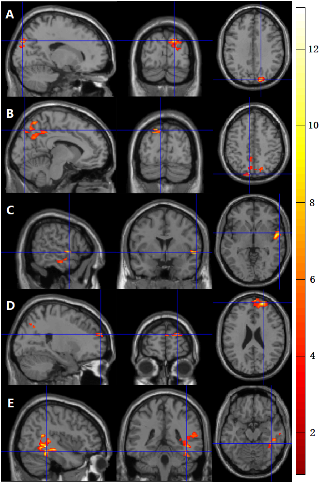
Figure 1. Significantly reduced functional activation was observed in
(A) the occipital region for the contrast of ‘X’; and more practice time was
associated with more reduced functional activation in the (B) precuneus, (C)
temporal/insular, and (D) mediofrontal regions for the contrast of ‘X’, and in
(E) the temporal for the contrast of ‘X-O’.
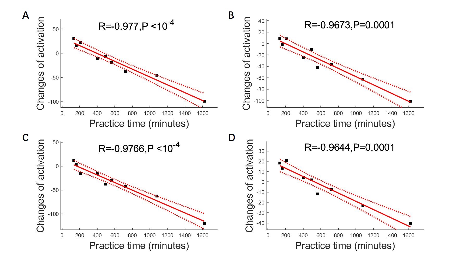
Figure 2.
Relationship between the changes of functional activation (after a 2-month
meditation training) and meditation practice time. Meditation practice time was
significantly correlated with the changes of functional activation in the
clusters of (A) precuneus, (B) temporal/insular, and (C) mediofrontal regions
for the contrast ‘X’, and in the cluster of (D) temporal region for the
contrast ‘X-O’.
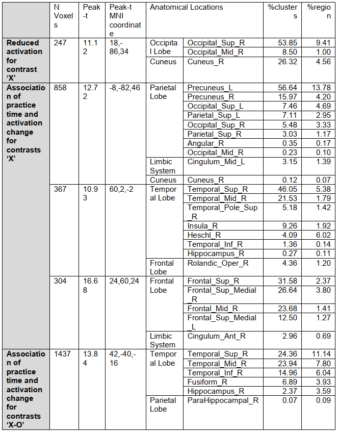
Table 1. Clusters showing significant changes
in functional activation following meditation practice and association between
meditation practice time and longitudinal changes of functional activation.
DOI: https://doi.org/10.58530/2022/4766