4747
Evaluation of k-space uniformity and blade number-optimized (K-B-optimized) binning method for abdominal 4D-DW-Propeller-EPI1Department of Diagnostic Radiology, University of Hong Kong, Hong Kong, China, 2Department of Health Technology and Informatics, The Hong Kong Polytechnic University, Hong Kong, China, 3Department of Biomedical Engineering, Chinese University of Hong Kong, Hong Kong, China
Synopsis
Four-dimensional diffusion-weighted imaging (4D-DWI) attracts lots of attention in MRI-guided radiotherapy since it can provide functional information and better delineate the tumor. However, the previously reported 4D-DW-Propeller-EPI technique used amplitude data binning with continuous interval to sort the blade data that might lead to non-uniform distribution of k-space sampling, therefore being suboptimal for subsequent Propeller-EPI reconstruction. Thus, we propose a new (K-B-optimized) binning method to consider the requirement of propeller reconstruction during the sorting of blade data, thereby improving the k-space distribution. Simulation and in-vivo studies demonstrated that the new binning method can improve the data quality of 4D-DW-Propeller-EPI.
Introduction
Four-dimensional magnetic resonance imaging (4D-MRI) is a promising technique due to its non-invasive property, high contrast in soft tissue1-3. Among the developed 4D-MRI techniques, four-dimensional diffusion-weighted imaging (4D-DWI) attracts lots of attention since it can provide functional information and better delineate the tumor 4,5. Yilin Liu et al. first demonstrated the feasibility of 4D-DWI using single-shot echo-planar imaging technique (4D-DW-ss-EPI), which can only provide limited data quality due to ssEPI-related artifacts.6 To enable high-quality 4D-DWI, our previous study proposed to use Propeller-EPI technique with golden angle for 4D-DWI data acquisition (4D-DW-Propeller-EPI).7 However, using amplitude data binning with continuous interval to sort the blade data can sometimes lead to non-uniform distribution of k-space sampling that is suboptimal for subsequent Propeller-EPI reconstruction. In this study, we developed a new binning method by optimizing the distribution of k-space data and blade number (K-B-optimized binning) during data sorting for improving image quality and robustness of 4D-DW-Propeller-EPI.Methods
K-B-optimized binningFig.1A shows the framework of 4D-DW-Propeller-EPI using K-B-optimized binning method. Because propeller-EPI has intrinsic reduction of motion present in acquired data, amplitude ranges of each bin can be extended to search the data with i) wide interval and ii) overlapping with adjacent bins. Thus, more blade data can be associated to the same bin, thereby alleviating the data insufficiency caused by the variations of respiratory waveform. Furthermore, a relatively uniform distribution of blades in k-space is desired for final image reconstruction of each bin that can be achieved by selecting proper data from all blades falling into the same bin. An index can be developed to assess k-space uniformity as
$$K_{uniformity}=1-\frac{N_{blank}}{N_{full}} (1) $$
Where Nblank represents the number of blank areas without sampling data, Nfull represents the number of expected sampling areas.
Simulation study
Image production: 2D abdominal images and respiratory waveform with multiple phase bins were generated by 4D Digital Extended Cardiac-Torso (XCAT) phantom with matrix size = 128x128, respiratory period = 3, 4 and 5s, diaphragm motion = +/-20 mm and chest wall motion = +/-10.2, 10.8, 11.4 and 12 mm.
Binning setting: Amplitude intervals were set as 0~1.97, 0.13~4.15, 0.97~6.45, 2.78~8.11, 5.07~9.68, 7.21~10 for K-B-optimized binning, and 0~0.63, 0.63~2.32, 2.32~4.61, 4.61~6.91, 6.91~8.41, 8.41~10 for conventional binning. The angle between neighbor blades was set as 20° for K-B-optimized binning.
Evaluation: Effects of the number of acquisition repeated cycles (Nacq) and sampling rate (Fs, i.e., 1/TR) on 4D-DW-Propeller-EPI using K-B-optimized and conventional binning were investigated. One-way ANOVA and Tukey-Kramer correction for multiple comparison were used for statistical analysis.
In-vivo study
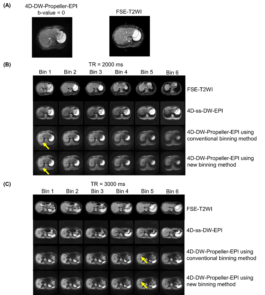
Image acquisition: 4D-DW-Propeller-EPI data were acquired from two healthy volunteers under free breathing with repeated acquisitions (Nacq=90 for each diffusion direction) at 1.5 T MRI (Explore, GE Healthcare). Two different data sampling rates were tested with (1) TR of 2000ms (maximum slice coverage=14; total scantime=720s), and (2) TR=3000ms (maximum slice coverage=21; total scantime=1080s). In addition, 4D-DW-ss-EPI data (TR=4000ms, slice coverage=35, total scantime=480s) and breath-holding FSE-T2 images (scantime=20s) were also acquired for comparison.
Respiratory signal recording: Respiratory belt transducer (25Hz) was used to record respiration during data acquisition.
Binning setting: Binning intervals were set as 1400~1900, 1700~2100, 2000~2500, 2200~2700, 2600~ 2900, 2800~3200 for K-B-optimized binning, and 1400~1700, 1700~2100, 2100~2300, 2300~2600, 2600~2900, 2900~3200 for conventional binning. The angle between neighbor blades was set as 20° for K-B-optimized binning.
Evaluation: Numbers of blades and k-space uniformity using K-B-optimized binning and conventional binning method were compared.
Results
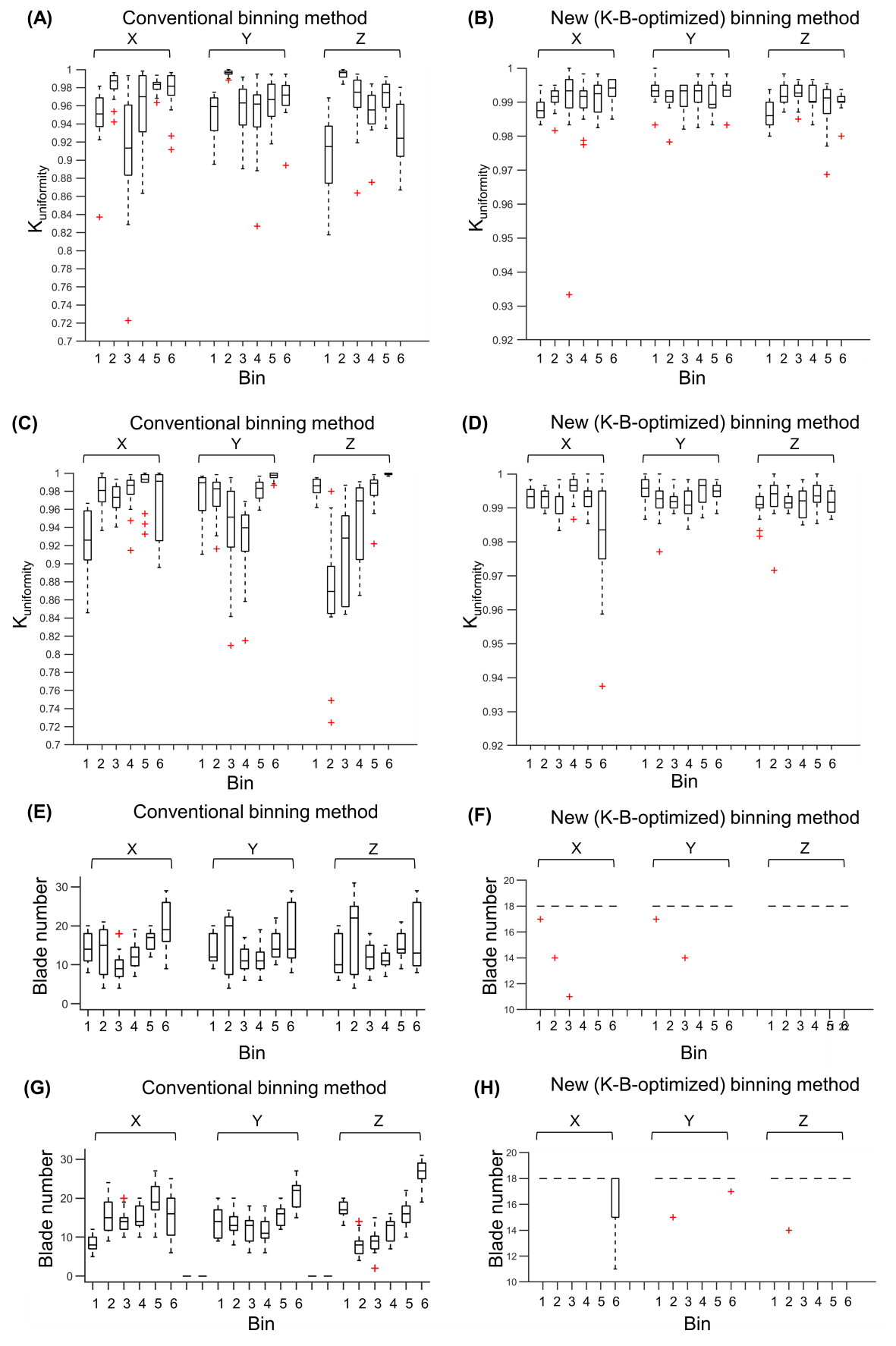
Fig.2 and Fig.3 show the assessment results of blade data distributions using K-B optimized binning and conventional binning associated with different sampling rate (Fs) and number of repeated acquisition (Nacq), respectively. Fig.4 shows the reconstructed images of from in-vivo 4D-DW-Propeller-EPI data acquired with two different sampling rates, using either K-B optimized binning or conventional binning method. Fig.5 shows the results of Kuniformity and blade number for in-vivo 4D-DW-Propeller-EPI data acquired with two different sampling rates, using either K-B optimized binning or conventional binning method.Discussion
Simulation study showed that the new proposed K-B-optimized binning method can help improve k-space uniformity of blades distribution after data sorting, thereby achieving better image quality for each bin data using Propeller-EPI reconstruction. Furthermore, the variations of blade number among different bins and different slice locations can also be reduced by using K-B-optimized binning. The tests for different TRs (Fig.2) and Nacqs(Fig.3) also demonstrated the similar improvement by using proposed K-B-optimized binning method, suggesting that K-B-optimized binning is highly robust for 4D-DW-Propeller-EPI technique. The performance was also verified with in-vivo experiments that shows good agreement with the simulation study. Compared with conventional binning, K-B-optimized binning method can improve in-vivo image quality (Fig.4B&C) of 4D-DWI-Propeller-EPI due to higher k space uniformity (Fig.5A-D) and with data sufficiency for each bins (Fig.5E-H). In addition, all images produced from 4D-DW-Propeller-EPI with K-B-optimized binning show high geometric fidelity (the comparison to FSE image in Fig.4A). To further decrease the acquisition time while maintaining high image quality, incorporating multi-band imaging8 or SCOPER9 reconstruction into 4D-DW-Propeller-EPI is desirable.Conclusion
In conclusion, the proposed binning method can improve the robustness of 4D-DW-Propeller-EPI, which may benefit the application of MRI-guided abdominal RT in future.Acknowledgements
The work was in part supported by grants from Hong Kong Research Grant Council (GRFs HKU17138616, HKU17121517 and HKU17106820) and Hong Kong Innovation and Technology Commission (ITS/403/18).
References
1. Cai, J.; Chang, Z.; Wang, Z.; Paul Segars, W.; Yin, F.-F. Four-Dimensional Magnetic Resonance Imaging (4D-MRI) Using Image-Based Respiratory Surrogate: A Feasibility Study. Medical physics 2011, 38 (12), 6384–6394.
2. Dinkel, J.; Hintze, C.; Tetzlaff, R.; Huber, P. E.; Herfarth, K.; Debus, J.; Kauczor, H. U.; Thieke, C. 4D-MRI Analysis of Lung Tumor Motion in Patients with Hemidiaphragmatic Paralysis. Radiotherapy and Oncology 2009, 91 (3), 449–454.
3. Liu, Y.; Yin, F.-F.; Chang, Z.; Czito, B. G.; Palta, M.; Bashir, M. R.; Qin, Y.; Cai, J. Investigation of Sagittal Image Acquisition for 4D-MRI with Body Area as Respiratory Surrogate. Medical physics 2014, 41 (10), 101902.
4. Hötker, A. M.; Mazaheri, Y.; Wibmer, A.; Zheng, J.; Moskowitz, C. S.; Tickoo, S. K.; Russo, P.; Hricak, H.; Akin, O. Use of DWI in the Differentiation of Renal Cortical Tumors. American Journal of Roentgenology 2016, 206 (1), 100–105.
5. Hayashida, Y.; Hirai, T.; Morishita, S.; Kitajima, M.; Murakami, R.; Korogi, Y.; Makino, K.; Nakamura, H.; Ikushima, I.; Yamura, M. Diffusion-Weighted Imaging of Metastatic Brain Tumors: Comparison with Histologic Type and Tumor Cellularity. American journal of neuroradiology 2006, 27 (7), 1419–1425.
6. Liu, Y.; Zhong, X.; Czito, B. G.; Palta, M.; Bashir, M. R.; Dale, B. M.; Yin, F.-F.; Cai, J. Four-Dimensional Diffusion-Weighted MR Imaging (4D-DWI): A Feasibility Study. Medical physics 2017, 44 (2), 397–406.
7. Lu Wang, Tian Li, Jing Cai, and Hing-Chiu Chang, Motion-Resolved Four-Dimensional Abdominal Diffusion-Weighted Imaging using Propeller Echo-Planar Imaging(4D-DW-Propeller-EPI), in International Society for Magnetic Resonance in Medicine Conference, Sydney, Australia, May 2021.
8. Cohen A D , Nencka A S , Lebel R M , et al. Multiband multi-echo imaging of simultaneous oxygenation and flow timeseries for resting state connectivity. Plos One, 2017, 12(3):e0169253.
9. Xiaoxi Liu, Di Cui, Erpeng Dai, Edward S. Hui, Queenie Chan, and Hing-Chiu Chang, Self-calibrated and Collaborative Propeller-EPI Reconstruction (SCOPER) for High-Quality Diffusion-Tensor Imaging, in International Society for Magnetic Resonance in Medicine Conference, Montréal, QC, Canada, May 2019.
Figures
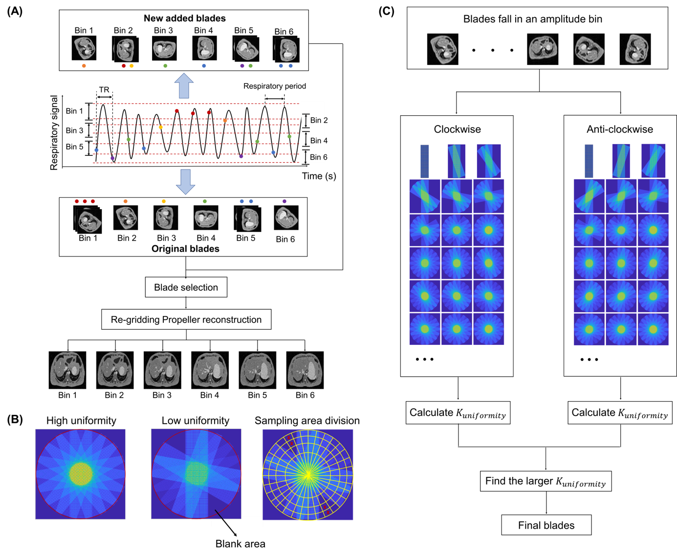
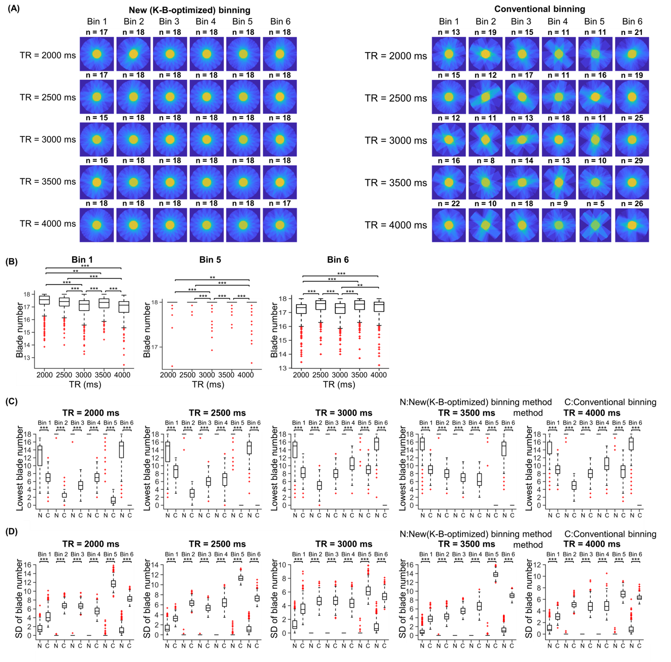
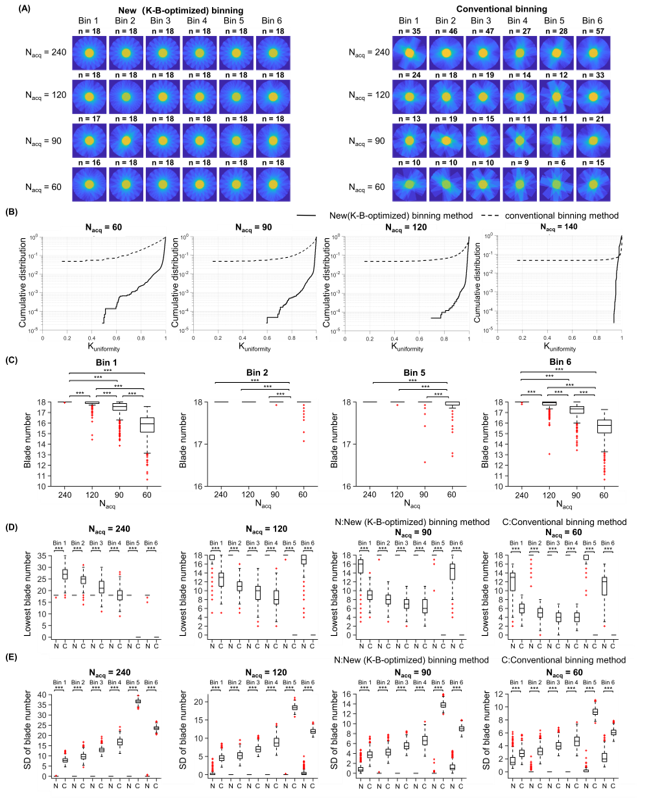
Figure 2. Results from 4D-DW-Propeller-EPI simulation experiments at different sampling rate (TR=2000, 2500, 3000, 3500, 4000ms) of 6 bins at Nacq= 90. (A) K-space data used for reconstruction of slice 1 using K-B-optimized and conventional method. (B) Statistical analysis of blade number using K-B-optimized method. Statistical analysis of lowest blade number (C) and standard variation of blade number (D) using K-B-optimized and conventional method. (:**: p<0.01, ***: p<0.001)