4742
Effects of Contrast Agents on Diffusion-Weighted Imaging and T2-Weighted Imaging for diagnosis of Breast tumor1Paul C. Lauterbur Research Center for Biomedical Imaging, Shenzhen Institute of Advanced Technology, Chinese Academy of Sciences, Shenzhen, China, 2Department of Radiology, The Second People’s Hospital of Yibin, Yibin, China, 3Department of Radiology,Pidu District People's Hospital, Chengdu, China, 4United Imaging Healthcare, Shanghai, China
Synopsis
Diffusion-Weighted Imaging (DWI) and T2-weighted imaging (T2WI) are two necessary sequences in breast MRI which of great significance for breast cancer. They are usually performed before contrast agent injection in the conventional breast MRI protocol. Recently, some studies have proposed simplified and fast breast scanning protocol, of which, DWI and T2WI are performed after contrast agent injection. This study evaluated the clinical value of DWI and T2WI after contrast agent in the diagnosis of breast cancer. The results showed that contrast injection has no effect on DWI, but it can improve the signal-to-noise ratio and contrast-to-noise ratio of T2WI.
Introduction
Breast cancer has the highest incidence rate among all tumors [1]. MRI is an important method for the diagnosis of breast cancer. Among them, Diffusion-Weighted Imaging (DWI) and T2-weighted imaging (T2WI) are two necessary sequences which of great significance for breast cancer [2-4]. In the conventional breast MRI examination protocol, DWI and T2WI are usually performed before intravenous injection of contrast agent for DCE-MRI. In order to shorten the breast tumor MRI acquisition time and hence improving the efficiency of breast examination, some studies have proposed simplified breast scanning protocol with ultra-fast DCE-MRI [5, 6]. In the simplified protocol, DWI and T2WI are performed after contrast agent injection. This study aims to investigate whether there is a significant difference of DWI and T2WI in the diagnosis of breast cancer between before and after contrast agent injection.Methods
The breast MRI images of 17 patients were acquired on a 3T MRI system (uMR 790, United Imaging Healthcare, Shanghai, China), including DWI and T2WI acquired before and after the contrast agent injection. The imaging parameters of DWI: TR/TE=3800.0ms/67.6ms, slice thickness: 5mm, slice number: 24, field of view: 200×360mm, spatial resolution: 98×176, b=50s/mm², b=800 s/mm²; and the dixon T2WI parameters: TR/TE=3700.0ms/75.6ms, slice thickness: 5mm, slice number: 24, field of view: 360×360mm, spatial resolution: 576×576. All the images were transferred to a dedicated workstation for analysis. The apparent diffusion coefficient (ADC) of DWI and the signal-to-noise ratio (SNR) and contrast-to-noise ratio (CNR) of T2WI were calculated for both images acquired from before and after the contrast agent as follows: SNR=SItumor/SDnoise , where SItumor is the signal intensity of the lesion, SDnoise is the standard deviation of the background noise; CNR=(SItumor-SInormal)/SDnoise, where SInormal is the signal intensity of the lesion’s adjacent normal gland tissue. The Wilcoxon signed-rank test was used to evaluate the significant difference of these calculated parameters between before and after the contrast agent. For the parameters with significant difference, further comparisons were performed according to the tumor types (invasive carcinoma, carcinoma in situ, and benign tumors). The Mann-Whitney U test was used to evaluate the diagnostic value of these parameters in differentiating tumor types.Results and Discussion
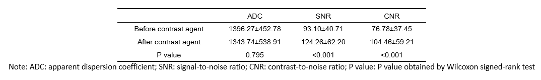
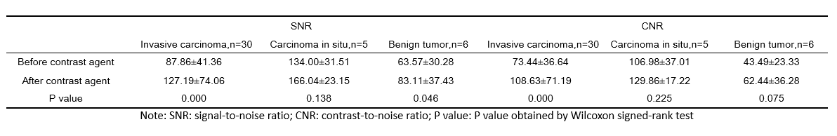
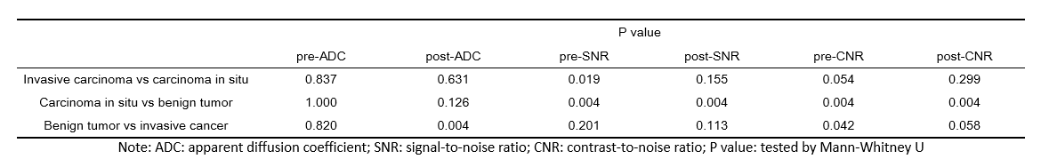
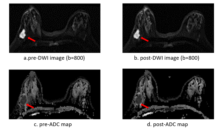
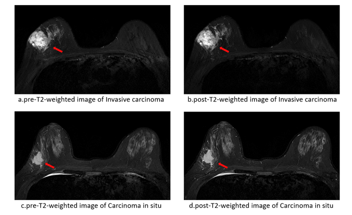
A total of 52 breast lesions were detected on the both DWI and T2WI images, including 30 invasive carcinoma, 5 carcinoma in situ, and 6 benign tumors. The ADC, SNR and CNR before and after contrast agent, and the corresponding comparison p values were summarized in Table 1. There was no significant difference in ADC value of breast lesions before and after contrast (P=0.795), indicating that the injection of contrast agent had no effect on DWI. However, both the SNR and CNR values of breast lesions on the T2WI image acquired after the contrast agent injection were significantly higher than those acquired before the contrast agent (P value<0.001). The SNR value measured after the contrast agent injection increased from 93.10±40.71 to 124.26± 62.20, and the CNR value increased from 76.78±37.45 to 104.46±59.21.Representative DWI images before and after contrast agent and corresponding ADC maps of a patient with Carcinoma in situ are shown in Figure 1. The comparison of the SNR and CNR of different types of tumors (invasive carcinoma, carcinoma in situ, and benign tumor lesions) before and after contrast agent injection were shown in Table 2. For carcinoma in situ, there is no significant difference of SNR and CNR between before and after contrast agent. For the other two types of tumors, the results showed the injection of contrast agent can improve the SNR and CNR of T2WI, and hence providing higher quality images for breast cancer diagnosis, which is conducive to clinical diagnosis. Figure 2 shows representative T2WI images before (pre) and after contrast agent (post) of invasive carcinoma and carcinoma in situ. The contrast of post-T2WI is visually better than pre-T2WI for invasive carcinoma. For carcinoma in situ, however, there is no visual difference of the lesion contrast in pre and post T2WI. The results of ADC, SNR and CNR used to distinguish different tumor types were shown in Table 3. Only the pre-contrast SNR can be used to differentiate the invasive carcinoma and carcinoma in situ. For distinguishing carcinoma-in-situ and benign tumors, both SNR and CNR before and after contrast agent were useful. Both post-contrast ADC and pre-contrast CNR could be used for identification of benign tumors and invasive carcinomas. This demonstrated that although the SNR and CNR increased after the contrast agent, it did not improve the ability to distinguish tumor types. This may also be due to the small sample size of carcinoma in situ and benign tumors in this study.Conclusion
Contrast injection has no effect on the acquisition of DWI images. It can improve the signal-to-noise ratio and contrast-to-noise ratio of T2WI images, and hence providing higher quality images for breast cancer diagnosis, which is conducive to clinical diagnosis.Acknowledgements
No acknowledgement found.References
[1]Turbow SD, White MC, Breslau ES, Sabatino SA. Mammography use and breast cancer incidence among older U.S. women. Breast Cancer Res Treat. 2021 Jul;188(1):307-316.
[2]Li Z, Li J, Lu X, Qu M, Tian J, Lei J. The diagnostic performance of diffusion-weighted imaging and dynamic contrast-enhanced magnetic resonance imaging in evaluating the pathological response of breast cancer to neoadjuvant chemotherapy: A meta-analysis. Eur J Radiol. 2021 Oct;143:109931.
[3]Sharma U, Agarwal K, Hari S, Mathur SR, Seenu V, Parshad R, Jagannathan NR. Role of diffusion weighted imaging and magnetic resonance spectroscopy in breast cancer patients with indeterminate dynamic contrast enhanced magnetic resonance imaging findings. Magn Reson Imaging. 2019 Sep;61:66-72.
[4]Yuen S, Monzawa S, Yanai S, Matsumoto H, Yata Y, Ichinose Y, Deai T, Hashimoto T, Tashiro T, Yamagami K. The association between MRI findings and breast cancer subtypes: focused on the combination patterns on diffusion-weighted and T2WI images. Breast Cancer. 2020 Sep;27(5):1029-1037.
[5]Kuhl CK. Abbreviated Magnetic Resonance Imaging (MRI) for Breast Cancer Screening: Rationale, Concept, and Transfer to Clinical Practice. Annu Rev Med. 2019 Jan 27;70:501-519.
[6]Weinstein SP, Korhonen K, Cirelli C, Schnall MD, McDonald ES, Pantel AR, Zuckerman S, Borthakur A, Conant EF. Abbreviated Breast Magnetic Resonance Imaging for Supplemental Screening of Women With Dense Breasts and Average Risk. J Clin Oncol. 2020 Nov 20;38(33):3874-3882.
Figures