4739
Differentiation of liver benign, primary malignant and secondary malignant tumors using MR DCE and mDIXON-Quant imaging1Department of Radiology, the First Affiliated Hospital of Dalian Medical University, Dalian, China, 2Philips Healthcare, Beijing, China
Synopsis
The quantitative MR dynamic contrast-enhanced (DCE) and mDIXON-Quant imaging were used in this study for differentiation between benign and malignant liver lesions. The combination of DCE and mDIXON-Quant showed significantly improved diagnostic efficacy between benign liver tumors and primary malignant tumors, as well as between primary and secondary malignant tumors.
Introduction
Hepatic hemangioma, hepatocellular carcinoma, cholangiocarcinoma and liver metastasis are common types of liver lesions, and the accurate diagnosis among them is important for treatment options. Several methods could be clinically used for differential diagnosis among these diseases including the laboratory examination, traditional computed tomography (CT), magnetic resonance imaging (MRI) and ultrasonography. But the accurate differential diagnosis still faces many challenges. MR dynamic contrast-enhanced (DCE) imaging is one of the main MRI perfusion techniques which calculates perfusion parameters by evaluating T1 shortening induced by a gadolinium-based contrast bolus passing through tissue, and it has been extensively explored for clinical diagnosis of tumors [1]. mDIXON Quant brings a fast and simple 3D procedure for non-invasive liver fat quantification by providing high quality 3D fat fraction (FF) maps of the whole liver, and the additional T2*/R2* maps can be provided for further diagnostic assessment [2]. The purpose of this study was to explore the value of MR dynamic contrast-enhanced (DCE) and mDIXON-Quant imaging in differentiation between benign and malignant liver lesions.Materials and Methods
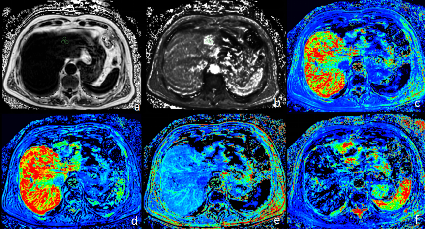
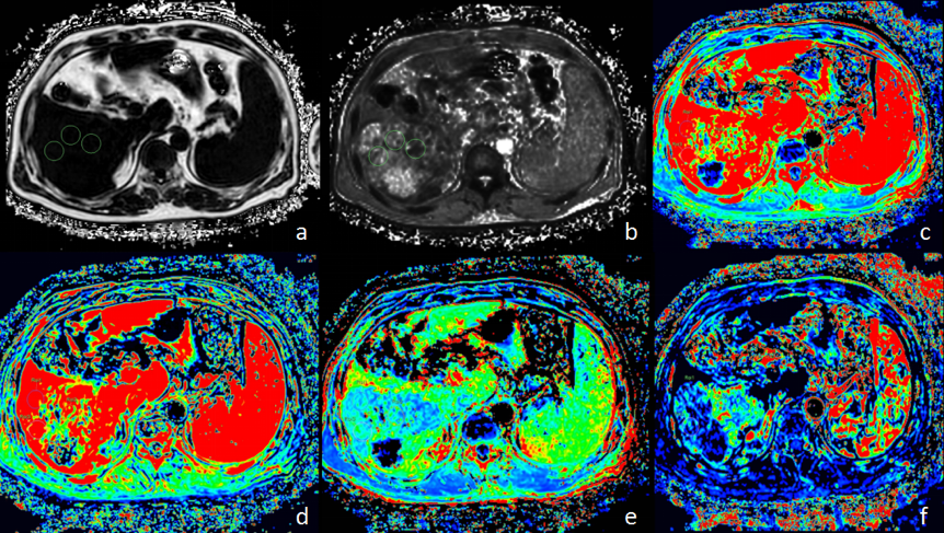
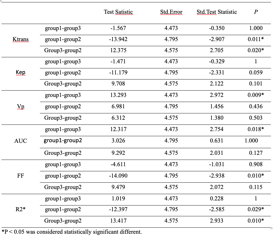
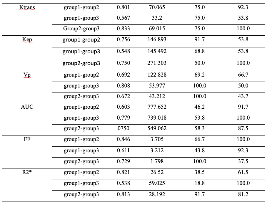
This study has been approved by the local IRB. 30 patients with 41 focal liver lesions in our hospital were retrospectively analyzed. According to the pathological results, three groups were divided as 10 patients with 13 hemangioma (6 male, mean age, 52 years; range, 30–75 years), 12 patients with hepatocellular carcinoma (HCC; 11 male, mean age, 59 years; range, 43–76 years), and 8 patients with hepatic metastasis (HM; 8 male, mean age, 58 years; range, 49–70 years). The MR protocol included DCE, mDIXON Quant and T2 weighted (T2WI) imaging sequences and detailed scan parameters is shown in Table 1. All patients performed the MR examination on a 3.0 T MR scanner (Ingenia CX, Philips Healthcare, the Netherlands). Referring to the anatomical location of lesion obtained on T2WI images, three circular regions of interest (ROI) with area of 100-200 mm2 were manually placed on the parenchyma tissue in the tumor on mDIXON Quant and DCE parameter images, respectively (Figures 1-2). Use Kruskal-Wallis test to compare whether there are statistical differences in DCE and mDIXON Quant parameters in benign liver lesions (group 1), primary malignant tumors (group 2) and secondary malignant tumors (group 3) with the SPSS 22.0 software (IBM, U.S.A). The receiver operating characteristic (ROC) curve was used to evaluated the diagnostic efficacy of DCE and mDIXON Quant parameters, as well as their combination, for differentiation between HCC and HM. Delong’s test was used to compare the area under the ROC curves (AUCs).Results
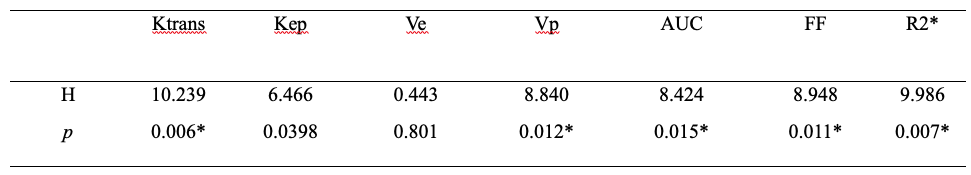
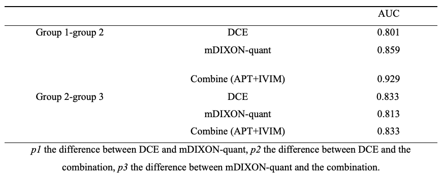
As shown in table 2 and 3, there is a difference in Ktrans, FF, R2* value between group 1 and group 2, Ktrans, R2* value between group 2 and group 3, and AUC value between group 1 and group 3. There is no statistical difference between other groups. As shown in table 4, the highest AUC is the FF value to discriminate group 1 and group 2. The combinational use of DCE and mDXION-quant imaging showed significantly improved differential diagnosis efficacy between benign and primary malignant tumors (Table 5).Discussion and Conclusion
Permeability parameters by DCE (Ktrans and AUC etc.) may reflect the hemodynamic changes of micro-vessels and characteristics of tumors from different perspectives [1]. FF calculated with the mDIXON Quant images can be used to assess fat content and has been proven sensitive to estimate hepatic triglyceride content [2]. In conclusion, the combinational use of DCE and mDIXON Quant showed significantly improved differential diagnosis efficacy among benign, primary and secondary malignant liver tumors, therefore, it may serve as a potential noninvasive method for clinical diagnosis of liver lesions.Acknowledgements
No acknowledgement found.References
[1] Li J, Xue F, Xu X, Wang Q, Zhang X. Dynamic contrast-enhanced MRI differentiates hepatocellular carcinoma from hepatic metastasis of rectal cancer by extracting pharmacokinetic parameters and radiomic features. Exp Ther Med. 2020;20(4):3643-3652.
[2] Chu C, Feng Q, Zhang H, Zhao S, Chen W, He J, Sun L, Zhou Z. Evaluation of salivary gland fat fraction values in patients with primary Sjögren's syndrome by mDIXON quant imaging: Initial findings. Eur J Radiol. 2020;123:108776.
Figures