4737
Time-reversed CENTRA-PLUS profile ordering (SULP-ARTNEC) for improvement of image contrast in contrast-enhanced brain metastasis screening1Radiology, Eastern Chiba Medical Center, Chiba, Japan, 2Diagnostic Radiology and Radiation Oncology, Graduate School of Medicine, Chiba University, Chiba, Japan, 3Philips Japan, Tokyo, Japan, 4Philips Healthcare, Best, Netherlands, 5Faculty of Health Sciences, Institute of Medical, Pharmaceutical and Health Sciences, Kanazawa University, Kanazawa, Japan, 6Orthopaedic Surgery, Eastern Chiba Medical Center, Chiba, Japan, 7General Medical Services, Graduate School of Medicine, Chiba University Graduate School of Medicine, Chiba, Japan
Synopsis
Shorter examination time might miss the appropriate timing for depicting the “delayed-enhanced” lesions, such as brain metastasis and multiple sclerosis plaques. We focused on the CENTRA-PLUS view-ordering framework and tried to modify the k-space profile ordering to enable completely timing-reversed (SLUP-ARTNEC). The purpose of this study is to demonstrate the feasibility of SULP-ARTNEC in the patients with brain metastasis. SLUP-ARTNEC is the unique approach in the post compressed sensing era that can improve the image contrast between delayed-enhanced brain metastasis lesions and background tissues in the contrast-enhanced studies without having the extra-waiting time, resulting in maintain the shorter examination time.
Purpose
Accelerated MRI using parallel imaging, compressed sensing or a combination of both (Compressed SENSE)1is widely used in routine clinical examination recently, and it could significantly shorten the total examination time even the contras-enhanced studies. Furthermore, accelerated MRI enables whole-brain 3D acquisition with clinically acceptable scan time. However, there would be a potential risk that such a shorter examination time might miss the appropriate timing for depicting the “delayed-enhanced” lesions, such as brain metastasis and multiple sclerosis plaques2,3. CENTRA-PLUS4 is an optimized centric k-space ordering which can be combined with fat-suppressed turbo field-echo (TFE) and keyhole imaging, it is typically used for abdominal dynamic contrast-enhanced studies to capture early timing after contrast injection. This is also utilized for free-breathing cardiac late-gadolinium enhancement MRI to minimize changes in the contrast kinetics due to washout over a potentially long 3D acquisition time on the order of several minutes5.We focused on the CENTRA-PLUS view-ordering framework and tried to modify the k-space profile ordering to enable completely timing-reversed. The central k-space part, most important part for image contrast determination, will be acquired at the almost last of acquisition. We hypothesized that the time-reversed CENTRA-PLUS (SLUP-ARTNEC) can improve the image contrast between delayed-enhanced lesions and background tissues in the contrast-enhanced studies without having the extra-waiting time, resulting in maintain the shorter examination time. The purpose of this study is to demonstrate the feasibility of SULP-ARTNEC in the patients with brain metastasis.Methods
Theory and pulse sequence:In 3D-TFE sequence, the fat suppression pulse is typically shared over a large number of TFE factors for efficient fat suppression while preventing prolongation of acquisition time. To that end, called low_high radial profile order is typically applied. In this order, from each Ky-Kz segment a profile is acquired per shot, starting with optimal fat suppression at the central Ky-Kz segment. Consequently, the acquisition of the image contrast determining central Ky-Kz profiles are distributed over the entire acquisition. This results in a non-distinct, time-averaged image contrast.In SULP-ARTNEC, k-space is basically segmented by two (central and peripheral) sectors in the Ky and Kz directions to efficiently obtain the effect of fat suppression pre-pulse. Actual data acquisition is started from the most peripheral k-space sector and then moves in toward centrically. After the complete filling of the peripheral sector, data acquisition is then continued toward central k-space with a centric profile-ordering. Actual k-space acquisition with each method is shown in Figure 1.
As aforementioned, low-high radial order could theoretically be affected by any timing of signals during the acquisition. Whereas, with SULP-ARTNEC, acquisition is started from the peripheral k-space sector and the most central k-space sector is acquired at a late stage. Thus, this may emphasize the increased signals of the brain metastasis due to delayed enhancement at the last of the imaging time (Figure 2).
Experiments: Eight patients with suspected brain metastasis underwent contrast-enhanced MRI on a 3.0T system (Ingenia CX, Philips Healthcare). The study was approved by the local IRB, and written informed consent was obtained from all subjects. We compared two types of 3D T1-TFE with conventional low_high radial order and SULP-ARTNEC. To evaluate the effect of the timing of contrast determination in k-space, each patient underwent three 3D TFE scans after contrast enhancement with either following two acquisition patterns.
Pattern A= Injection (immediately after)-> 1st Conventional-> SULP-ARTNEC-> 2nd Conventional
Pattern B= Injection (immediately after)-> 1st SULP-ARTNEC-> Conventional-> 2nd SULP-ARTNEC
The obtained images were evaluated visually and quantitatively using contrast ration (CR) of neoplastic lesions and cerebral white matter. The scan parameters were as follows: axial, voxel size=0.57x0.57x1.4 mm3 (recon =0.43x0.43x0.7 mm3), FOV=220x220 mm, C-SENSE factor=1.6, SPIR, TR/TE=6.0/2.9ms, and acquisition time=4:40.
Results and Discussion
Representative images of conventional 3D T1-TFE and SULP-ARTNEC without contrast-enhancement in original and reformatted orientations were shown in Fig.3. These images showed similar contrast and image quality.Fig. 4 shows the example images of pattern A. Bone metastases in the clivus and multiple metastases in the brain were identified. Lesion detection was visually and quantitatively improved over time. SULP-ARTNEC and 2nd Conventional showed close CR because the contrast determination time were close, but 1st Conventional showed lower CR. In addition, there were some brain metastases that enhanced early, but the ring enhancement was more pronounced in the late timing. On the other hand, representative images of pattern B are shown in Fig. 5. A single metastasis in the brain was observed. The CR of the 1st SULP-ARTNEC and Conventional sequences was close, and the CR of the 2nd SULP-ARTNEC sequence was higher. These results suggest that the contrast between metastatic tumors and brain parenchyma was improved over time after contrast enhancement, it is important to take some waiting time. SULP-ARTNEC showed sufficient contrast even it started immediately after the contrast injection, which suggests it is not necessary such an extra waiting time, otherwise, it can further increase the contrast if the waiting time can still be applied.
Conclusion
SLUP-ARTNEC is the unique approach in the post compressed sensing era that can improve the image contrast between delayed-enhanced brain metastasis lesions and background tissues in the contrast-enhanced studies without having the extra-waiting time, resulting in maintain the shorter examination time.Acknowledgements
No acknowledgement found.References
1. Geerts-Ossevoort L, et al. Compressed SENSE Speed done right. Every time. The Netherlands: Philips Healthcare; 2018 Jan. Report No: 4522 991 31821. https://www.philips.de/content/dam/b2bhc/de/resourcecatalog/landingpages/ingeniaelition/White_Paper_Compressed_SENSE-opt.pdf.
2. Yuh WT et al. The effect of contrast dose, imaging time, and lesion size in the MR detection of intracerebral metastasis. AJNR Am J Neuroradiol. 1995 Feb;16(2):373-80.
3. Silver NC. Sensitivity of contrast enhanced MRI in multiple sclerosis. Effects of gadolinium dose, magnetization transfer contrast and delayed imaging. Brain. 1997 Jul;120 (Pt 7):1149-61.
4. Beck GM, et al. Contrast-enhanced timing robust acquisition order with a preparation of the longitudinal signal component (CENTRA plus) for 3D contrast-enhanced abdominal imaging. J Magn Reson Imaging. 2008 Jun;27(6):1461-7. doi: 10.1002/jmri.21393.
5. Kawaji K, et al. 3D late gadolinium enhanced cardiovascular MR with CENTRA-PLUS profile/view ordering: Feasibility of right ventricular myocardial damage assessment using a swine animal model. Magn Reson Imaging. 2017 Jun; 39: 7–14. doi: 10.1016/j.mri.2017.01.015.
Figures
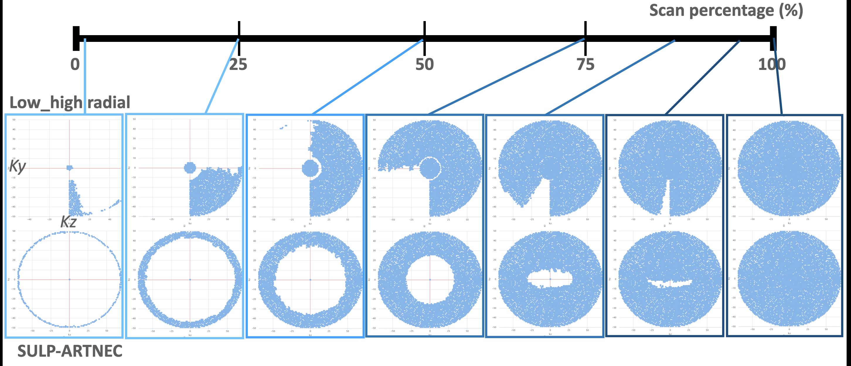
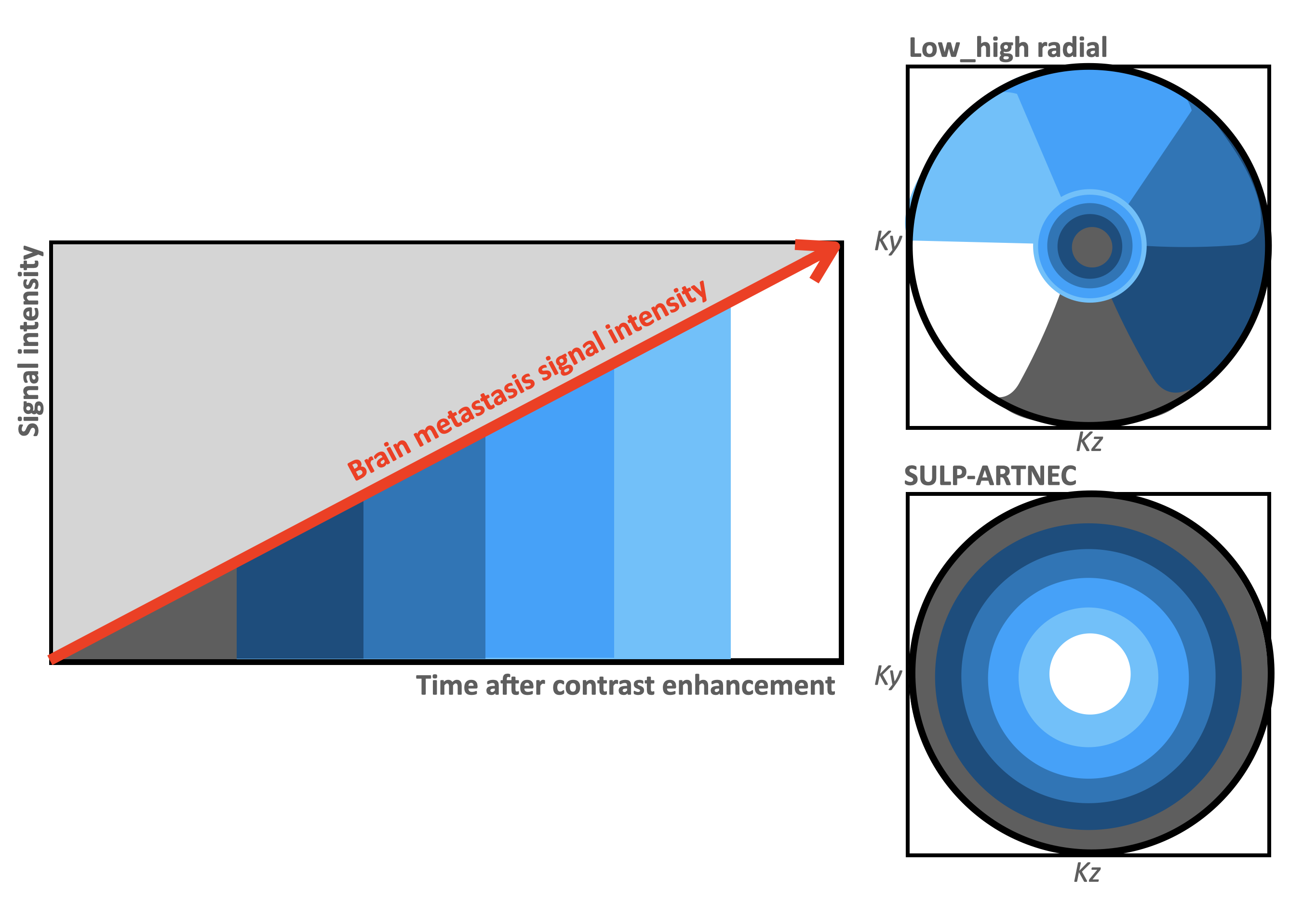
Fig.2. Hypothesis for capturing delayed enhanced lesions in respective k-space orders.
Low-high radial order could be affected by any timing of signals during the acquisition because the central Ky-Kz profiles are distributed over the entire acquisition. SULP-ARTNEC is started from the peripheral k-space sector and the most central k-space sector is acquired at a late stage. It may emphasize the increased signals of the brain metastasis due to delayed enhancement at the last of the imaging time
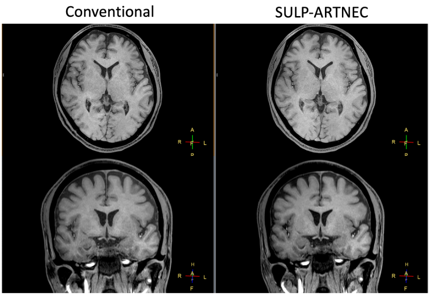
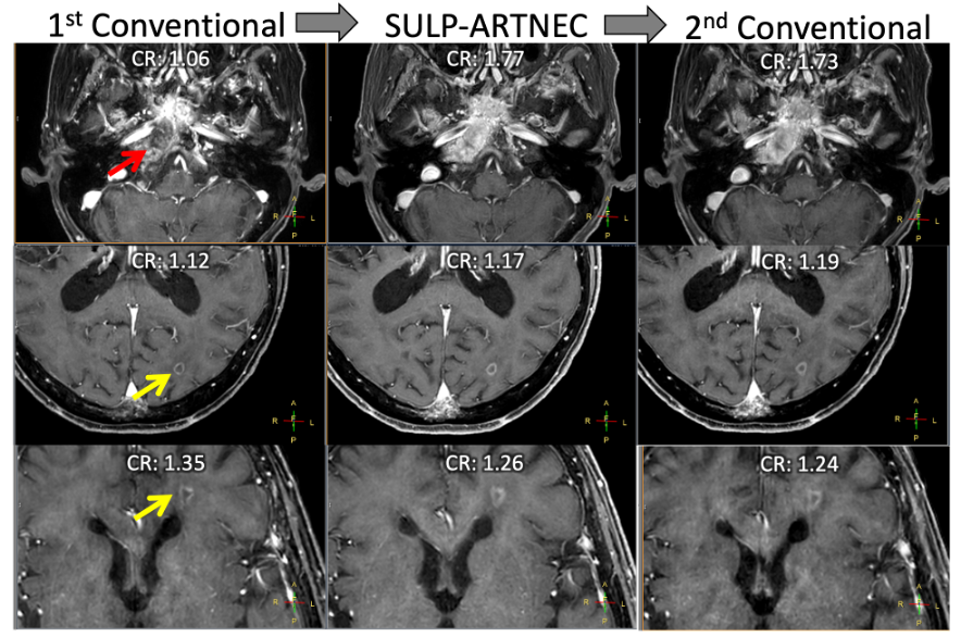
Fig.4. A representative example of pattern A. Age 71, Female. Lung cancer. After gamma knife treatment for multiple metastatic brain tumors.
Bone metastases in the clivus (red arrow) and brain multiple metastases (yellow arrow).
Contrast ratio (CR): neoplastic lesions / cerebral white matter.
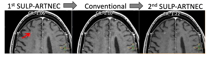
Fig.5. A representative example of pattern B. Age 70, Male. Lung cancer.
Follow-up of metastatic brain tumors (red arrow).
Contrast ratio (CR): neoplastic lesions / cerebral white matter.