4735
Accuracy of fat fraction when using IDEAL-IQ with a liver nonspecific extracellular contrast agent1Radiology, Guangdong Hospital of Traditional Chinese Medicine, Zhuhai, Zhu Hai, China, 2MR Research, GE Healthcare, Beijing, China, Beijing, China, 3Hepatology, Guangdong Hospital of Traditional Chinese Medicine, Zhuhai, Zhu Hai, China
Synopsis
To long-term track patients with liver disease is often carried out in clinics. It is also noted that nonspecific contrast agents change the relaxation time of tissue components, but few studies focus on the strong or weak FF alteration reflected by IDEAL-IQ derived measurements acquired before and after the contrast injection. In our study, we discovered significantly altered T1 values and fat fraction but not R2* even after the non-specific contrast agent achieved balanced condition at 3 minutes later of post-injection. Therefore, the liver non-specific contrast agent had an impact on the fat fraction measurements computed by IDEAL-IQ, and the utility of IDEAL-IQ should be performed before the injection of contrast agents to ensure the accuracy of fat fraction.
Introduction
IDEAL-IQ is well-known to offer whole-liver fat fraction (FF) map and R2* map in reflection of in vivo liver condition. Both map-derived values are in good agreement with liver biopsy[1]. To long-term track patients with liver disease is often carried out in clinics. It is also noted that nonspecific contrast agents change the relaxation time of tissue components[2], but few studies focus on the strong or weak FF alteration reflected by acquiring IDEAL-IQ before and after the contrast injection. Liver non-specific contrast agents are widely used in the liver and can affect T1WI signal intensity contrast by shortening T1 relaxation time and not affecting T2 effect. Follow-ups for patient with liver diseases have routinely applied IDEAL-IQ; therefore, the T1-shortening contrast agent was used to acquire FF and R2* before and after the injection. In this study, we aimed to explore the feasibility and reproducibility of R2* and FF before and after the injection for future follow-ups in responses to treatments or physiological progression.Materials and methods
21 patients with chronic hepatitis B were recruited and underwent IDEAL-IQ and variable-flip-angle T1mapping (FA = 3.6.9.12.15) imaging before injection of non-specific contrast agent (Gd-BT-DO3A) and at the equilibrium phase, around 3-min post-injection, for the upper abdominal enhancement examinations. Three ROIs of 80-100 mm2 were placed in each segment of liver S5-8 in fat fraction map, R2* relaxation, and T1 mapping, avoiding large vessels and intrahepatic bile ducts. FF, R2* and T1 values were measured. All statistics was carried out using SPSS 21.0, including paired T test or nonparametric Wilcoxon signed test depending on data distribution normality.Results
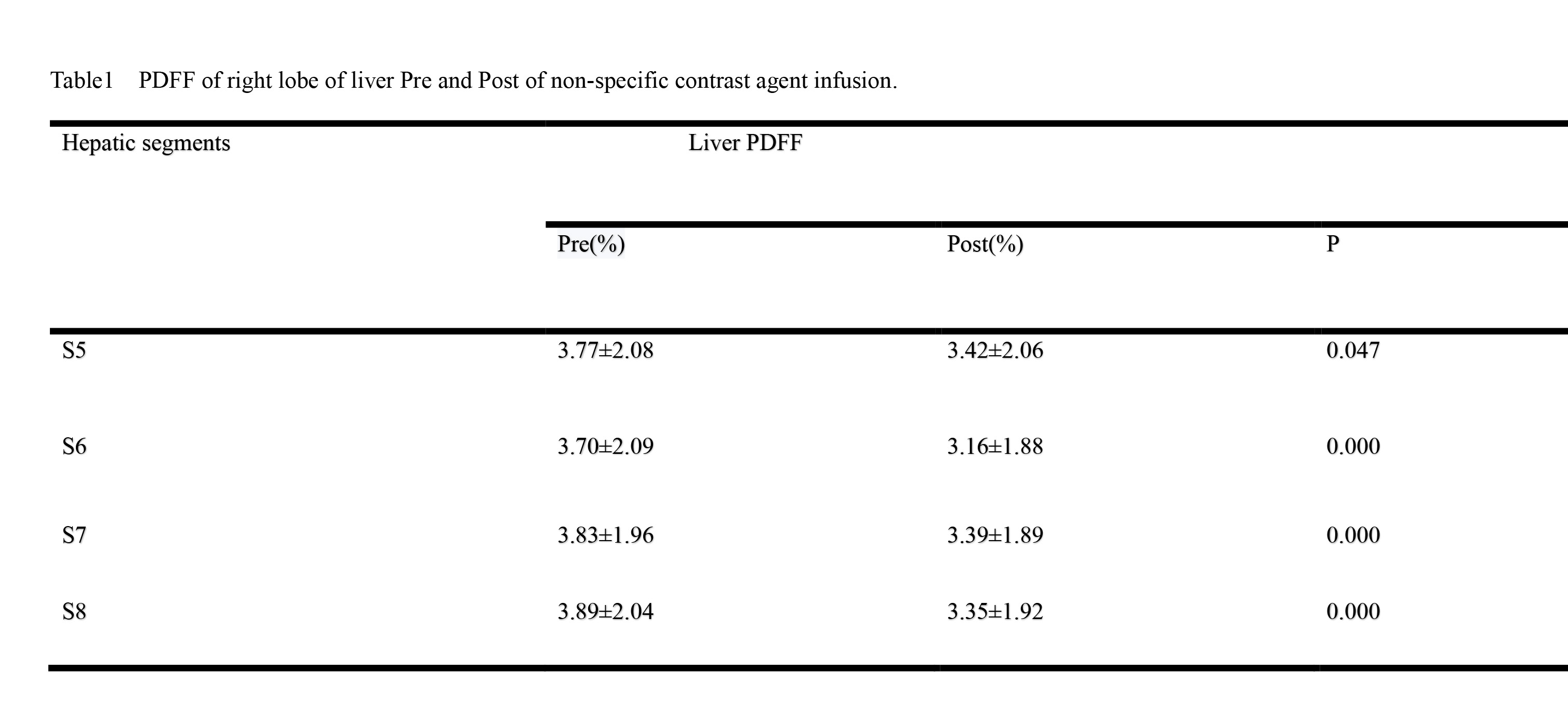
T1 value significantly decreased after liver S5-8 enhanced (p<0.05) (Table 1). No statistically significant change of R2* value was found before and after hepatic S5-8 enhancement (Table 2). The difference of FF before and after hepatic S5-8 enhancement was statistically significant (Table 3and Figure 1).Discussion
We discovered significantly altered fat fraction but no R2* change after the non-specific contrast agent achieved balanced condition at 3 minutes later of post-injection. Gd-BT-DO3A is a kind of non-specific extracellular contrast agent with the feature of shortening the T1 relaxation time and accelerating the signal recovery of tissues. It assists clinics to better determine properties of lesions via lesion enhancement[2]. Liau [3]et al found that R2* changed and PDFF remained unchanged after SPIO injection via drawing only one ROI for the measurement of the whole liver. In contrast, measurements were performed on each segment of the right lobe of the liver in our study and thus it could avoid the interference of intrahepatic large vessels and bile ducts. Most importantly, fat fraction was different between pre- and post-injection of Gd-based contrast agent. Time for scanning with IDEAL-IQ sequence is important especially when this kind of contrast agent was used.Conclusion
The liver non-specific contrast agent had an impact on the fat fraction measurements of IDEAL-IQ, and the utility of IDEAL-IQ was recommended to perform before the injection of contrast agents to ensure the accuracy of fat fraction.Acknowledgements
No fundingReferences
[1].Tang A, Tan J, Sun M, Hamilton G, Bydder M, Wolfson T, Gamst AC, Middleton M, Brunt EM, Loomba R, Lavine JE, Schwimmer JB, Sirlin CB. Nonalcoholic fatty liver disease: MR imaging of liver proton density fat fraction to assess hepatic steatosis. Radiology. 2013 May;267(2):422-31. doi: 10.1148/radiol.12120896. Epub 2013 Feb 4. PMID: 23382291; PMCID: PMC3632805.
[2].Ibrahim MA, Hazhirkarzar B, Dublin AB. Gadolinium Magnetic Resonance Imaging. 2021 Jul 9. In: StatPearls [Internet]. Treasure Island (FL): StatPearls Publishing; 2021 Jan–. PMID: 29494094.
[3].Liau J, Shiehmorteza M, Girard OM, Sirlin CB, Bydder M. Evaluation of MRI fat fraction in the liver and spine pre and post SPIO infusion. Magn Reson Imaging. 2013 Jul;31(6):1012-6. doi: 10.1016/j.mri.2013.01.016. Epub 2013 Apr 18. PMID: 23602721; PMCID: PMC3676690.
Figures