4734
The effect of contrast agent injection in MRCP under respiratory-triggered 3D-SPACE1Xi'an Daxing Hospital, Xi'an, China, 2Siemens Healthineers, Ltd., Xi'an, China
Synopsis
This study compared the image quality of MRCP before and after contrast agent injection on a 3T MRI scanner using the same respiratory-triggered 3D-SPACE sequence. Our results showed that enhancement of contrast-to-noise ratio (CNR) and background suppression will make the contour of pancreaticobiliary system more clear, which indirectly improves the imaging effect of MRCP. We performed MRCP scanning during the delayed waiting period for enhanced scans, which shortens the overall scan time for such patients. In summary, respiratory-triggered 3D-SPACE MRCP imaging with enhanced contrast injection significantly improved the imaging quality of the pancreaticobiliary duct and shorten the overall acquisition time. It is worthy of promotion and application in clinical practice.
Introduction/Purpose
Respiratory-triggered 3D magnetic resonance cholangiopancreatography sequence has become the principal method for obtaining high-resolution isotropic images in the last 10 years[1]. A sampling perfection with application-optimized contrasts using different flip angle evolutions (SPACE) sequences has also been noted to improve image quality and the contrast-to-noise ratio(CNR)[2]. This study aim to investigate the effect of contrast agent injection in MRCP under respiratory-triggered 3D-SPACE.Method
Twenty-three patients (13 males and 10 females, age range 49-97 years) were enrolled in this study. All the patients underwent respiratory-triggered 3D-SPACE MRCP examination before and after contrast agent injection on a 3T MR scanner (MAGNETOM Prisma, Siemens Healthineers, Erlangen, Germany). All patients completed a respiratory-triggered 3D-SPACE scanning before injecting contrast agent. After injecting contrast agent, technicians firstly collected the arterial phase and portal phase of abdominal dynamic enhancement scanning, after that, the second respiratory-triggered 3D-SPACE sequence with the same parameters was performed. The scanning parameters of 3D-SPACE sequence were as follows: TR variable, TE = 570ms, FOV = 320mm×320mm, Flip angle = 110°, slice thickness = 3 mm, slices =128. Maximum intensity projection (MIP) was used to process the collected original images. The image quality of the two sequences was analyzed statistically. The paired-sample t test was used to compare the pancreaticobiliary system scores of the two groups before and after contrast agent injection. Used the Wilcoxon rank test to compare the image quality of the two groups. P<0.05 indicated that the difference was statistically significant.Result
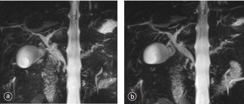
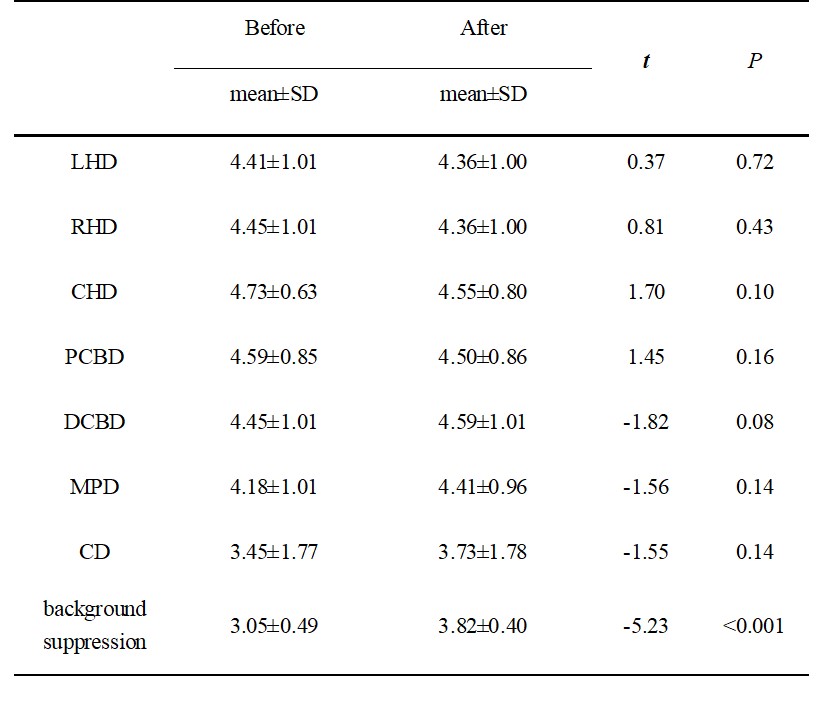
The pancreaticobiliary system scores and background suppression results of the two groups of MRCP images before and after contrast agent injection were shown in Table 1. Contrast noise ratio (t=-4.17, P<0.001) and background suppression score (t=-5.23, P<0.001) were different between the respiratory-triggered 3D-SPACE sequence before and after contrast agent injection(Figure 1), with the overall acquisition time shortened.Discussion/Conclusion
Due to the particularity of pancreaticobiliary diseases, enhancement scanning usually requires a delayed waiting period after arterial phase and portal phase scanning. In the past, we performed enhancement sequence scanning after completing MRCP scan. If we performed MRCP scanning during the delayed waiting period, we can save the sequence acquisition time. The background suppression effect in 3D-SPACE MRCP with contrast agent was more significant than respiratory-triggered 3D-SPACE MRCP without contrast agent which increased CNR of pancreaticobiliary duct. In addition, although there was no significant difference in the acquisition time of MRCP itself, the overall scanning time for patients was shortened due to the use of the fragment time in the delayed waiting period of enhanced scanning. The results of this study indicated that respiratory-triggered 3D-SPACE MRCP with contrast agent had a great value in clinical practice. For patients undergoing MRCP and abdominal enhancement scanning at the same time, they can firstly collect the images of arterial phase and portal vein phase of abdominal dynamic enhancement scanning, and then conduct 3D-SPACE MRCP sequence scanning during the waiting period, so as to save one sequence scan time and improve the examination pass rate.Acknowledgements
We thanks Shaoyu Wang from Siemens Healthineers, Ltd., Xi’an, China, for technical support.References
1. Zins M. Breath-holding 3D MRCP: the time is now[J]? Eur Radiol 2018;28:3719-3720.
2. Takeshi N, Masafumi K, Natsuki M, et al. Usefulness of the SPACE pulse sequence at 1.5T MR cholangiography: comparison of image quality and image acquisition time with conventional 3D-TSE sequence[J]. J Magn Reson Imaging 2013;38:1014-1019.
Figures