4724
To Evaluate Values of Six Cancer-Related DWI Models in Predicting the Levels of HER-2 Expression in Invasive Ductal Carcinoma of the Breast1Department of CTMRI, The Fourth Hospital of Hebei Medical University, Shijiazhuang, China, 2MR Scientific Marketing, Siemens Healthineers, Beijing, China, 3Shanghai Key Laboratory of Magnetic Resonance, East China Normal University, Shanghai, China
Synopsis
In this study, we evaluated and compared the values of quantitative parameters derived from the six DWI models in the diagnosis of HER-2 expression levels in patients with IDC of the breast, including conventional mono-exponential model, IVIM, DKI, SEM, FROC, and CTRW models. We found that the CTRW model showed higher sensitivity in the predicting HER-2 expression levels than the other models. Meanwhile, a logistic regression model was built by combining parameters of the advanced DWI models, which showed a high diagnostic performance and may potentially be used in clinical practice.
Purpose
To compare the values of quantitative parameters derived from multiple cancer-related DWI model in predicting the levels of HER-2 expression in invasive ductal carcinoma (IDC) of breast, which includes conventional mono-exponential model derived parameter ADCs, intro-voxel incoherent movement (IVIM) derived parameters (D, Dstar, f), diffusion kurtosis imaging (DKI) derived parameters (D, K), stretched exponential model (SEM) derived parameters (DDC, α), fractional order calculus (FROC) derived parameters (DDC, α), continuous-time random-walk diffusion model (CTRW) derived parameters (Dm, α, β). The comprehensive diagnostic value of these models is also studied.Materials and Methods
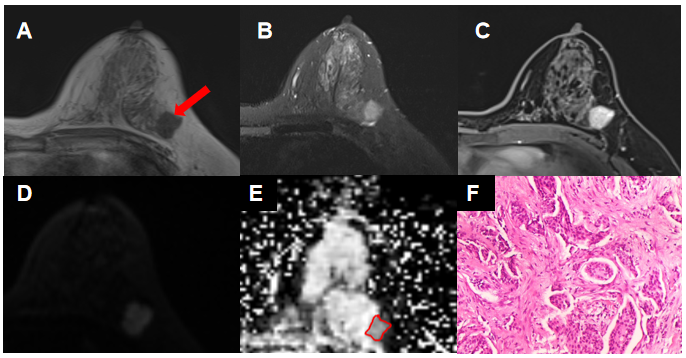
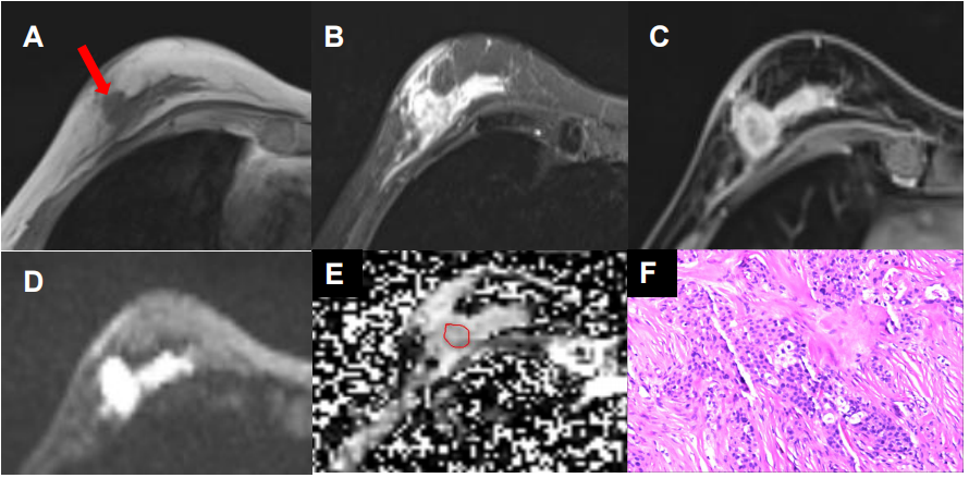
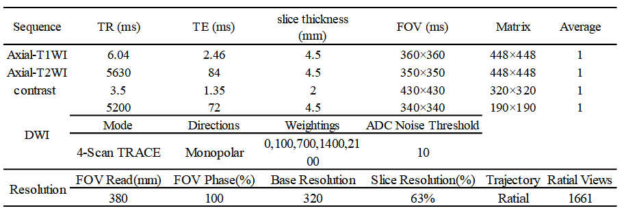
The institutional review board of The Fourth Hospital of Hebei Medical University approved this retrospective study. From June 2020 to October 2020, we enrolled 36 consecutive women with primary IDC of breast and all with biopsy-confirmed. The examination was underwent at a 3T scanner ((MAGNETOM Prisma, Siemens, Erlangen, Germany), the details of parameters can be found in Table 1. All the levels of IDC HER-2 were confirmed using an immunohistochemical method, and according to the effect of HER-2 expression to therapeutic schedule, the patients were divided into three groups: Group1 [HER-2(0) and HER-2(1+)]; Group2 [HER-2(2+)]; Group3 [HER-2(3+)]. The parametric maps of all 6 DWI models (conventional DWI, IVIM, DKI, SEM, FROC, CTRW) were calculated using a unified in-house developed software called BoDiLab, which is based on Python 3.7. Typical cases were showed in Figure 1,2.All region-of-interest (ROI) measurements were done on the tumor’s slice with the largest area using MRIcron software (https://www.nitrc.org/projects/mricron). The normality and variance homogeneity of the measured parameters were tested, and the difference among the three groups was analyzed using the ANOVA methods, the difference between one group and the other two groups was analyzed by using the Mann-Whitney-U test. In order to study the diagnostic performance of these DWI parameters and their combinations, a logistic regression (LR) model was established using a step-by-step method. All the analysis was performed using the software SPSS 22.0 and R language.
Results
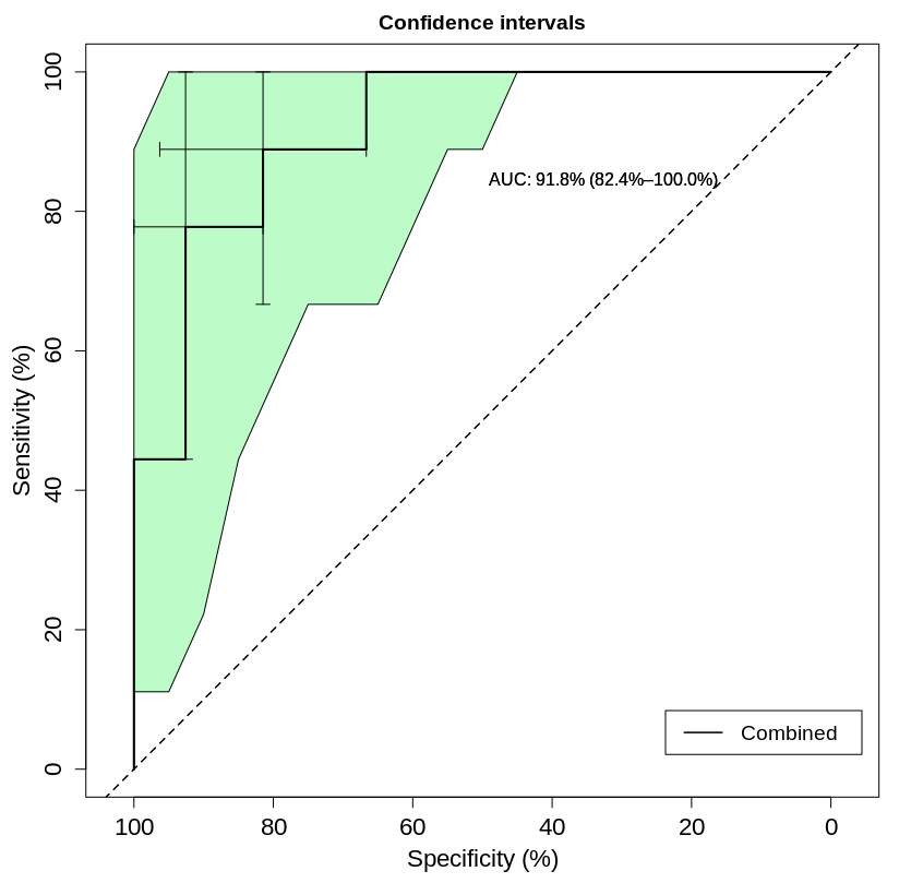
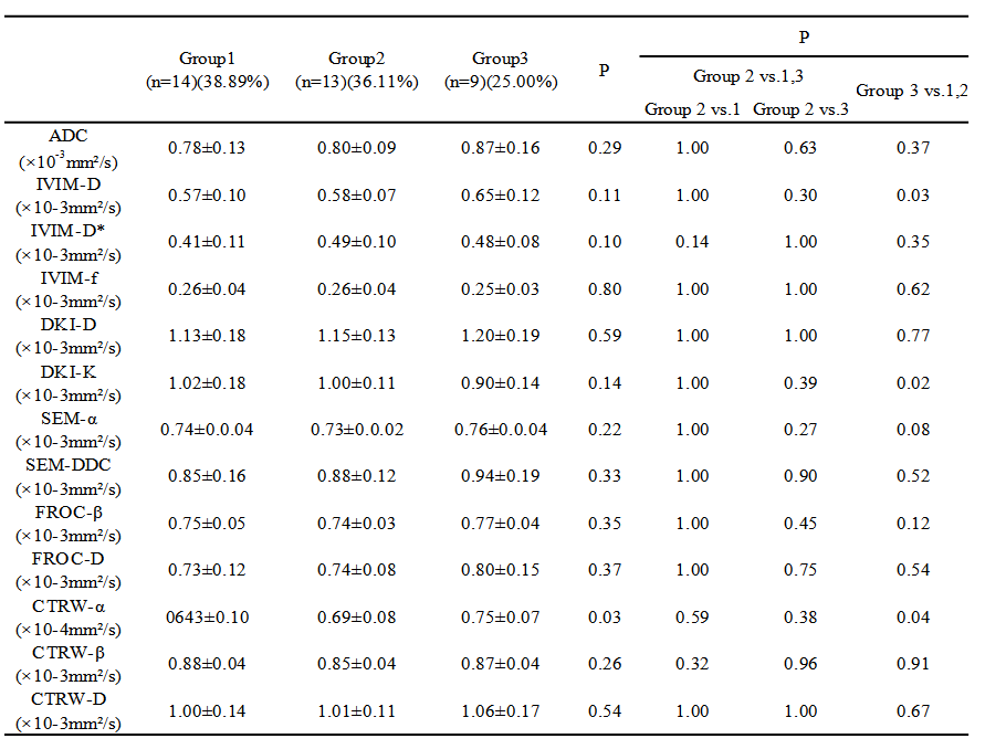
The parameter CTRW_α demonstrated significant difference (F=3.883, P=0.031) among three HER-2 groups. In differentiating Group 3 from the other two groups, the CTRW_α, DKI_K and IVIM_D showed significant differences (all P<0.05)(Table 2). After filtering the parameters in establishing the LR model, the parameters of CTRW_α, DKI_D, DKI_K, FROC_D, IVIM_D, IVIM_D*, SEM_DDC, and Mono_ADC were enrolled. The established model included the comprehensive enrolled parameters that demonstrated a high diagnostic efficiency with an AUC/sensitivity/specificity/NPV/PPV of 0.918/0.889/0.815/0.615/0.956. Figure 3).Discussions
Human epidermal growth factor receptor-2 (HER-2) is an important proto-oncogene and the positive expression of HER-2 is closely related to early metastasis, recurrence, and prognosis of patients. As some literatures report, the level of HER-2 expression has a relationship with the heterogeneity of the IDC, and the level of this factor also determines the choice of clinical therapy schedule. So, a non-invasive biomarker that can diagnose the expression levels of HER-2 is extremely important in clinical practice.In these years, advanced diffusion models showed high diagnosis value for cancer. There are some popular cancer related diffusion models, such as IVIM, diffusion kurtosis imaging (DKI), stretched-exponential (SEM), FROC and recently proposed continuous-time random walk (CTRW) models. Unlike the DKI model reflecting non-Gaussian diffusion behavior, the SEM, FROC and CTRW models were more focus on spatial heterogeneity of diffusion within voxel, which could be commonly found in high grade tumor. While the CTRW is an extension of the FROC diffusion model, which reflects intravoxel diffusion heterogeneity in both time and space, thus could be a potential biomarker for changes of tissue complexity and microenvironment.
In this study, these models were applied in predicting level of HER-2 expression, and their individual and combined performance was evaluated and compared. we have found the CTRW_α outperformed the other diffusion parameters in predicting of HER-2 expression levels. The probabilistic nature of the CTRW theory relaxes a priori distributions of waiting times and distance increments of water molecular diffusion, providing a more realistic description of the complex structures of biological tissues. The parameter CTRW_α is different to other diffusion parameters by reflecting diffusion heterogeneity in time. Meanwhile, a LR model was built and combined different DWI parameters to achieve a high diagnostic power, which may be used as a non-invasive biomarker in clinical practice in future.
Conclusions
CTRW may possess a high value in the diagnosis of HER-2 expression levels in patients with IDC of the breast. The LR model combined different quantitative parameters of the advanced DWI model may be used as a non-invasive biomarker in clinical practice in the future.Acknowledgements
No acknowledgement found.References
[1] Karaman, MM (Karaman, M. Muge)1;Sui, Y (Sui, Yi)1,2;Wang, H (Wang, He)3;Magin, RL (Magin, Richard L.);Li, YH (Li, Yuhua)4;Zhou, XJ (Zhou, Xiaohong Joe)1,2,5,6.Differentiating low- and high-grade pediatric brain tumors using a continuous-time random-walk diffusion model at high b-values[J]. Magnetic Resonance in Medicine.2016.
[2] Liu, F (Liu, Fei)1;Wang, M (Wang, Mei)1;Li, HG (Li, Haige)1.Role of perfusion parameters on DCE-MRI and ADC values on DWMRI for invasive ductal carcinoma at 3.0 Tesla.[J]. World Journal of Surgical Oncology.2018.
[3] Niendorf T, Dijkhuizen RM, Norris DG, van Lookeren Campagne M, Nicolay K. Biexponential diffusion attenuation in various states of brain tissue: implications for diffusion-weighted imaging. Magn Reson Med 1996;36:847–857.
[4] Bennett KM, Schmainda KM, Bennett R, Rowe DB, Lu H, Hyde JS. Characterization of continuously distributed cortical water diffusion rates with a stretched-exponential model. Magn Reson Med 2003;50: 727–734.
[5] Jensen JH, Helpern JA, Ramani A, Lu H, Kaczynski K. Diffusional kurtosis imaging: the quantification of non-Gaussian water diffusion by means of magnetic resonance imaging. Magn Reson Med 2005;53: 1432–1440.
[6] Zhou XJ, Gao Q, Abdullah O, Magin RL. Studies of anomalous diffusion in the human brain using fractional order calculus. Magn Reason Med 2010;63:562–569.
[7] Ingo C, Magin RL, Colon-Perez L, Triplett W, Mareci TH. On random walks and entropy in diffusion-weighted magnetic resonance imaging studies of neural tissue. Magn Reson Med 2013;627:617–627.
Figures