4716
Super-resolution MR Vessel Wall Images Using deep learning1Paul C. Lauterbur Research Center for Biomedical Imaging, Shenzhen Institute of Advanced Technology, Chinese Academy of Sciences, Shenzhen, China, 2Faculty of Information Technology, Beijing University of Technology, Beijing, China, 3Department of computing,Imperial College London, London, United Kingdom, 4Department of Radiology, Northern Jiangsu People's Hospital, Jiangsu, China
Synopsis
To develop a super-resolution method based on the 3D high-resolution MR vessel wall images for generating high-resolution images from low-resolution, a 3D complex-valued super resolution (CVSR) neural network was proposed, which maintained complex algebraic structure of the original acquired images. CVSR was trained on 20 pairs of data sets and tested on 5 pairs. Ground truth with 0.44 mm were compared with Fourier interpolation method, EDSR with two real-valued channels and CVSR. Evaluations were performed using structural similarity (SSIM), peak signal-to-noise ratio (PSNR), and error map quality metrics. The CVSR achieved the best performance when compared with the other methods.
INTRODUCTION
MR vessel wall imaging can directly image and evaluate arterial vessel walls and plaques. Since the intracranial arteries, especially the perforator arteries are very fine tissues. MR vessel wall imaging requires very high spatial resolution to accurately detect and evaluate vessel walls and plaques. However, MRI spatial resolution, image signal-to-noise ratio and scan time are mutually restricted. High spatial resolution imaging requires a very long scan time to obtain acceptable image quality for clinical diagnosis. Long scanning time is prone to image motion artifacts and could not satisfy clinical diagnosis. Recently, many studies have proposed the use of deep learning for high resolution reconstruction of MRI images. As we know, in MRI, k-space data are complex values with a real and an imaginary component [1]. Most of these studies do not really use the complex-valued characteristics of k-space [2]. This work proposed a complex value based deep convolution neural network (CNN) which preserves both the magnitude information and phase information of the data for super-resolution reconstruction of MR vessel wall images.METHODS
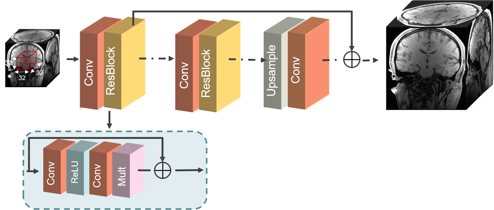
The 0.44 mm isotropic 3D high-resolution MR vessel wall images were acquired from 25 subjects on a 3T whole-body MR system (TrioTim, Siemens, Germany). The matrix size is 448×448×352 or 416 depending on the head size. Then the corresponding 0.88 mm isotropic low-resolution images were reconstructed by truncating the acquired k-space of the high-resolution images. The 25 pairs of data sets were devided into 20 pairs of data sets for training and 5 pairs for testing. Due to the different matrix sizes of the acquired data sets, all datasets were uniformly cropped to a consistent size of 448×448×352 and used as input for training the network. Each data set was further divided into smaller patches of size 64×64×64 with a stride of 32 to adapt to the limited computation ability of our equipment. A 3D complex value based super resolution CNN entitled CVSR was proposed and trained on the training dataset including low-resolution and high-resolution data pairs to transform low-resolution MR vessel wall images into high-resolution images. The architecture of the proposed CVSR is displayed in Figure 1. The training of CVSR was performed utilizing 8 residual blocks with 64 feature channels, and the size of each kernel is 5 × 5 × 5. Structural similarity (SSIM), peak signal-to-noise ratio (PSNR) and root mean square error (RMSE) were used to evaluate the performance of the proposed CVSR model.RESULTS
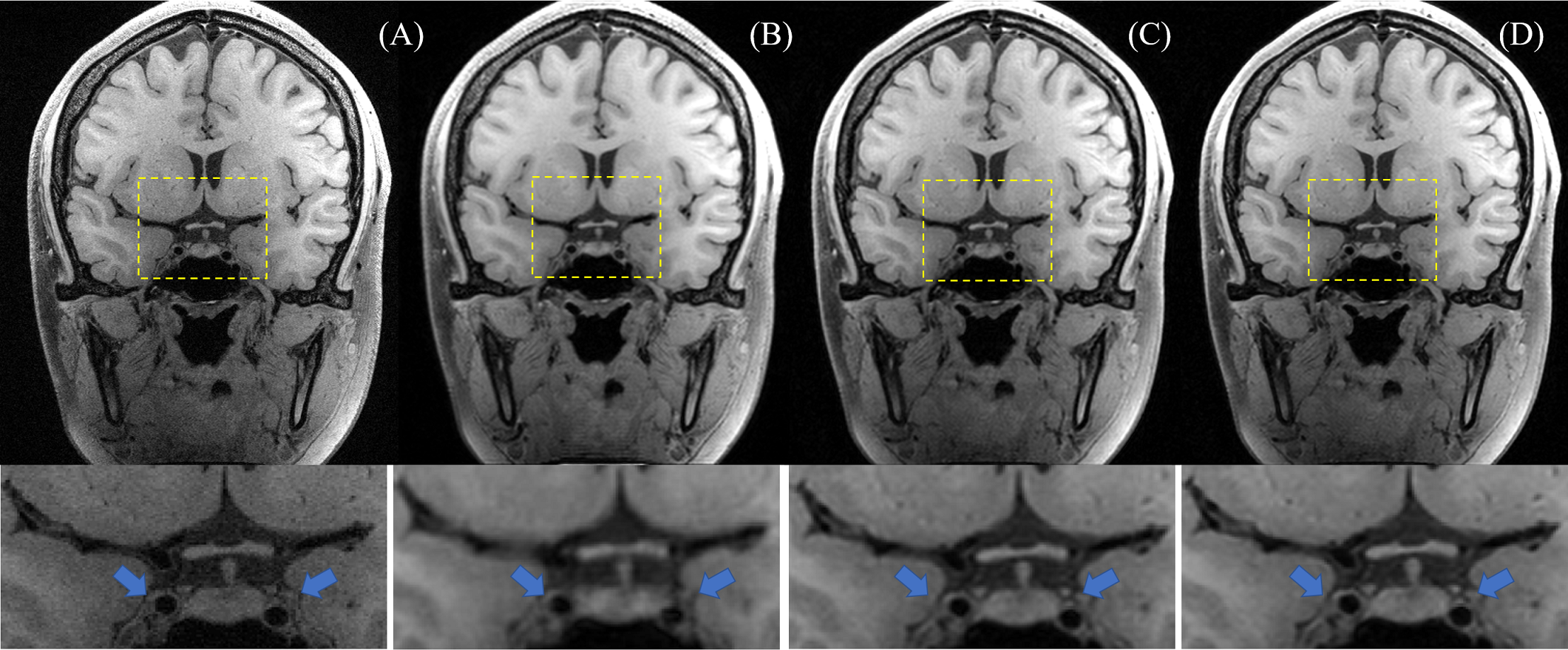
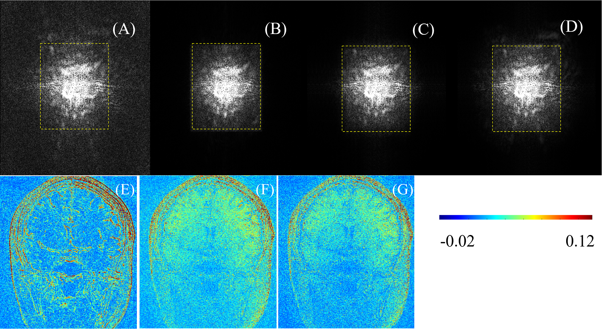
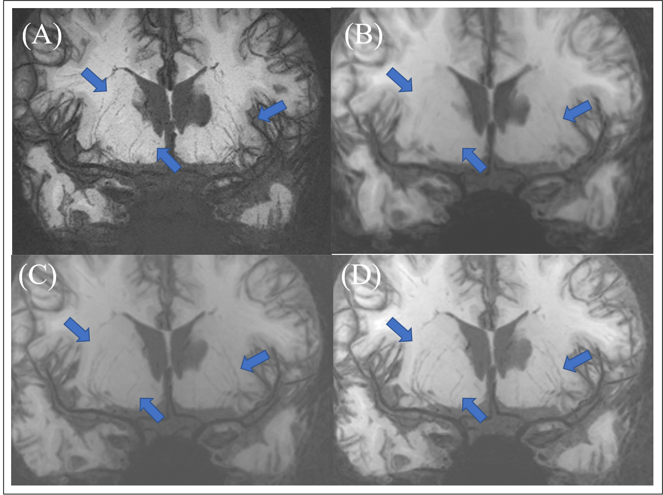
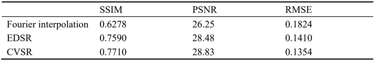
The quantitative analysis results of performance of the Fourier interpolation, EDSR with two real-valued channels and the proposed CVSR for super-resolution reconstruction of MR vessel wall images were summarized in Table1. The proposed CVSR achieved a SSIM of 0.7710, PSNR of 28.83, and RMSE of 0.1354, which were higher than those achieved by EDSR and Fourier interpolation. These evaluation metrics achieved by Fourier interpolation were the lowest. Figure 2 shows a representative coronal slice of the acquired 0.44 mm isotropic high-resolution images (ground truth), and the corresponding super-resolution images by Fourier interpolation, EDSR and CVSR. The super-resolution image obtained by CVSR is visually comparable to the ground truth for the depiction of vessel wall, and is significantly better than the image obtained by Fourier interpolation, and is also better than the super-resolution image obtained by EDSR. The reason is CVSR uses both real and imaginary information of the data and hence can recover more fine textural details and a higher signal-to-noise ratio than the other methods. Figure 3 shows the corresponding k-space data of the Figure 2 and the difference of Fourier interpolation, EDSR and CVSR relative to the ground truth. As can be seen from Figure 3 (D), there are more high-frequency signal of CVSR than the other two methods. The minMIP images for visualizing perforator arteries obtained from ground truth,Fourier interpolation, EDSR and CVSR are shown in Figure 4. Compared with the Fourier interpolation and EDSR, CVSR shows more perforator arteries details. There are discontinuous and missing perforator arterial segments of the images obtained by Fourier interpolation and EDSR, while images obtained by CVSR displays perforating vessels more clearly and closer to the ground truth.DISCUSSION
In this study, a complex value based CNN (CVSR) which preserves both the magnitude information and phase information of the data was proposed for super resolution reconstruction of MR Vessel Wall Images. It was capable of transforming low-resolution images into high-resolution images and be able to observe the fine arteries more clearly, particularly for the perforator arteries. The CVSR achieved the best performance in super-resolution reconstruction of MR Vessel Wall Images when compared with Fourier interpolation and EDSR with two separate real-valued channels.CONCLUSION
The proposed CVSR can transform low-resolution images into high-resolution and obtain satisfactory super-resolution MR vessel wall images. It not only shows the arterial vessel wall more clearly but also shows more details and the numbers of perforator arteries, which is of great significance for assisting clinically accurate diagnosis of fine tissues, especially arterial vessel wall and plaque.Acknowledgements
The study was partially support by National Natural Science Foundation of China (81830056), Key Laboratory for Magnetic Resonance and Multimodality Imaging of Guangdong Province (2020B1212060051), Shenzhen Basic Research Program (JCYJ20180302145700745 and KCXFZ202002011010360), and Guangdong Innovation Platform of Translational Research for Cerebrovascular Diseases.References
[1] Zhang Z , Wang H , Feng X , et al. Complex-Valued Convolutional Neural Network and Its Application in Polarimetric SAR Image Classification[J]. IEEE Transactions on Geoscience and Remote Sensing, 2017, PP(12):1-12.
[2] Eun D I , Jang R , Ha W S , et al. Deep-learning-based image quality enhancement of compressed sensing magnetic resonance imaging of vessel wall: comparison of self-supervised and unsupervised approaches[J]. Scientific Reports, 2020, 10(1):13950.
Figures