4715
Low field multi-contrast MRI denoising using robust PCA1Center for Biomedical Imaging Research, Department of Biomedical Engineering, School of Medicine, Tsinghua University, Beijing, China, 2Marvel Stone Healthcare Co., Ltd.,, Wuxi, China
Synopsis
Owing to hardware advancements, a resurgence of interest has emerged in low field MRI systems recently. However, the intrinsic low signal to noise ratio (SNR) due to the low magnetic field strength has hindered the acquisition of high-resolution images on low field MRI systems. Here we propose a non-local robust PCA-based method to jointly denoise multi-contrast images acquired on a 0.5 T MRI system. In-vivo brain data are used to test the proposed method. The results show that the proposed method outperforms BM3D and BM4D denoising methods.
Introduction
Recently, increasing interest has emerged in the MR community to revisit the methods of using low-field MRI for clinical scans as it provides a lower-cost alternate for the commonly used 1.5T and 3T systems 1-2. Also, low field systems lead to reduced specific absorption rate (SAR) limitations, smaller magnetic susceptibility artifacts and reduced acoustic noise levels 3. Nevertheless, considering the fact that the signal to noise ratio (SNR) for the MRI scans depends on the magnetic field strength, the image quality decreases as the field strength becomes lower. Thus, a robust denoising method is needed and efforts have been made 4-6. Since MRI images of different contrasts share similar anatomical information, here we propose a robust PCA (rPCA)-based denoising method by jointly using the multi-contrast information to improve the image quality of low-field MRI images.Theory
rPCA denoising methodMRI images of different contrasts share similar information and group sparsity exists between different contrasts. In this study we extract similar patches across different contrasts to form a low-rank plus sparse matrix and then use an rPCA-based method to extract the signals from noisy data.
For the proposed denoising method, we use the following matrix completion formulation:
$$\min_{L,S} \| L \|_{*}+ \|\phi(S) \| _{1} +\| L+S-M\|_{F}^{2}$$
Here $$$L$$$ refers to the low-rank component, $$$S$$$ refers to the sparse component, $$$M$$$ refers to the noise-corrupted signals and $$$\phi$$$ refers to wavelet transform.
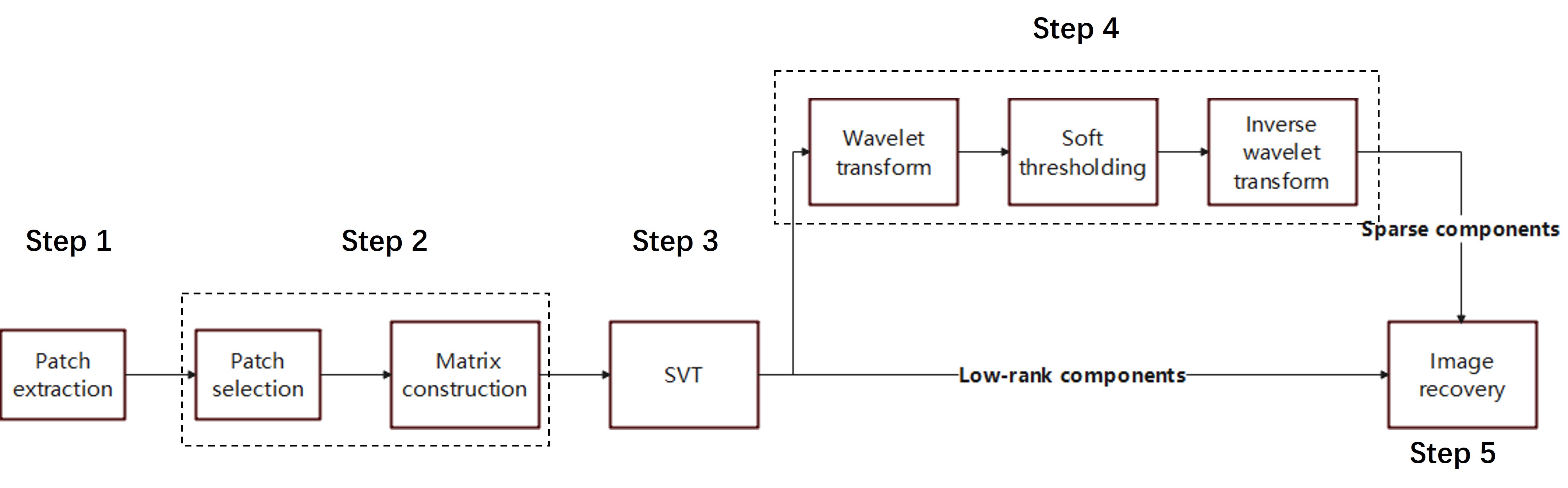
The denoising method consists of the following 5 steps shown in Fig. 1. 1) Patch extraction: extract patches using a sliding 3D window across different contrasts at the same spatial locations. 2) Matrix formation: for each patch, select K nearest patches based on the Euclidian distance and stack the patches into a matrix. 3) Low-rank estimation: use singular value shrinkage to estimate the low-rank components. 4) Sparse component estimation: remove the low rank components from the matrix and then apply wavelet transform and soft-thresholding to the residual signal matrix. 5) Image recovery: recover multi-contrast images from low-rank and sparse patch matrices.
Evaluation
In-vivo brain T1W, T2W and FLAIR images with the same FOV were acquired on a Marvel Stone 0.5T scanner (Marvel Stone Healthcare Co., Ltd., China), using an 8-channel head coil. The three acquisitions with one average lasted for 8 min 30s. The scan was repeated for 4 times to provide references. The multi-contrast images with single average were used as noisy image sets for denoising.
In the study, 8*8 window size was used and K was chosen as 196 for the proposed method. BM3D 7 and BM4D 8 were also performed for comparison.
Results
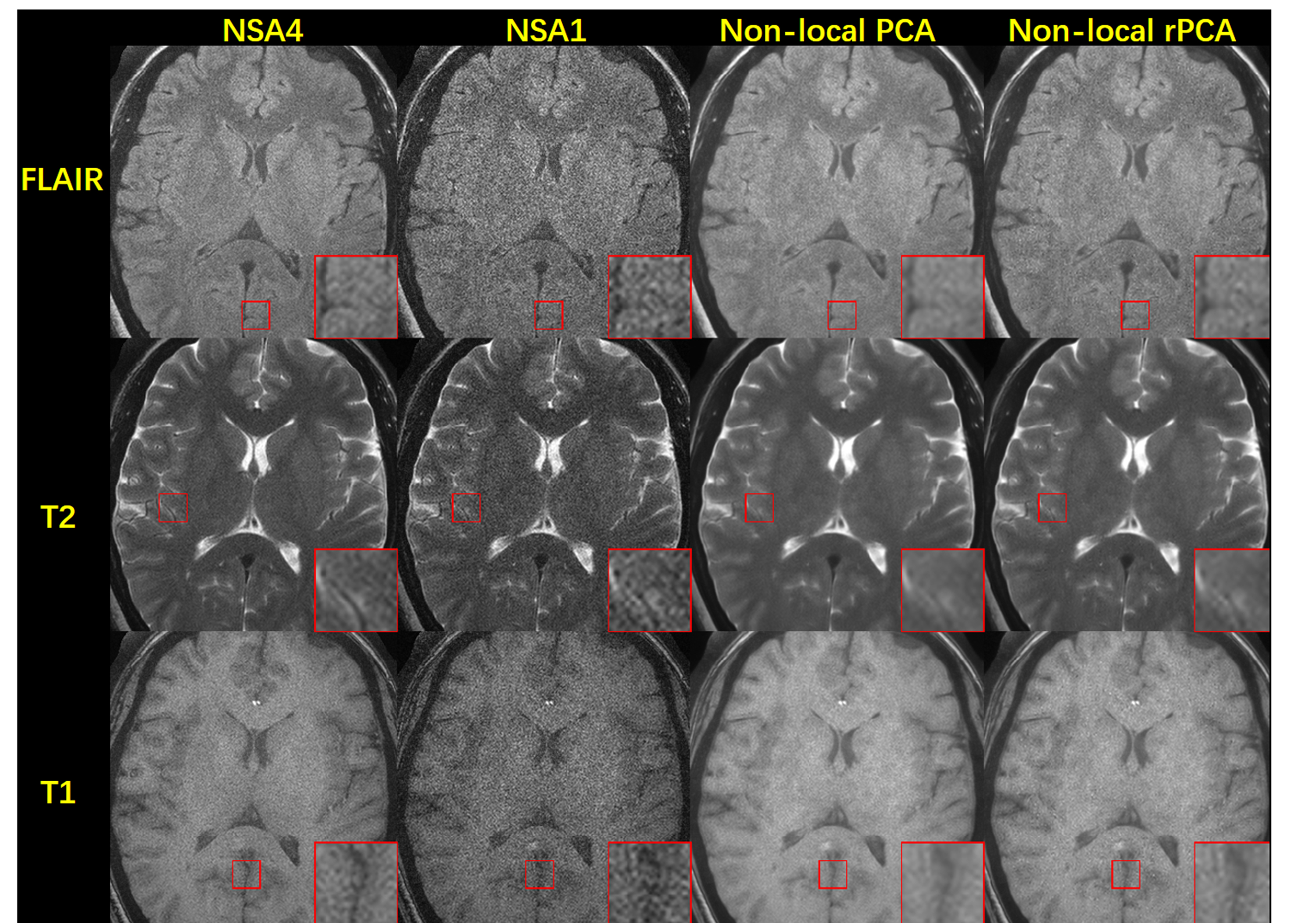
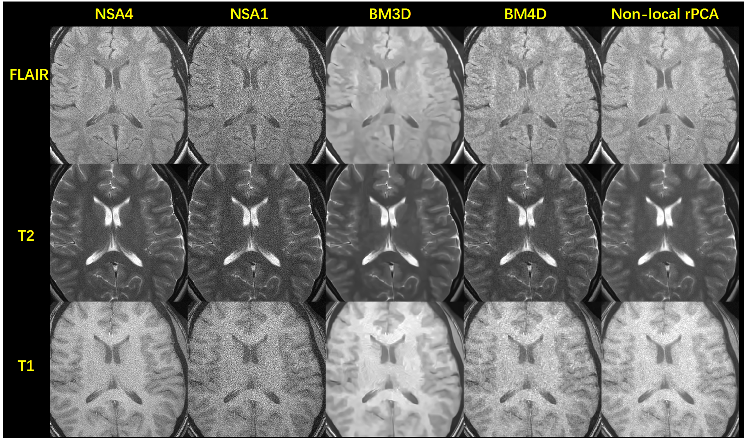
Figure 2 shows the denoising results from one subject acquired with 0.7$$$\times$$$ 0.7$$$\times$$$ 5mm3 resolution using non-local PCA and non-local rPCA methods respectively. Compared to the NSA1 noisy images, both methods could suppress the noise in the FLAIR, T2W and T1W images, producing denoised images that are visually similar to the NSA4 reference images. As shown in the zoomed-in images, compared to the results from the PCA method, the structure edges are clearer in the rPCA results, which indicates that the anatomical information is better preserved with the assistance of sparse components introduced by rPCA.Figure 3 shows the comparison between different denoising methods for another subject acquired with 0.7 $$$\times$$$0.7$$$\times$$$ 5mm3 resolution. BM3D produces over-smoothed results, leading to loss of structure information. Meanwhile, BM4D results have visible nonuniform noise patterns, especially in the center part of the images. However, the proposed method shows reduced noise levels and consistent noise patterns in the denoised images which are similar to the reference images. Additionally, the edges are clearly seen and easy to distinguish.
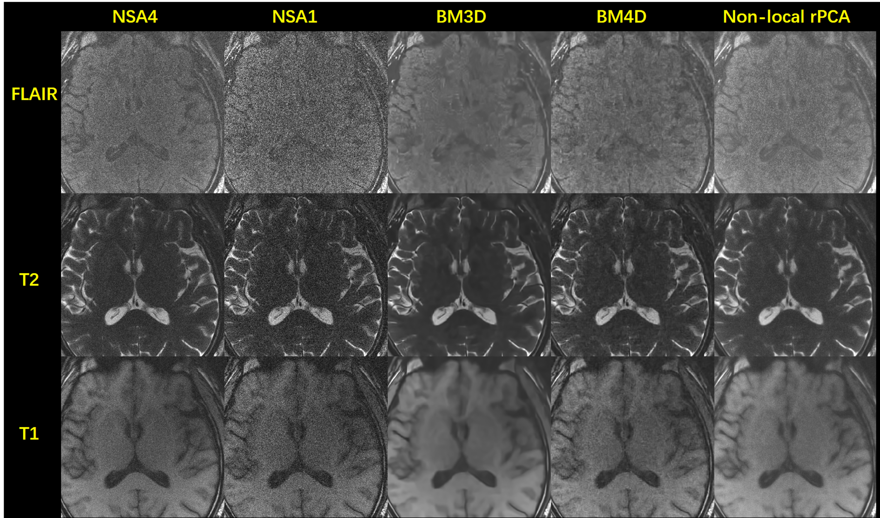
The denoising results for another subject acquired with 0.7$$$\times$$$ 0.7$$$\times$$$ 3mm3 resolution are shown in Figure 4. One representative slice is shown. For this case, the SNR is lower especially for the FLAIR images. BM3D produces blurry results and BM4D leads to artifacts when suppressing noise. The proposed method still manages to remove the noise and the image quality is comparable to that acquired with 4 averages. The noise distribution is visually uniform, indicating that the noise variance has been reduced by the proposed method. For T1W images, though the proposed method produces smoothed images, the results are still better than BM3D.
Conclusion
We propose a non-local robust PCA-based method to jointly denoise 0.5T FLAIR, T1W and T2W images. We demonstrate that the proposed method can suppress noise while preserving the anatomical structures. Based on the results, the proposed method may perform better compared to BM3D and BM4D, enabling significant image quality improvement for the low field MRI images. As such, the high-resolution images could be acquired without adding extra scan time for averaging on low field systems.Acknowledgements
No acknowledgement found.References
1.Bhat SS, Fernandes TT, Poojar P, et al. Low-Field MRI of Stroke: Challenges and Opportunities. J Magn Reson Imaging 2021;54(2):372-390.
2.Campbell-Washburn AE, Ramasawmy R, Restivo MC, et al. Opportunities in Interventional and Diagnostic Imaging by Using High-Performance Low-Field-Strength MRI. Radiology 2019;293(2):384-393.
3.Marques JP, Simonis FFJ, Webb AG. Low-field MRI: An MR physics perspective. J Magn Reson Imaging 2019;49(6):1528-1542.
4.Ishak NF, Gangeh MJ, Logeswaran R. Comparison of Denoising Techniques Applied on Low-field MR Brain Images. I C Comp Graph Im Vi 2008:345-+.
5.Koonjoo N, Zhu B, Bagnall GC, Bhutto D, Rosen MS. Boosting the signal-to-noise of low-field MRI with deep learning image reconstruction. Sci Rep-Uk 2021;11(1).
6.Waddington DEJ, Boele T, Maschmeyer R, Kuncic Z, Rosen MS. High-sensitivity in vivo contrast for ultra-low field magnetic resonance imaging using superparamagnetic iron oxide nanoparticles. Sci Adv 2020;6(29).
7.Dabov K, Foi A, Katkovnik, V, Egiazarian, K. Image denoising by sparse 3-D transform-domain collaborative filtering. Ieee T Image Process 2007;16(8): 2080-2095.
8.Maggioni M, Katkovnik V, Egiazarian K, Foi A. Nonlocal Transform-Domain Filter for Volumetric Data Denoising and Reconstruction. Ieee T Image Process 2013;22(1):119-133.
Figures