4703
Investigation of cerebral asymmetry in full-term newborns and its relationship with neurobehavioural assessment1The First Affiliated Hospital of Xi’an Jiaotong University, Xi’an, China, 710061, Xi'an, China, 2Department of Research and Development, Shanghai United Imaging Intelligence Co., Ltd., Shanghai, China,200232, Shanghai, China, 3GE Healthcare,Beijing, China,100176, Beijing, China
Synopsis
The specialization of cerebral lateralization and hemispheric function is common in adults. Existing studies have shown that there is already left and right asymmetry in the fetal brain [1]. The study of normal brain structure and development information helps to identify abnormal developmental trajectories. This study takes healthy full-term newborns as the research object, aiming to explore the situation of cerebral asymmetry in the neonatal period and the relationship with neurobehavioral. Meanwhile expand our understanding of normal early brain development, and provide important insights for further revealing the inner connection between brain lateralization and certain diseases in the future.
Summary of Main Finding
Brain asymmetry has already occurred in the full-term neonatal period, significantly on the left-biased. The laterality of occipital lobe, hippocampus, Cingulate gyrus posterior part WM showed a strong correlation with neurobehavioral scores.Introduction
Being a very complex organ, the brain has a high degree of symmetry between the left and right hemispheres. However, in recent years, with the wide application of non-invasive examination techniques, the differences in the structure and function of the left-right hemisphere have become widely known. The laterality of brain structure and function can make each hemisphere dominate when processing specific cognitive tasks. For example, the right hemisphere plays a special role in spatial attention and some neurological diseases are closely related to cerebral lateralization defects, including dyslexia, schizophrenia, bipolar disorder and autism [2]. At present, there are relatively few studies on neonatal cerebral asymmetry. This study aims to study the correlation between brain lateralization and neurobehavioral in the neonatal period, deepen our understanding of brain development during this period, and furthermore, provide a starting point for future research on the changes of laterality in brain development with age. In the end we can provides important insights into some neurodevelopmental disorders whose pathogenesis is a lateral factor.Materials and Methods
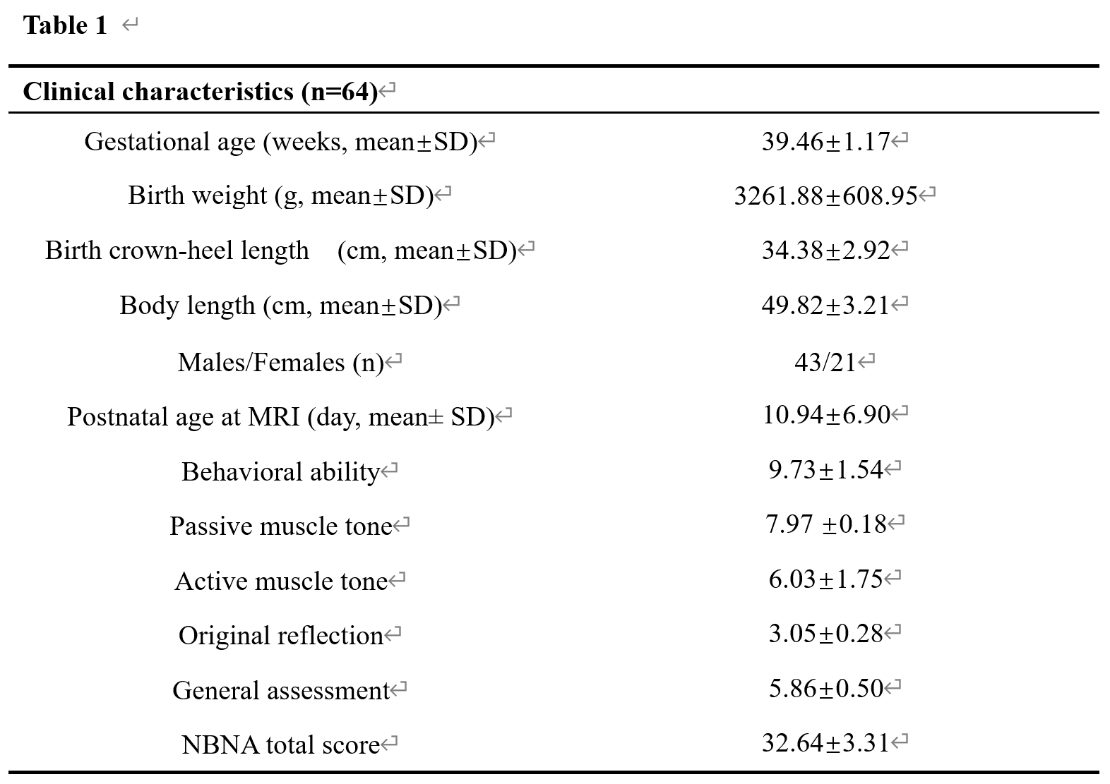
We retrospectively collected 65 newborns who underwent cranial MRI screening for craniocerebral lesions in our hospital from November 2010 to September 2017. Inclusion criteria: full-term newborns; complete MRI imaging data, including 3D-T1WI and T2WI data; complete NBNA data ; complete newborn birth indicators (birth weight, head circumference). Exclusion criteria: intracerebral hemorrhage; focal white matter damage; hypoxic-ischemic encephalopathy; diffuse white matter hyperintensity; cerebral softening; subependymal cyst degeneration; other conventional MRI abnormalities; image artifacts.Data acquisition: Use the American GE Signa HDxt 3.0 T MRI equipment, with 8-channel neonatal special head coil for scanning. The responsible nurse followed the instructions of the clinician in charge and gave 10% chloral hydrate 0.5 ml/kg orally 30 minutes before the MRI examination. The selection of subjects and vital signs related to the use of chloral hydrate were strictly followed in strict accordance with the relevant guidelines. Monitored for follow-up observation and follow-up [3]. The acquired MRI sequence and parameters are as follows: (A) 3D-T1WI was acquired using fast spoiler gradient echo sequence: TR 10.28 ms, TE 4.62 ms, FOV 240 mm×240 mm, voxel size 1 mm×1 mm×1 mm. (B) Using fast spin echo sequence acquisition T2WI: TR 4 200 ms, TE 118.91 ms, layer thickness 4 mm, FOV 180 mm × 180 mm, acquisition matrix 256 × 256. The demographic data of the included newborns are shown in Table 1.
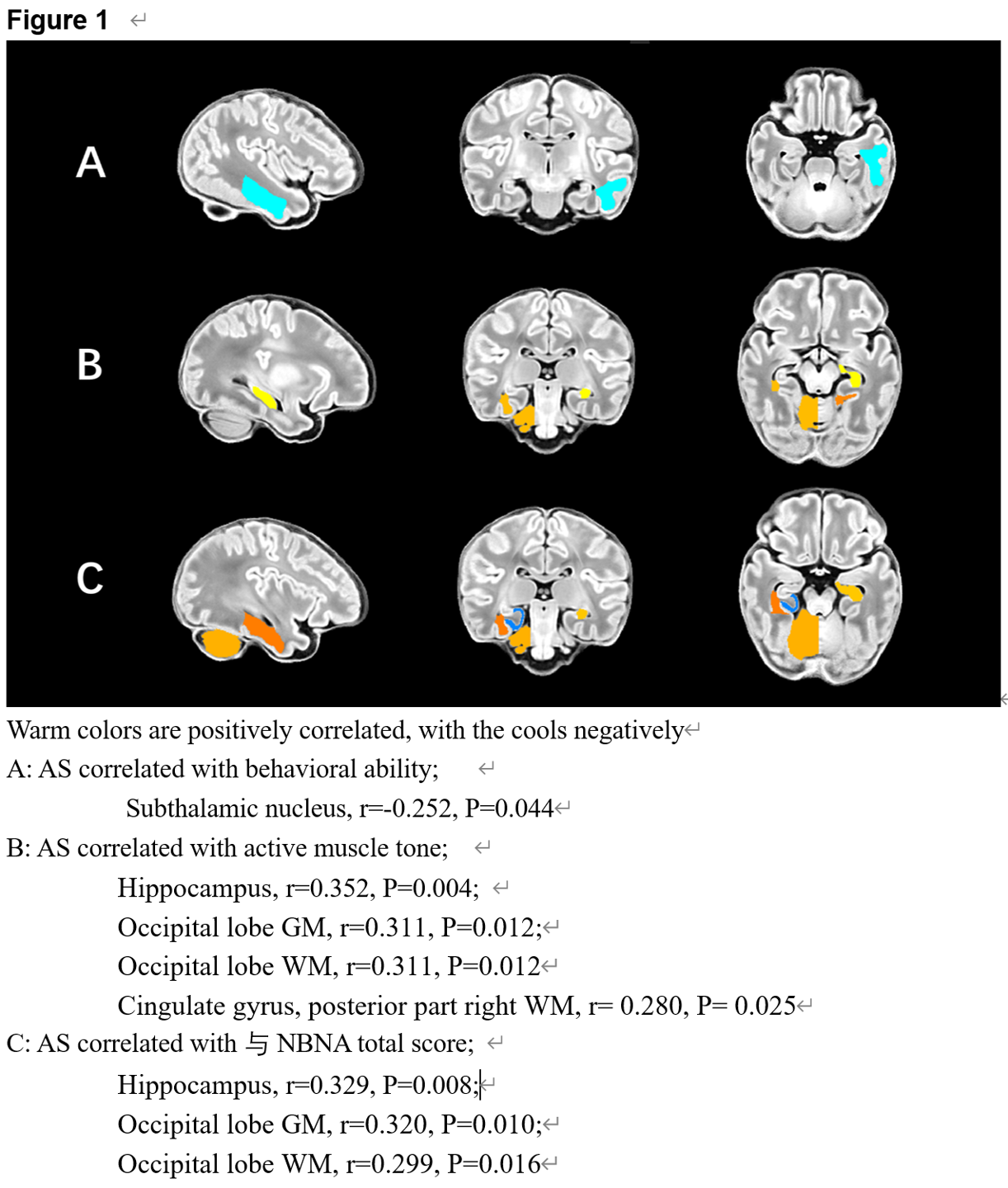
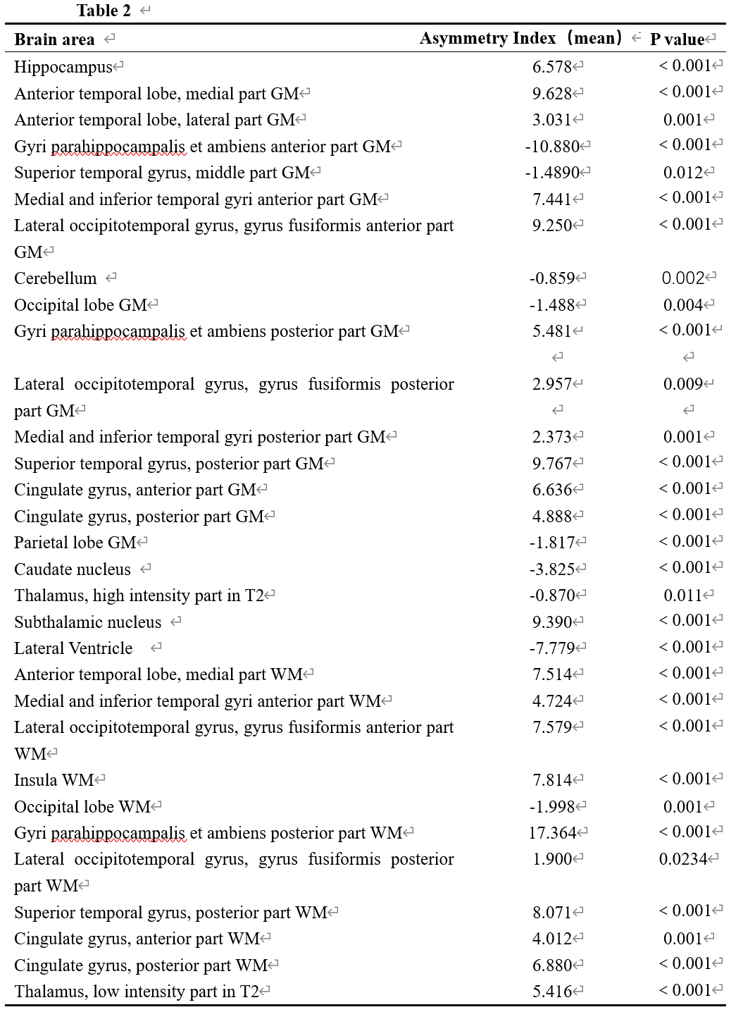
Data processing: Use the dHCP brain segmentation standard to train a deep learning model, use V-Net with a bottleneck layer to segment the brain into 87 regions. Calculate the asymmetry Index (AS). AS=100×(left-right)/(left+ right). The statistical significance of hemispherical asymmetry uses a one-sample t test (testing the reference value of 0.00). If asymmetry is found, further correlative research with neurobehavior scores is done(Table 2). The statistically different projections to the corresponding brain areas are displayed on the software FSL (Figure 1). All analyses were performed using SPSS software (SPSS version 18) and MATLAB software (MATLAB version R2016b). P<0.05 indicates that the correlation is statistically significant.
Results
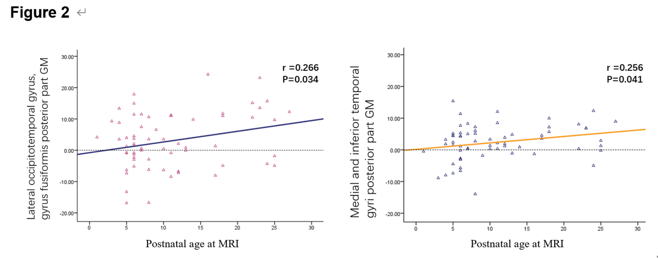
Totally 64 full-term newborns were enrolled, and the results showed that cerebral asymmetry had occurred in the full-term newborn period, significantly on the left-biased. (Table 2). This is consistent with previous conclusion of John H. Gilmore et al. 2007 [1] and. The laterality of Occipital lobe, Hippocampus, Cingulate gyrus posterior part WM showed a strong correlation with neurobehavioral scores. (Figure 1). Postnatal age at MRI is correlated with individual brain regions (Figure 2).Discussion and conclusion:
The results of this study show that full-term neonates have lateralization in the brain at this stage, significantly on the left-biased, which is consistent with the conclusion of John H. Gilmore et al (2007) [4] and Bisiacchi P et al (2021) [5]. Studies have shown that the brain asymmetry pattern of newborns is opposite to that of older children and adults [4], which indicates that the cerebral asymmetry pattern changes with age, but how it changes with age needs further research. The further research may provide important insights for revealing the pathogenesis of certain diseases. The left deflection of the occipital lobe and hippocampus shows a positive correlation with neurobehavioral scores, indicating that the left occipital lobe and hippocampus participate in and play a huge role in behavioral motor functions in the early postnatal period.Acknowledgements
This study was supported by National Natural Science Foundation of China (81901516, 81901823, 81971581 and 82101815), Shaanxi Provincial Innovation Team (2019TD-018).
* Correspondence: Chao Jin, Ph.D., Professor Department of Radiology The First Affiliated Hospital of Xi’an Jiaotong University, Xi’an, Shaanxi, China E-mail: jinny.369@163.com
References
(1). Rajagopalan V , Scott J, Habas PA, Kim K, Rousseau F, Glenn OA, Studholme C (2011) Mapping directionality specific volume changes using tensor based morphometry: an application to the study of gyrogenesis and lateralization of the human fetal brain. NeuroImage 63(2):947–958. https ://doi.org/10.1016/j.neuro image .2012.03.092
(2). Alqadah A, Hsieh YW, Morrissey ZD, Chuang CF. Asymmetric development of the nervous system. Dev Dyn. 2018 Jan;247(1):124-137. doi: 10.1002/dvdy.24595. Epub 2017 Oct 13. PMID: 28940676; PMCID: PMC5743440.
(3). Kauffman R E, Banner, W., Berlin, C.M. Guidelines for monitoring and management of pediatric patients during and after sedation for diagnostic and therapeutic procedures. Pediatrics, 1992, 89: 1110-1115.
(4). Gilmore JH, Lin W, Prastawa MW, Looney CB, Vetsa YS, Knickmeyer RC, Evans DD, Smith JK, Hamer RM, Lieberman JA, Gerig G. Regional gray matter growth, sexual dimorphism, and cerebral asymmetry in the neonatal brain. J Neurosci. 2007 Feb 7;27(6):1255-60. doi: 10.1523/jneurosci.3339-06.2007. PMID: 17287499; PMCID: PMC2886661.
(5). Bisiacchi P, Cainelli E. Structural and functional brain asymmetries in the early phases of life: a scoping review. Brain Struct Funct. 2021 Mar 18. doi: 10.1007/s00429-021-02256-1. Epub ahead of print. PMID: 33738578.
Figures
Brain regions with the asymmetry index meaningful
Asymmetry Index(AS)=100×(left-right)/(left+ right)