4702
Comparison of conventional and spiral TOF-MRA in clinical applications1Magnetic Resonance Center, Zhongshan Hospital Affiliated to Xiamen University, Xiamen, China, 2Philips Healthcare, Beijing, China, 3Philips Healthcare, Shenzhen, China, 4Department of Electronic Science, Xiamen University, Xiamen, China
Synopsis
TOF-MRA is a routine noninvasive method that does not require intravenous contrast agents or exposure to radiation for brain artery assessment. However, artifacts are prone to appear in conventional TOF-MRA under situations of complicated blood vessels or changes in blood flow; and the display of distal arterioles is also not good. The spiral acquisition technique has been previously introduced for scan acceleration of TOF-MRA, while here we compared the performance of conventional and spiral TOF-MRA under similar scan duration. Results indicated that spiral TOF-MRA provide better quality MRA images, and may help improve the clinical diagnosis of vascular problems.
Introduction
Brain artery imaging is a mainstay of the diagnostic approach in cerebrovascular diseases that help make the choice of treatment and allows for patient counseling and prognostication. The common imaging modalities for evaluation of arterial pathologies include digital subtraction angiography (DSA), computed tomography angiography (CTA), and magnetic resonance angiography (MRA). DSA, as the gold standard, is associated with limitations of (a) the invasive nature of the procedure, (b) the use of iodinated contrast agents, and (c) the exposure to radiation. CTA can be fast and accurate; however, in addition to the abovementioned drawbacks (b and c) of DSA, its diagnostic accuracy is impaired in calcified vasculature. Time-of-flight MRA (TOF-MRA) is a non-invasive method that does not require exposure to radiation or application of contrast agents1-2. However, artifacts are prone to appear in conventional TOF-MRA under situations of complicated blood vessels or changes in blood flow; and the display of distal arterioles is also as good as DSA. The spiral acquisition technique has been previously introduced for scan acceleration of TOF-MRA. Actually, in addition to the speed acceleration, the spiral acquisition can also bring the TOF-MRA with improved image contrast and enhanced robustness to motion artifacts3-4. Here, we presented a clear comparison about the performance of conventional and spiral TOF-MRA under similar scan duration.Methods
70 patients who were suspected with cerebrovascular diseases and underwent the MRA examinations were included in this study. MR scans were carried out on a 3.0T scanner (Ingenia CX, Philips Healthcare, Best, the Netherlands) using a 20-channel head and neck joint coil. The conventional TOF-MRA with Cartesian acquisition was accelerated with a SENSE factor of 3. All the other parameters for the conventional and spiral TOF-MRA were with similar settings as following: TR/TE, 19/3.5; FOV, 180X180X120 mm3, voxel size, 0.8X0.8X1.2 mm3, flip angle, 18°, and the scan time of 3min40 s and 3min44s, respectively. Data for two sequences were reconstructed in the identical way by using the maximum intensity projection.Results
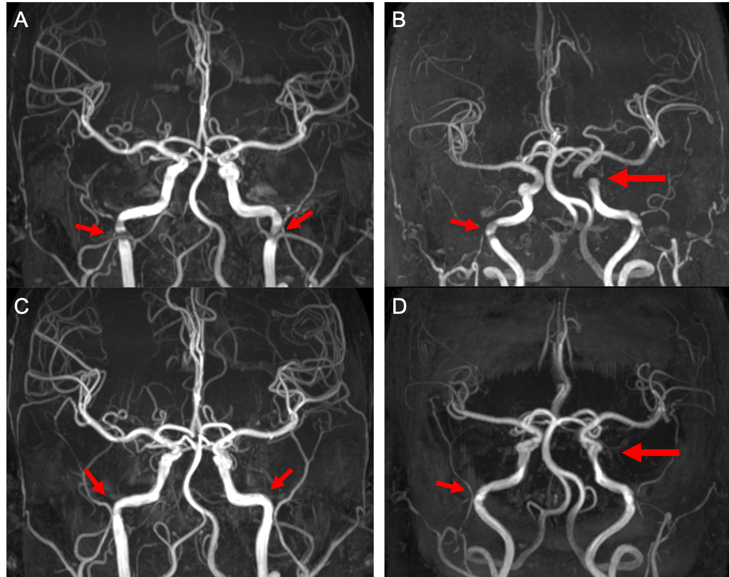
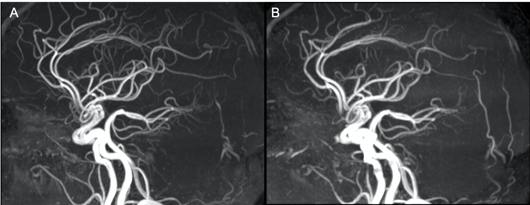
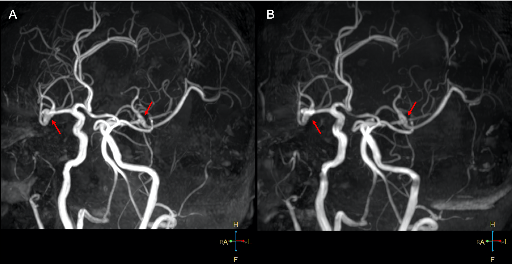
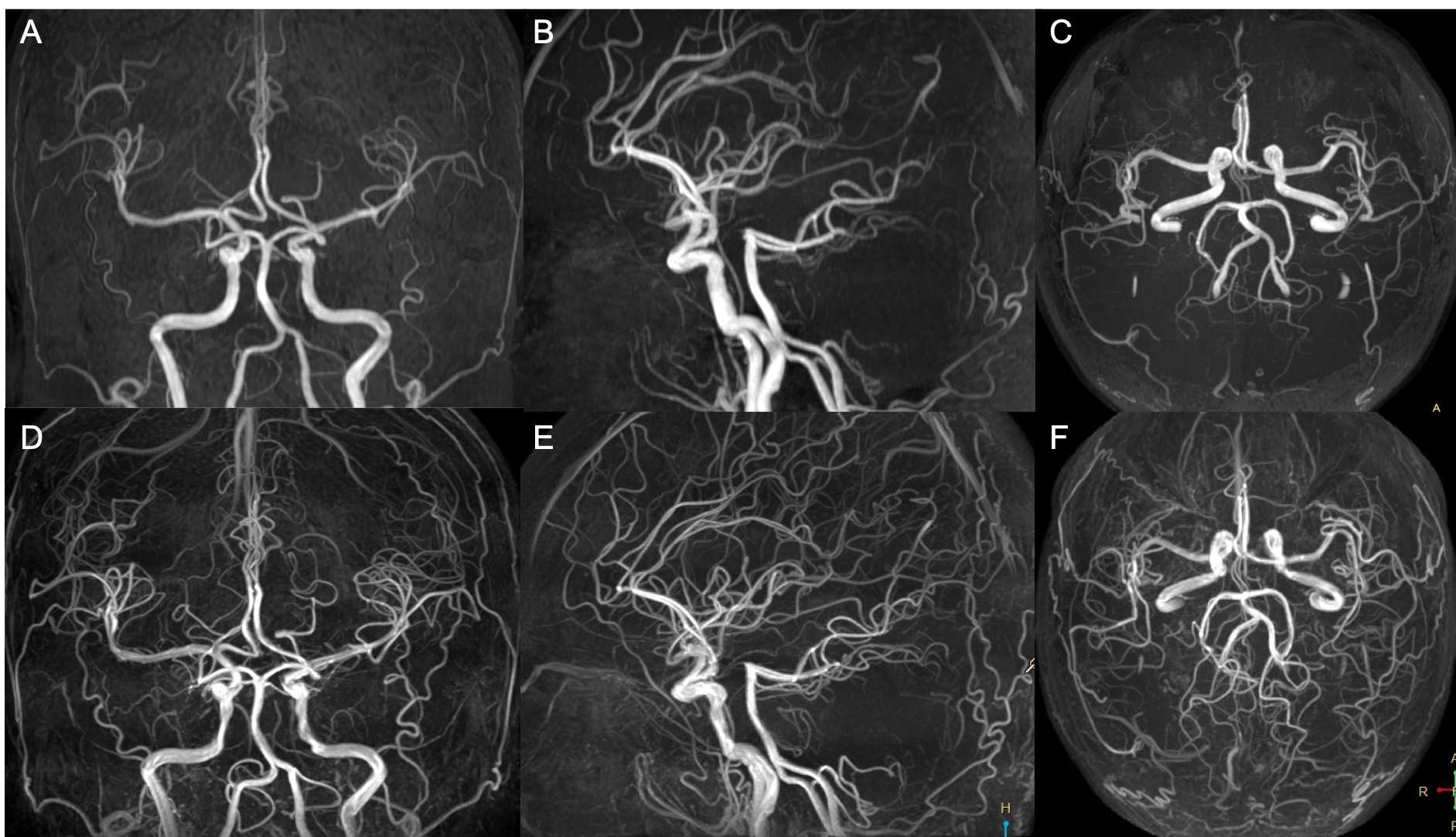
For the overall comparison across all images, we found the superior performance of spiral over the conventional TOF-MRA in different aspects. And we choose to present some specific cases to support our observations. The artifacts introduced by the seriously curved blood vessels and blood turbulence in conventional TOF-MRA can be significantly reduced in spiral TOF-MRA (Figure 2). Figure 2 shows that spiral TOF-MRA can give a better display of the distal arterioles. The tiny aneurysms at the cavernous sinus segment (Figure 3) can also be better recognized on the spiral TOF-MRA which has been confirmed by CTA. Finally, the spiral TOF-MRA can be further optimized with image quality comparable to that by DSA but at the cost of increased scan time (about 8 mins).Discussion and conclusion
Under similar scan times, the spiral TOF-MRA overperform conventional TOF-MRA in different aspects, including the display of brain artery structure, the reduction of image artifacts, and the recognition of lesions.Acknowledgements
No acknowledgement found.References
1. Miyazaki M, Akahane M. Non‐contrast enhanced MR angiography: Established techniques. Journal of magnetic resonance imaging, 2012, 35(1): 1-19.
2. Laub G A. Time-of-flight method of MR angiography. Magnetic resonance imaging clinics of North America, 1995, 3(3): 391-398.
3. Greve T, Sollmann N, Hock A, et al. Novel Ultrafast Spiral Head MR Angiography Compared to Standard MR and CT Angiography. Journal of Neuroimaging, 2021, 31(1): 45-56.
4. Greve T, Sollmann N, Hock A, et al. Highly accelerated time-of-flight magnetic resonance angiography using spiral imaging improves conspicuity of intracranial arterial branches while reducing scan time. European radiology, 2020, 30(2): 855-865.
Figures