4700
Ultra-Fast T1-Weighted Imaging with Wave-CAIPI MPRAGE for Clinical Pediatric Brain MR imaging: A Prospective Quantitative Assessment Study
Yung-Chieh Chen1, Duen-Pang Kuo1,2, and Cheng-Yu Chen1
1Department of Medical Imaging, Taipei Medical University Hospital, Taipei, Taiwan, 2Translational Imaging Research Center, Taipei Medical University Hospital, Taipei, Taiwan
1Department of Medical Imaging, Taipei Medical University Hospital, Taipei, Taiwan, 2Translational Imaging Research Center, Taipei Medical University Hospital, Taipei, Taiwan
Synopsis
Morphologic neuroimaging in children can be challenging and time-sensitive during routine clinical imaging workflow due to patient discomfort, susceptibility to motion, and frequent need of sedation. Our study quantitatively assessed the imaging performance of ultra-fast T1 3D magnetization-prepared rapid acquisition gradient echo (MPRAGE) using wave-controlled aliasing with parallel imaging (wave-CAIPI) sequence for accelerated pediatric brain MR imaging via whole brain wide analysis under different MR imaging parameter settings with and without contrast administration. Our results show that wave-CAIPI can deliver comparable image quality to standard MPRAGE with significantly reduced scan times with optimized imaging parameters.
Introduction
Good morphologic neuroimaging with magnetic resonan0ce imaging (MRI) in children is essential for clinical assessment of various neuropathological diseases, structural lesions or congenital anomalies. Patient discomfort and motion are frequent undesirable situations during routine clinical imaging practice that can often lead to suboptimal or even undiagnostic image quality, meanwhile standard structural imaging sequence such as magnetization-prepared rapid acquisition gradient echo (MPRAGE) often require prolonged scan time to obtain acceptable T1-weighted contrast, therefore image acquisition techniques such accelerated parallel imaging are commonly employed to reduce scan time1. Wave-controlled aliasing with parallel imaging (wave-CAIPI) is an emerging imaging encoding technique for highly accelerated parallel imaging that can significantly reduce scant time while retaining sufficient spatial resolution2. In this study, we quantitatively assessed the imaging performance of ultra-fast T1 3D MPRAGE scans using wave-CAIPI in comparison with conventional MPRAGE for pediatric brain imaging, in consideration of both contrast enhancement effect and different MR imaging parameter settings.Methods
50 pediatric patients under 18 of age underwent sagittal T1-weighted imaging with MPRAGE, wave-CAIPI MPRAGE with TR of 2000ms (wave-CAIPI2000), and wave-CAIPI MPRAGE with TR of 2300ms (wave-CAIPI2300) consecutively in both pre- and post-contrast imaging settings in a 3T MRI scanner using a 64-channel coil for this IRB-approved study. Signal intensity (SI) and standard deviation (SD) measurements were performed within whole-brain gray matter (GM), white matter (WM) and cerebral spinal fluid (CSF), which were segmented via a standard FSL post-processing pipeline. Signal-to-noise ratio (SNR) and contrast-to-noise ratio (CNR) were calculated based on the aforementioned parameters. The differences in SNR and CNR values among the three sequences were analyzed via ANOVA test.Results
Structural imaging with wave-CAIPI MPRAGE provided up to 76% reduction in scan time. Before contrast enhancement, the SNRs of wave-CAIPI2000 and wave-CAIPI2300 were both lower than those of MPRAGE (all p<0.01). The CNRs of wave-CAIPI2000 were lower than those of MPRAGE (all p<0.02), while most CNRs of wave-CAIPI2300 were not inferior to those of MPRAGE. After contrast enhancement, MPRAGE retained better SNRs over wave-CAIPI2000 and wave-CAIPI2300 (all p<0.01), however the CNRs were comparable among all three sequences (p=0.32-1). There were no significant differences in SNR or CNR between wave-CAIPI2000 and wave-CAIPI2300 (p = 0.39–1) regardless of contrast usage.Discussion
Pediatric structural brain imaging remains a challenging clinical task due to patient discompliance partly contributed by prolonged scan time, whereas sedation is not always available on site or even contraindicated. Therefore, methods to shorten MRI scan time are warranted and under constant improvement3. Wave-CAIPI can greatly reduce scan time through combination of characteristic corkscrew k-space trajectories and voxel aliasing patterning to minimize g-factor and artifact penalties2, thus can be applied for pediatric brain imaging with good qualitative performance4,5. On the other hand, the limitations of wave-CAIPI technique should be considered during pediatric neuroimaging since optimized imaging parameters are necessary to obtain sufficient imaging contrast and signal quality to maintain diagnostic accuracy. Nonetheless, Wave-CAIPI is a promising approach to accelerate pediatric neuroimaging and improve the clinical imaging workflow.Conclusion
Wave-CAIPI MPRAGE can facilitate rapid pediatric brain T1-Weighted structural imaging and deliver comparable image quality to conventional MPRAGE sequence with use of imaging contrast and sequence parameter optimization.Acknowledgements
No acknowledgement found.References
- Barkovich MJ, Xu D., Desikan RS, et al. Pediatric neuro MRI: Tricks to minimize sedation. Pediatr. Radiol. 48, 50–55.
- Polak D, Setsompop K, Cauley SF, et al. Wave-CAIPI for highly accelerated MP-RAGE imaging. Magn Reson Med. 2018;79(1):401-406.
- Tekes A, Senglaub SS, Ahn E S, et al. Ultrafast brain MRI can be used for indications beyond shunted hydrocephalus in pediatric patients. J. Am. J. Neuroradiol. 39, 1515–1518.
- Yim Y, Chung MS, Kim SY, et al. Wave-controlled aliasing in parallel imaging magnetization-prepared gradient echo (wave-CAIPI MPRAGE) accelerates speed for pediatric brain MRI with comparable diagnostic performance. Sci Rep. 2021;11(1):13296.
- Tabari A, Conklin J, Figueiro Longo MG, et al. Comparison of ultrafast wave-controlled aliasing in parallel imaging (CAIPI) magnetization-prepared rapid acquisition gradient echo (MP-RAGE) and standard MP-RAGE in non-sedated children: initial clinical experience. Pediatr Radiol. 2021;51(11):2009-2017.
Figures

Example of MPRAGE, wave-CAIPI and tissue
masks of WM, GM and CSF on axial image

Results of SNR and CNR analyses
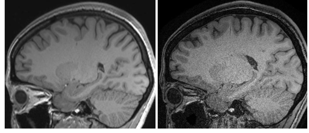
Sagittal image of MPRAGE & wave-CAIPI
DOI: https://doi.org/10.58530/2022/4700