4698
Spin-Echo Revisited: Improved Visualization of Accelerated T2W Spin-Echo Spiral Imaging with Compressed Sensing with SENSE (CS-Spiral)
Kosuke Morita1, Masami Yoneyama2, Hiroshi Hamano2, Shogo Fukuda1, Hiroyuki Uetani3, Akira Sasao3, Takeshi Nakaura3, Seitaro Oda3, Masahiro Hatemura1, and Toshinori Hirai3
1Radiology, Kumamoto university, kumamoto-shi, Japan, 2Philips Japan, minato-ku, Japan, 3Diagnostic Radiology, Graduate School of Medical Sciences, Kumamoto university, kumamoto-shi, Japan
1Radiology, Kumamoto university, kumamoto-shi, Japan, 2Philips Japan, minato-ku, Japan, 3Diagnostic Radiology, Graduate School of Medical Sciences, Kumamoto university, kumamoto-shi, Japan
Synopsis
CS-Spiral T2WI was good image quality for midbrain. We used 3 sequences; CS-Spiral, conventional-Turbo Spine Echo and neuromelanin image for six healthy volunteers. In this study, we used Compressed Sensing with Sensitivity Encoding (CS-Spiral) technology for SPIRAL acquisition to perform midbrain T2WI for neurodegenerative diseases with clinically usefulness acquisition time. It is expected that parameter settings will provide more clinically useful image quality.
INTRODUCTION
In recent years, much research has been done on neurodegenerative diseases, but its exact mechanism is still unknown. Parkinson's disease (PD) takes place after the disease has progressed and many dopamine neurons in the substantia nigra (SN) have already died. While therapies are currently being developed to prevent this neuronal death, it is essential to diagnose the disease at an earlier stage and monitor the response to therapies. Recent studies suggest that this can be accomplished by using MRI to detect neuromelanin (NM), a characteristic pigment of SN dopaminergic and nucleus pallidus noradrenergic neurons.In addition, dentatorubral-pallidoluysian atrophy (DRPLA) was characterized by myoclonus, epilepsy, and mental retardation in young patients at the age of onset, and cerebellar ataxia, ataxia, and dementia in adults and senile patients. Previous reports have shown that T2WI MRI of patients with senile onset showed symmetrical high-signal lesions in the cerebral white matter, globus pallidus, thalamus, midbrain, and pyramid, but MRI of younger patients did not show such lesions, and CT did not show lesions in the globus pallidus or brainstem. Thus, MRI findings of the midbrain are very important.Proton density-weighted imaging contrast, such as neuromelanin imaging, is often used for midbrain MRI diagnosis. The reason for this is that turbo spin-echo (TSE) techniques have been established and have shortened the acquisition time, but the T2 contrast has become insufficient. However, pure spin echo (non-TSE) acquisition is clinically difficult to obtain because of its very long acquisition time. Spiral SE sequence, which is capable for acquiring pure spin echoes while enabling a short acquisition time by using long acquis ion window (readout time) is promising to obtain. However, since long acquis ion window greatly increases magnetic susceptibility effects, conventional Spiral should apply shorter acquisition window, resulting in a long acquisition time. In this study, we attempt to combine Compressed Sensing with Sensitivity Encoding (CS-SENSE) technology for Spiral SE acquisition (CS-Spiral) to perform midbrain pure spin-echo T2WI for neurodegenerative diseases with clinically feasible acquisition time.METHODS
The subjects were six healthy volunteers who consented to the study. We used 3 sequences: CS-Spiral, conventional TSE and neuromelanin image (NMI). All sequences were 2D transverse acquisition in a cross section centered on the midbrain and orthogonal to the brainstem. Scan parameters of CS-Spiral was as follows: TR/TE = 3000/80 ms, slices thickness = 3 mm, voxel size = 0.74 × 0.74 × 3.00mm, CS factor = 2.0, acquisition window = 19.9 ms, spiral interleaves = 22, 3point dixon and acquisition time = 3:21. Scan parameters of TSE was as follows: TR/TE = 4416/80 ms, slices thickness = 3 mm, voxel size = 0.45 × 0.68 × 3.00mm, SENSE factor = 2.0, TSE factor = 9, NSA = 2 and acquisition time = 2:16. Scan parameters of NMI was as follows: TR/TE = 600/7.4 ms, slices thickness = 3 mm, voxel size = 0.69 × 1.00 × 3.00mm, SENSE factor = no, TSE factor = 2, NSA = 3 and acquisition time = 10:10. CS-SPIRAL were compared to conventional TSE images for image quality, especially for the reduction and contrast of image noise. ROIs were placed on left and right gray matter (WM). We calculated the Contrast Ratio (CR) of signal intensities (SI) of white matter (WM) and SI of red nucleus (RN) and SN on T2W image in respective volunteers as follows: CR = SI of WM / SI of RN or SI of SN.RESULTS & DISCUSSION
Figure 1 shows results of CRs. A volunteer images are shown in Figure 2 and Figure 3. CRs of CS-Spiral and TSE for SN and RN were 1.78 ± 0.11, 1.57 ± 0.12, 1.58 ± 0.13 and 1.47 ± 0.09. Figure 2 shows good image quality for a volunteer. There was not susceptibility artifact and motion artifact. On the other hands, figure 3 shows poor image quality. In this case, volunteer’s motion was occurred blurred image. In figure 2 and 3, image quality was appeared CS-Spiral images were better than TSE images. Using the Spiral SE sequence, we were able to acquire a pure T2 spin echo signal. Furthermore, CS technique was applied to prevent prolongation of the acquisition time even with a short acquisition window. However, reducing the susceptibility artifact and motion artifact in the CS-Spiral sequence is still challenging, further sequence improvements would be needed.CONCLUSION
CS-Spiral provided pure T2 spin echo signal in the midbrain images within clinically feasible scan time. Revisiting spin-echo images might be useful to obtain further important findings of midbrain structures.Acknowledgements
No acknowledgement found.References
- Thomas Sartoretti, Elisabeth Sartoretti, Luuk van Smoorenburg, et al. Spiral 3-Dimensional T1-Weighted Turbo Field Echo: Increased Speed for Magnetization-Prepared Gradient Echo Brain Magnetic Resonance Imaging, Invest Radiol. 2020;55:775-784.
- Tobias Greve, Nico Sollmann, Andreas Hock, et al. Novel Ultrafast Spiral Head MR Angiography Compared to Standard MR and CT Angiography, J Neuroimaging 2021;31:45-56.
- Meyer CH, Hu BS, Nishimura DG, et al. Fast spiral coronary artery imaging. Magn Reson Med 1992;28:202-213.
- Amann M, Bock M, Floemer F, et al. Three-dimensional spiral MR imaging: application to renal multiphase contrast-enhanced angiography. Magn Reson Med 2002;48:290-296.
- Block KT, Frahm J. Spiral imaging: a critical appraisal. J Magn Reson Imaging 2005;21:657-668.
- BornertP,SchombergH,AldefeldB,etal.Improvementsinspiral MR imaging. MAGMA 1999;9:29-41.
Figures
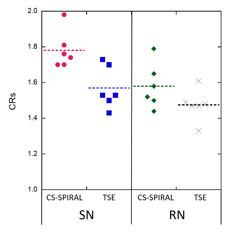
Comparison of CRs between CS-SPIRAL and TSE for SN and RN. Dot lines show median value for each sequence.

Representative volunteer images. T2W images on midbrain show CS-SPIRAL (left), TSE (center) and NMI (right). SN and RN on CS-SPIRAL image were better than on TSE image.

Representative volunteer images. T2W images on midbrain show CS-SPIRAL (left), TSE (center) and NMI (right). In CS-SPIRAL image, the signal intensities of the red nucleus and substantia nigra were similar to those of TSE image.
DOI: https://doi.org/10.58530/2022/4698