4697
Comparison of integrated slice-specific dynamic shimming (ishim) and single shot EPI techniques in patients with Crohn’s disease1Department of Radiology,The Second Xiangya Hospital, Central South University, Changsha, China, 2MR Scientific Marketing, Siemens Healthineers, Wuhan, China, 3MR Application, Siemens Healthineers, Changsha, China, 4MR Application Predevelopment, Siemens Healthcare GmbH, Erlangen, Germany
Synopsis
In this study, image quality and apparent diffusion coefficient (ADC) values of conventional single-shot spin-echo echo-planar imaging (SS-EPI) and prototype integrated slice-specific dynamic shimming EPI (ishim_EPI) diffusion-weighted imaging in patients with Crohn’s disease were compared. Results showed that ishim_EPI had better image quality, including fewer distortion artifacts and clearer edges of the lesions. High correlation and good agreement of ADC value were found between the two techniques. Ishim_EPI DWI technique is recommended to replace conventional SS_EPI for the detection of lesions in patients with Crohn’s disease and further in routine intestinal examination.
Introduction
Crohn’s disease (CD) is an inflammatory bowel disorder characterized by noninfectious chronic inflammation of the gastrointestinal tract. Diffusion-weighted imaging (DWI) has been shown useful for detecting active bowel inflammation in Crohn’s disease without MRI contrast.1-3 However, conventional single-shot spin-echo echo-planar imaging (SS_EPI) suffers from geometric distortion due to changes of susceptibility, especially for the organs with interfaces of large different susceptible tissues, such as intestinal tracts. Therefore, the performance of DWI in lesion detection is doubtful sometimes. In previous studies on Crohn’s disease, DWI had large heterogeneity of diagnostic accuracy, which may be caused partially by DWI distortion. Recently, a novel DWI technique, integrated slice-specific dynamic shimming (iShim)_EPI, has been developed that can reduce distortion artifacts and improve image quality compared with SS_EPI DWI.4-6 However, the application of ishim_EPI in the intestinal tract has not yet been assessed. The aim of this study is to compare image quality and detection capability of lesions between SS_EPI and iShim_EPI in patients with CD.Materials and Methods
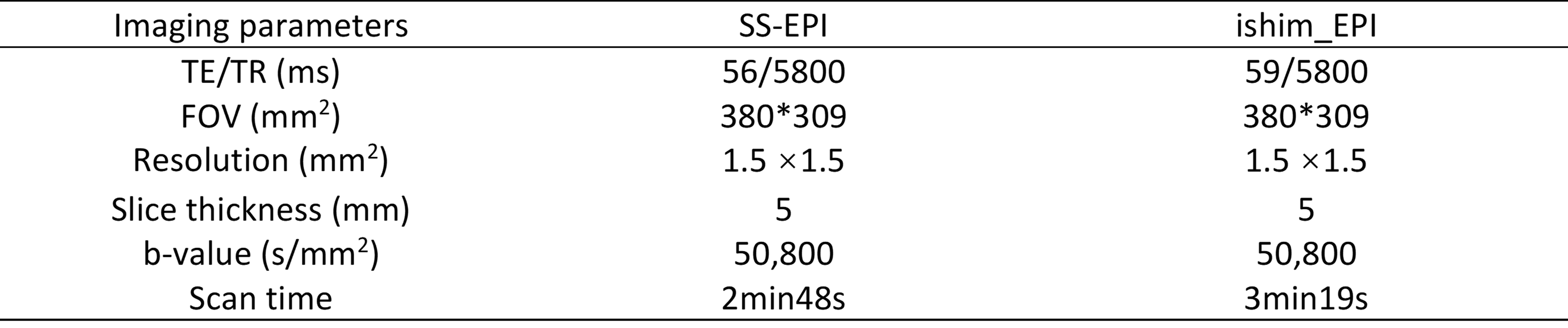
This prospective study was approved by the Ethics Committee. Eleven patients with CD (n=11, 5 females, age = 53 +/- 4.6 years) were enrolled, and all patients signed an informed written consent. All patients underwent intestinal MR examinations on a 3T MRI scanner (MAGNETOM Skyra, Siemens Healthcare, Erlangen, Germany) with 18-channel abdominal phase array coil. Besides to conventional MR sequences, SS_EPI and prototypical ishim_EPI DWI sequences were included, and their imaging parameters are listed in Table 1. The distortion artifacts, the edge of lesion, and overall image quality of DWI images were assessed using a 5-point Likert scale by one blinded gastrointestinal radiologist with 8-10 years of experience. In addition, several regions of interest (ROI), located on the lesions and normal intestinal wall, were drawn on the b=800 s/mm2 DWI image, and then copied to apparent diffusion coefficient (ADC) map for both SS_EPI and ishim_EPI DWI. Mean ADC value of each ROI was obtained. Pearson and Bland-Altman analysis were adopted to assess the correlation and agreement of ADC between the two methods. p value <0.05 was considered statically significant.Results
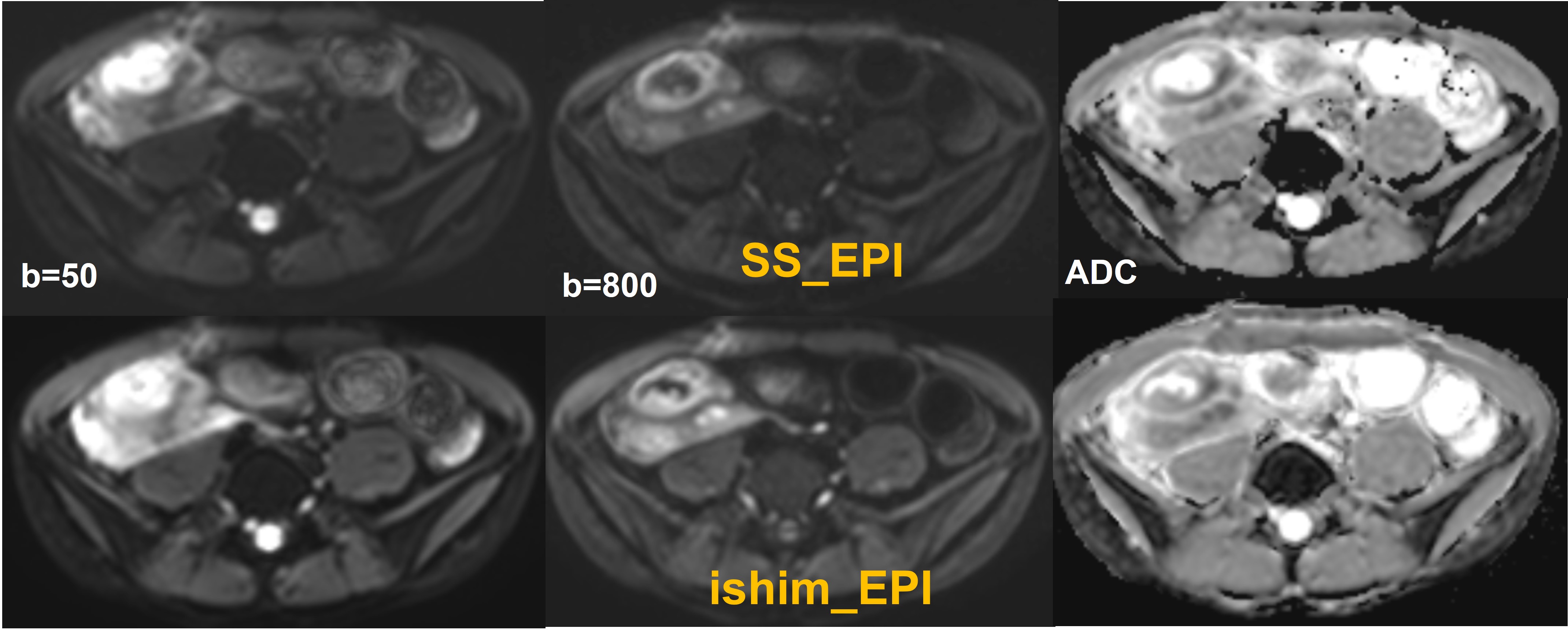
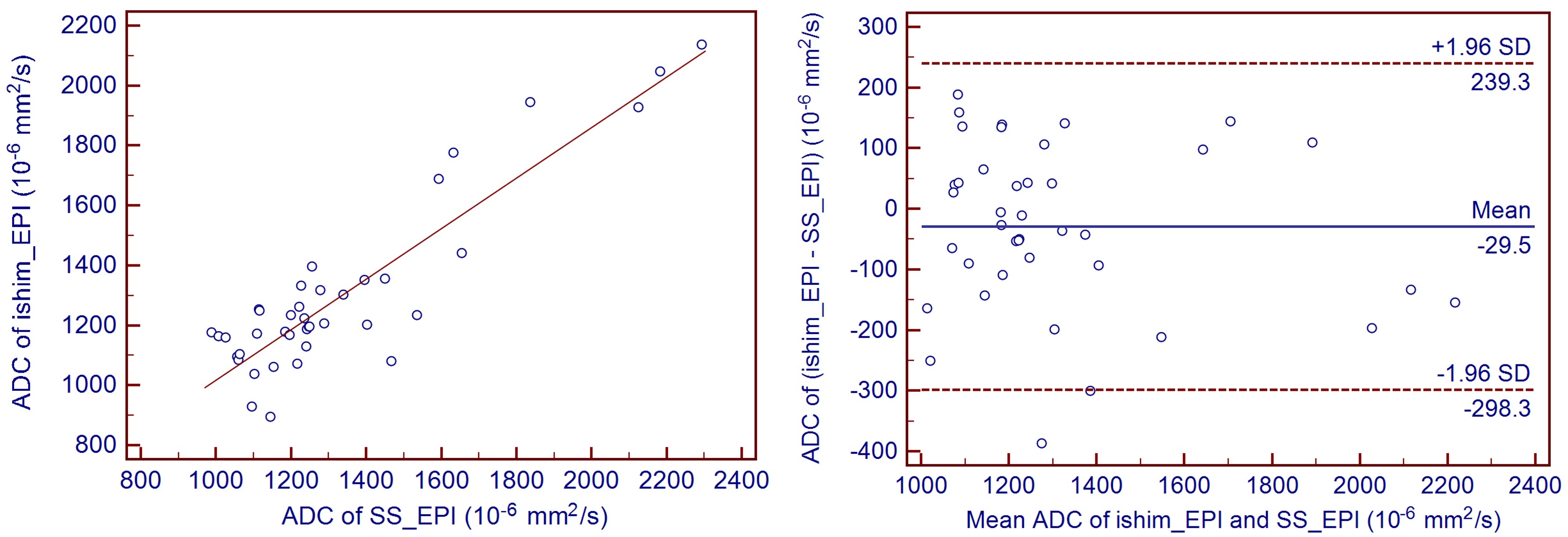
Table 2 shows the subjective scores of image quality. Ishim_EPI had significantly higher image quality scores than those of SS_EPI (p<0.05). Ishim_EPI showed less blur, increased sharpness of boundaries, and fewer distortion artifacts (Figure 1). ADC value of ishim_EPI had excellent correlation (r=0.901, p<0.001) and good agreement with SS_EPI, and Bland-Altman analysis showed that ADC values of ishim_EPI were lower than that of SS_EPI, with a bias of 29.5 ×10⁻⁶ s/mm2 (Figure 2). No significant difference was found between the two methods (p>0.05).Discussion
As an important MRI technique, DWI can increase the speed and accuracy of lesion detection and localization for whole body applications. However, due to the conventional SS_EPI which is prone to susceptibility artifacts, DWI’s applications were limited. In this preliminary study, ishim_EPI outperformed conventional SS_EPI with better image quality, less distortion and clearer edges of lesions. The main reason is that the ishim_EPI technique combines integrated slice-specific dynamic shimming and pixel-wise unwrapping distortion correction. Therefore, it adjusts local B0 inhomogeneity effects and reduces image distortions and signal voids. In addition, high correlations and good agreements of ADC were found between the two methods, which are consistent with findings in previous studies [4-6]. ADC is an important parameter in lesion identification and prediction. Our results for ADC vales mean that ishim_EPI had the same performance in the quantitative analysis of lesions and normal tissue for Crohn’s disease. In the future, due to the improved image quality, ishim_EPI technique may replace conventional SS_EPI in routine examinations for Crohn’s disease.Conclusions
IShim_EPI technique is a potential technique to reduce the distortion and improve image quality, and hence useful for the detection of intestinal lesions in patients with Crohn’s disease.Acknowledgements
No acknowledgement found.References
1. Hordonneau C, Buisson A, Scanzi J, et al. Diffusion-weighted magnetic resonance imaging in ileocolonic Crohn’s disease: Validation of quantitative index of activity. Am J Gastroenterol 2014;109(1):89-98.
2. Ream JM, Dillman JR, Adler J, et al. MRI diffusion-weighted imaging (DWI) in pediatric small bowel Crohn disease: Correlation with MRI findings of active bowel wall inflammation. Pediatr Radiol 2013;43(9):1077-1085.
3. Chang HC, Chen G, Chung HW, et al. Multi-shot Diffusion-Weighted MRI With Multiplexed Sensitivity Encoding (MUSE) in the Assessment of Active Inflammation in Crohn's Disease. J Magn Reson Imaging. 2021. doi: 10.1002/jmri.27801.
4. Stocker D, Manoliu A, Anton SB, et al. Image Quality and Geometric Distortion of Modern Diffusion-Weighted Imaging Sequences in Magnetic Resonance Imaging of the Prostate, Investigative Radiology, 2017.
5. Li H, Liu L, Shi Q, et al. Bladder cancer: detection and image quality compared among iShim, RESOLVE, and ss-EPI diffusion-weighted MR imaging with high b value at 3.0 T MRI, Medicine, 2017,96: 50.
6. Chen L, Sun P, Hao Q, et al. Diffusion-weighted MRI in the evaluation of the thyroid nodule: Comparison between integrated-shimming EPI and conventional 3D-shimming EPI techniques, Oncotarget, 2018, 9: 26209-26216.
Figures