4696
Performance of PROPELLER FSE T2WI in reducing metal artifacts for patients with various material porcelain fused to metal1Department of Imaging, The Second Hospital of Shanxi Medical University, Taiyuan, China
Synopsis
The metal artifacts caused by metal copings in porcelain fused to metal (PFM) always impair MRI quality. This study aimed to compare MRI quality between common FSE T2WI with PROPELLER FSE T2WI for patients with various metal copings. Fifty-nine subjects with the most commonly used PFM were recruited and imaged at 1.5T MRI system. By comparing the image quality and artifact areas, we found PROPELLER technique was less sensitive to metal artifacts than common FSE. It can be proposed the PROPELLER FSE T2WI to be used as clinical MRI examination of oral cavity and maxillofacial part for patients with PFM.
INTRODUCTION
Porcelain fused to metal (PFM) restorations are increasingly used in prosthetic dentistry. The metal artifacts caused by metal copings in PFM always impair magnetic resonance image (MRI) quality of oral cavity and maxillofacial. It is important to identify preferable material compositions of metal coping in PFM and investigate methods to reduce or avoid metal artifacts caused in patients with PFM, so as to guide clinicians to making individualized treatment and MRI scanning regimens.Although several MRI sequences have developed to reduce susceptibility artifacts, the clinical use of these methods were restricted due to complex principle and higher demand of MRI equipment in hardware and software1-3. The recent study confirmed that the PROPELLER sequence could decreases metallic artifacts apart from motion artifact4. Therefore, we aimed to compare MR image quality between common fast spin echo T2 weighted imaging (FSE T2WI) with periodically rotated overlapping parallel lines with enhanced reconstruction (PROPELLER) FSE T2WI for patients with PFM of various metal copings and investigate the value of PROPELLER technique in reducing metal artifacts.METHODS
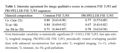
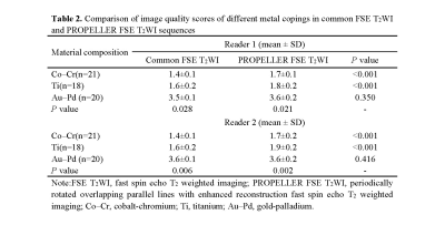
This prospective study enrolled participants with PFM of three different metal copings (cobalt-chromium (Co-Cr)alloy, pure titanium(Ti), gold-palladium(Au-Pd) alloy) between July 2020 and March 2021. Common FSE T2WI and PROPELLER FSE T2WI sequences were applied in participants for axial imaging of head, including maxillofacial region. Two radiologists evaluated overall image quality of section in PFM using a 5-point scale qualitatively and measured the maximum artifact area and artifact signal-to-noise ratio (SNR) quantitatively. The image quality scores and artifact areas in different material compositions and sequences were compared respectively. Statistical evaluations were performed by using the Wilcoxon and the Friedman test, with inter-reader variability calculated using Cohen’s kappa. Differences with P < 0.05 were regarded as statistically significant.RESULTS
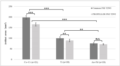
Fifty-nine participants (24 male and 35 female;mean age ± SD, 63 ± 5.2 years) were evaluated. The metal coping with the least artifacts and the optimum image quality shown in common FSE T2WI and PROPELLER FSE T2WI were in Au–Pd alloy, Ti, and Co–Cr alloy order. Performance of PROPELLER FSE T2WI in terms of artifact reduction was dependent on the material composition. PROPELLER FSE T2WI was superior to common FSE T2WI in improving image quality and reducing artifact area for Co-Cr alloy (17.0±0.2% smaller artifact area, p<0.001) and Ti (11.6± 0.7 % smaller artifact area, p=0.005), but performed as well as common FSE T2WI for Au-Pd alloy. For all PFMs, PROPELLER FSE T2WI significantly reduced signal-to-noise ratio (SNR) of artifact (393.57±89.75 VS. 214.05±70.45, p < 0.001) when compared to common FSE T2WI.DISCUSSION
Our study showed that the metal coping with the least artifacts and the optimum image quality shown in common FSE T2WI and PROPELLER FSE T2WI were in Au–Pd alloy, Ti, and Co–Cr alloy order. The reason are most likely due to the specific ferromagnetic compositions of these alloys. Cobalt and chromium are ferromagnetic metals, they distort local magnetic fields, causing large artifacts that make image interpretation impossible. Titanium itself has ferromagnetic properties but has a lower magnetic susceptibility. Although gold is a diamagnetic substance, gold alloys contain traces of other ferromagnetic metals could also explain the ability to degrade MRI images5,6.Our qualitative and quantitative studies confirmed that PROPELLER FSE T2WI significantly improved imaging quality and reduced artifact areas compared to the common FSE T2WI sequence in patients with the metal coping of Co–Cr alloy and pure Ti. These findings can be attributed to 1) PROPELLER’s unique radial k-space acquisitionsequencing, combined with a fast-spin echo (FSE) technique, which diminishes artifact in the phase-encoding direction 7. 2) Susceptibility effects primarily affect T2*signal decay, by inducing local distortions in the static magnetic field. Therefore, the PROPELLER refocuses T2 with the use of a spin-echo pulse prior to each read out, and removes the distorted T2 * signal component from the signal, so as to reduce the subsequent image distortion caused by magnetic susceptibility 4.
CONCLUSION
The different metal coping of PFM generated varying degrees of metal artifact areas in MR imaging. Compared with common FSE T2WI, the PROPELLER can effectively reduce metal artifacts in dental MR imaging especially in the metal coping of Co-Cr alloy in PFM. It can be proposed the PROPELLER FSE T2WI to be used as clinical MRI examination of oral cavity and maxillofacial part for patients with PFM restorations.Acknowledgements
No acknowledgement found.References
1. Tran LTX, Sakamoto J, Kuribayashi A, et al. Quantitative evaluation of artefact reduction from metallic dental materials in short tau inversion recovery imaging: efficacy of syngo WARP at 3.0 tesla. Dentomaxillofac Radiol. 2019;48(7):20190036.
2. Liebl H, Heilmeier U, Lee S, et al. In vitro assessment of knee MRI in the presence of metal implants comparing MAVRIC-SL and conventional fast spin echo sequences at 1.5 and 3 T field strength. J Magn Reson Imaging. 2015;41(5):1291-9.
3. Hilgenfeld T, Prager M, Schwindling FS, et al. MSVAT-SPACE-STIR and SEMAC-STIR for Reduction of Metallic Artifacts in 3T Head and Neck MRI. Am J Neuroradiol. 2018;39(7):1322-1329.
4. Czarniecki M, Caglic I, Grist JT, et al. Role of PROPELLER-DWI of the prostate in reducing distortion and artefact from total hip replacement metalwork. Eur J Radiol. 2018;102:213-219.
5.Murakami S, Verdonschot RG, Kataoka M, et al. A standardized evaluation of artefacts from metallic compounds during fast MR imaging. Dentomaxillofac Radiol. 2016;45(8):20160094.
6.Tymofiyeva O, Vaegler S, Rottner K, et al. Influence of dental materials on dental MRI. Dentomaxillofac Radiol. 2013;42(6):20120271.
7. Smeets R, Schöllchen M, Gauer T, et al. Artefacts in multimodal imaging of titanium, zirconium and binary titanium-zirconium alloy dental implants: an in vitro study. Dentomaxillofac Radiol. 2017;46(2):20160267.
Figures

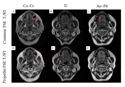
Figure 1. Images of common FSE T2WI (A,B,C) and PROPELLER FSE T2WI(D,E,F) in different participants. Images of the metal coping of Co-Cr alloy in participant 1 (A,D); Images of the metal coping of pure Ti in participant 2 (B,E); Images of the metal coping of Au–Pd alloy in participant 3 (C,F). The metal coping with the least artifacts shown in common FSE T2WI and PROPELLER FSE T2WI are in Au–Pd, Ti, and Co–Cr order.The PROPELLER FSE T2WI had reduced artifact areas and improved image quality compared to the common FSE T2WI.

Figure 2. Comparison of artifact areas of common FSE T2WI and PROPELLER FSE T2WI sequences caused by the PFM of different metal coping. Highly significant difference between common FSE T2WI and PROPELLER FSE T2WI (P < .001).n.s,not significant; **, P < .01;***, P < .001.


