4695
Highly Accelerated Volumetric Brain Imaging with META and Deep Learning Reconstruction
Naoyuki Takei1, R Marc Lebel2, Suryanarayanan Kaushik3, Shohei Fujita4,5, Issei Fukunaga4, Shigeki Aoki4, Suchandrima Banerjee6, and Tetsuya Wakayama1
1GE Healthcare, Tokyo, Japan, 2GE Healthcare, Calagary, AB, Canada, 3GE Healthcare, Waukesha, WI, United States, 4Radiology, Juntendo University School of Medicine, Tokyo, Japan, 5Radiology, The University of Tokyo Graduate School of Medicine, Tokyo, Japan, 6GE Healthcare, Menlo Park, CA, United States
1GE Healthcare, Tokyo, Japan, 2GE Healthcare, Calagary, AB, Canada, 3GE Healthcare, Waukesha, WI, United States, 4Radiology, Juntendo University School of Medicine, Tokyo, Japan, 5Radiology, The University of Tokyo Graduate School of Medicine, Tokyo, Japan, 6GE Healthcare, Menlo Park, CA, United States
Synopsis
We combine advanced acquisition and reconstruction techniques for highly accelerated 3D brain imaging. A hybrid acquisition called META (Mixed Echo Train Acquisition) generates 3D T1W, T2W and FLAIR contrasts simultaneously and with high SNR efficiency. A 3D deep learning reconstruction (AIR Recon DL) reduces image noise, reduces artifacts, and enhances resolution relative to conventional reconstructions, solving the trade-off between SNR, scan time, and resolution. META with the DL Recon achieved 1mm isotropic T1W, T2W and FLAIR images within 4 minutes. These combinations are well suited in high resolution volumetric brain imaging.
Introduction
MR volumetric imaging provides the acquisition of thin contiguous slices for multiplanar reformatting to depict detailed anatomical disease with high sensitivity which is why it is widely adopted in brain protocols. 3D imaging also has an inherent SNR efficiency advantage over 2D since each RF pulse excites the entire imaging volume and each acquisition represents an average of the entire sampled volume during two-dimensional phase encodings. To maximize this advantage, it is preferable not only to use large number of phase encodings, but also to increase data acquisition efficiency, which defines the total data acquisition window per TR, i.e., to reduce the dead time of pulse sequence1. Mixed Echo Train Acquisition (META)2 is an efficient 3D acquisition technique for multi-contrast imaging using hybrid acquisitions of GRE and FSE in one TR to generate T1W, T2W, and T2-FLAIR images simultaneously. Deep learning reconstruction (DL Recon)3 provides an opportunity for increased SNR, reduced artifacts, and enhanced resolution. We propose using a META acquisition with DL Recon to obtain 1mm isotropic resolution of T1W, T2W, and T2-FLAIR for highly accelerated brain imaging.Methods
Figure.1 shows the META pulse sequence diagram and reconstruction pipeline of the proposed method. The first FSE with variable refocusing flip angle builds T2 weighted contrast. Followed by the first inversion pulse to null fluid signal, the second variable refocused FSE provides FLAIR contrast. Then the second inversion pulse to make T1 weighted contrast is followed by GRE. Echo train length (ETL) is the same in both FSE and GRE. Three red bold lines indicate data acquisition window in TR. The 3D segmented acquisition continues until it fills all the necessary data in the k-space. The image reconstruction uses Cartesian parallel imaging, ARC4 and compressed sensing, HyperSense5. The 3D DL Recon used in our study (AIR Recon DL, GE Healthcare)3 is a deep convolutional residual encoder network trained to reconstruct images from MR data with reduced noise, reduced Gibbs ringing, and enhanced resolution. The convolutional network is embedded in the image reconstruction, operating on raw complex image volumes. To compare with the conventional 3D acquisition of each T1w, T2w, and FLAIR scan, healthy volunteer scan was performed under site IRB approval with the scan protocol in Table 1. A 3.0 T System (Discovery MR 750w, GE Healthcare, Waukesha, WI, U.S.A.) with 32 channel coils (MR instrument) was used. For META scan parameter, the same variable refocusing flip angle of FSE was used in T2w and FLAIR. Sequential view ordering and centric view ordering were used in FSE and GRE, respectively. Spoiled-GRE (SPGR) was used in GRE with flip angle 15 degree. The same ETL of 200 was used to complete all the data acquisition at the same time. ROI measurement was performed to calculate image contrast of white matter (WM)/gray matter (GM) for T1W, gray matter (GM)/white matter (WM) for T2W and FLAIR , and gray matter (GM)/CSF for FLAIR.Results
The scan time of META, conventional 3DT1W, 3DT2W, and 3DFLAIR was 3:42, 2:10, 2:46, and 4:18, respectively. Figure.2 shows in-plane sagittal images of META and conventional T1W, T2W, and FLAIR. META had the comparable visualization to conventional images in terms of SNR, image contrast, and image artifact for T1W and T2W. FLAIR of META appeared to be stronger T2 weighted image than conventional FLAIR and sub-optimal CSF nulling. Figure.3 shows the reformatted axial images of META and conventional T1W, T2W, and FLAIR providing reduced blurring because of isotropic spatial resolution. T1W in META exhibited improved SNR with the DL Recon compared to conventional T1W under the same conditions of BW/Pixel and parallel imaging factor. For the ROI measurement, WM / GM was 1.35 and 1.38 for META-T1W and conventional T1W. GM/WM was 1.66 and 1.49 for META-T2W and conventional T2W. GM/WM was 1.43 and 1.35 for META-FLAIR and conventional FLAIR. GM/CSF was 1.81 and 8.94 for META-FLAIR and conventional FLAIR.Discussions and conclusion
We have demonstrated that META with DL Recon achieved 1mm isotropic T1W, T2W, and FLAIR images within 4 minutes and 2.5 times faster than the total scan time of conventional 3D T1W, T2W, and FLAIR that are acquired under the reasonable scan parameters about TR, ETL, and parallel imaging factor 2x2 for the comparison. DL Recon provided much higher SNR, cleaner, and sharper image on META images. It was noted that more than 3x3 acceleration factor with parallel imaging alone is needed in the conventional scans to meet the scan time of META. Additionally, META generates images from three commonly prescribed sequences (T1, T2, T2FLAIR) in one scan button push, which helps in workflow simplification by reducing scan prescription time, prescan time as well as the actual scan time compared to the T1W, T2W, and FLAIR images being acquired individually.In conclusion, the combination of advanced techniques using META for increased 3D SNR efficiency and DL Recon for reduced image noise provided highly accelerated scan time, which be well suited for brain volumetric imaging by leveraging the benefit of DL Recon that solves the trade-off between SNR, scan time, and image quality.Acknowledgements
No acknowledgement found.References
- Johnson G, Wadghiri YZ, Turnbull DH. 2D multislice and 3D MRI sequences are often equally sensitive. Magnetic Resonance in Medicine. 1999;41(4):824–8
- Takei, N. et al. Simultaneous 3D T1 weighted, T2 weighted and FLAIR imaging using Serial Connection of Echo Train Acquisitions (SCETA) for Multi- contrast imaging. in Proc. Intl. Soc. Mag. Reson. Med 1246 (2021).
- Lebel RM. Performance characterization of a novel deep learning-based MR image reconstruction pipeline. 2020. arXiv preprint, arXiv:2008.06559.
- Brau AC, Beatty PJ, Skare S, Bammer R. Comparison of reconstruction accuracy and efficiency among autocalibrating data‐driven parallel imaging methods. Magn Reson Med 2008; 59: 382– 395.
- King et al, Proc. Intl. Soc. Mag. Reson. Med. 18 (2010), 4881
Figures
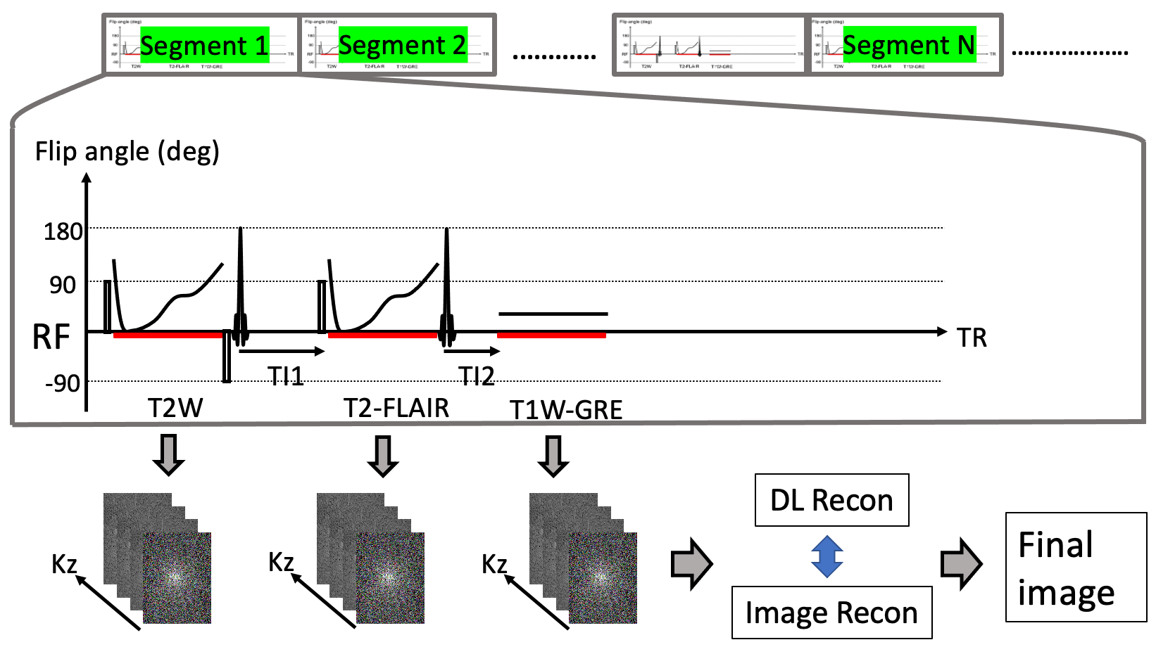
Figure.1. A schematic
pulse sequence diagram of META (Mixed Echo Train Acquisition) and image
reconstruction of DL Recon to acquire T1W, T2W, and FLAIR using the hybrid
acquisition of FSE and GRE. Based on the segmented 3D acquisition, efficient 3D
data acquisition is used to increase the total data acquisition window per TR.
After obtaining the data of 3D k-space, the recon pipeline including DL recon embedded
in the image reconstruction creates the final image
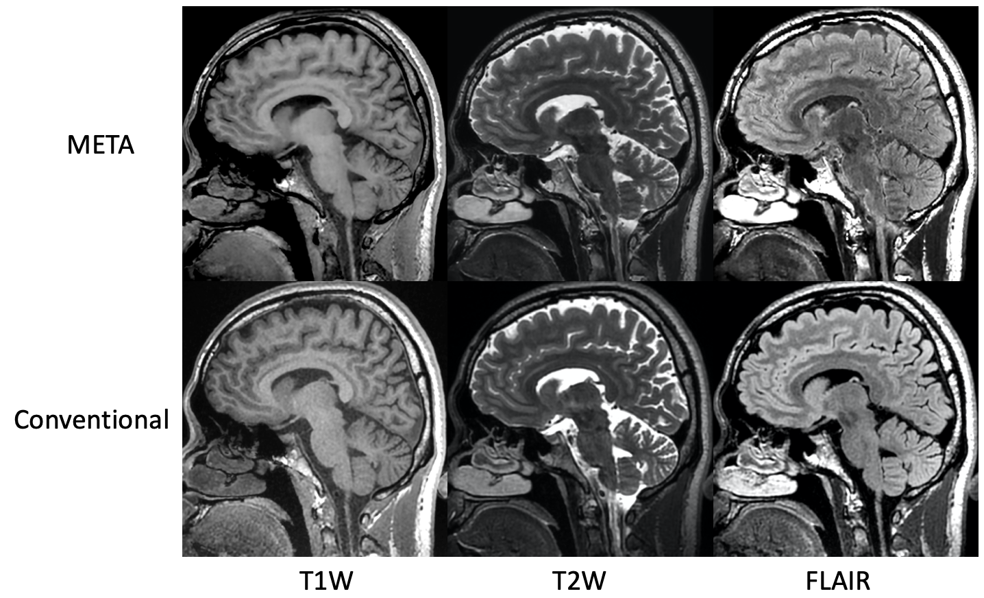
Figure.2. In-plane
sagittal images of META and conventional T1W, T2W, and FLAIR.
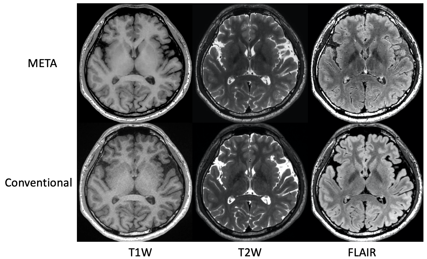
Figure.3. Reformatted
coronal images of META and conventional T1W, T2W, and FLAIR.
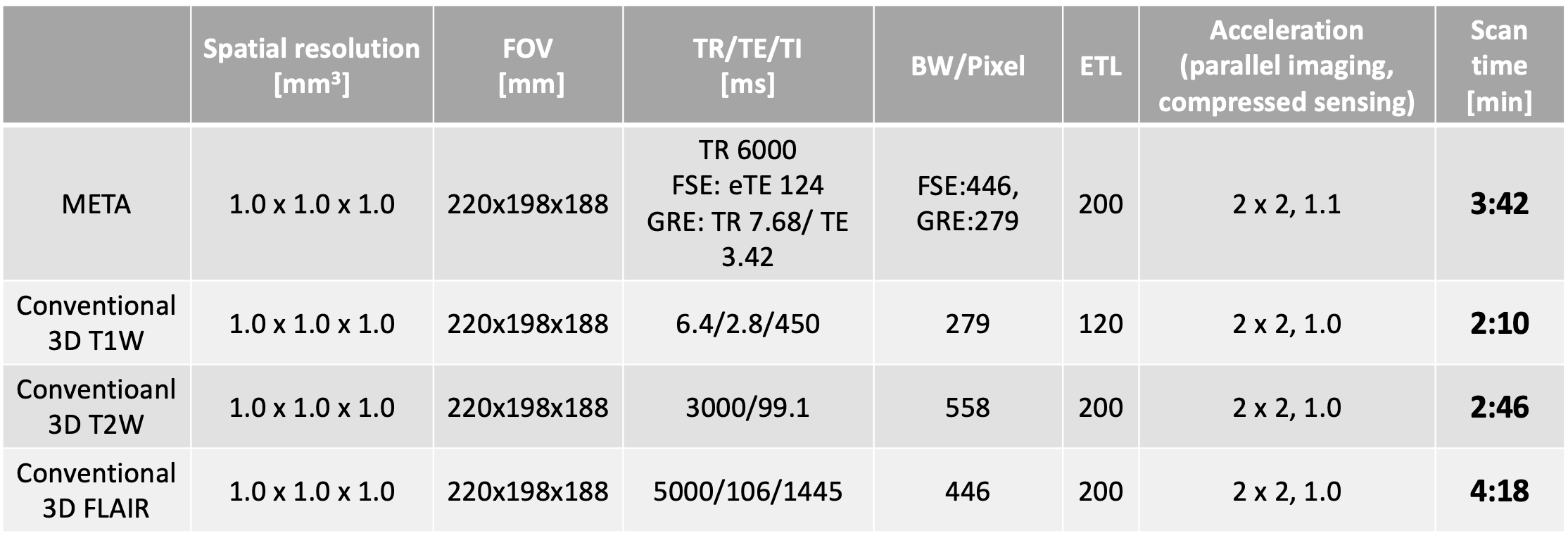
Table.1 Scan parameters
for META (Mixed Echo Train
Acquisition) and conventional
individually acquired image contrasts.
DOI: https://doi.org/10.58530/2022/4695