4694
Simultaneous Multi-slab DWI Reconstruction with Intrinsic Distortion Correction using Blip-up/down Acquisitions1Center for Biomedical Imaging Research, Department of Biomedical Engineering, School of Medicine, Tsinghua University, Beijing, China, 2Department of Radiology, Stanford, CA, United States, 3Department of Radiology and Nuclear Medicine, Xuanwu Hospital, Capital Medical University, Beijing, China, 4Beijing Key Laboratory of Magnetic Resonance Imaging and Brain Informatics, Beijing, China, 5MR Clinical Science, Philips Healthcare, Suzhou, China
Synopsis
Simultaneous multi-slab imaging (SMSlab) can achieve high-resolution DWI with high SNR efficiency, but image distortions remain a problem. Joint reconstruction of blip-up/down EPI data can correct for distortion and reduce g-factors simultaneously. This study proposed a joint reconstruction framework with intrinsic distortion correction for SMSlab. Three sampling patterns were compared in terms of levels of distortions and g-factors. According to the results, a multi-shot acquisition should be used to reduce the distortion. Combined with the controlled aliasing in parallel imaging (CAIPI) sampling pattern, it can further reduce the g-factors.
Introduction
Simultaneous multi-slab imaging (SMSlab) 1-3 combines simultaneous multislice (SMS) 4-10 and 3D multi-slab 11-13 sampling. It can achieve high-resolution whole-brain DWI with high SNR efficiency 1-3,14,15, but image distortions caused by B0 inhomogeneities remain a problem 16. Typically, two acquisitions with reversed phase-encoding (PE) directions are used to estimate the off-resonance maps and correct for distortions 17. Rather than correcting the images after reconstruction, joint reconstruction utilizing the blip-up/down data can correct for distortions and reduce g-factors intrinsically 18,19. This study aims to propose such a framework to conduct joint reconstruction for SMSlab DWI.Materials & Methods
Joint Reconstruction with Intrinsic Distortion CorrectionThe generation of a distorted under-sampled 3D image can be formulated as 18,19
$$F_{u} W S \Phi x=A x=d, (1)$$
where Fu represents the 2D FFT operator with undersampling along ky and kz directions. W describes off-resonance-induced phase accrual causing image distortions. S represents coil sensitivities. Φ is the background phase difference between the blip-up/down acquisitions 19. x is the full-sampled distortion-corrected image. d is the acquired signals transformed to the x-ky-kz hybrid space 20. For diffusion imaging, motion-induced inter-shot phase variations Θ should be included:
$$F_{u} W S \Phi \Theta x=d. (2)$$
The reconstruction framework is shown in Fig. 1. In step 1, motion-induced phase variations Θ are extracted from the reconstructed multi-band navigators. Then Θ is used to reconstruct the blip-up/down data separately via 3D SENSE 21. In step 2, the reconstructed b=0 s/mm2 images are used to estimate the off-resonance maps using topup 17 from the FSL 22, which generate W. In step 3, background phase differences Φ between the blip-up/down acquisitions are extracted from the results of step 1. Both Φ and Θ are combined with S for the joint reconstruction 19,23, which is done by computing the least-squares solutions of Eq. 1 or 2.
Blip-up/down Acquisitions for SMSlab
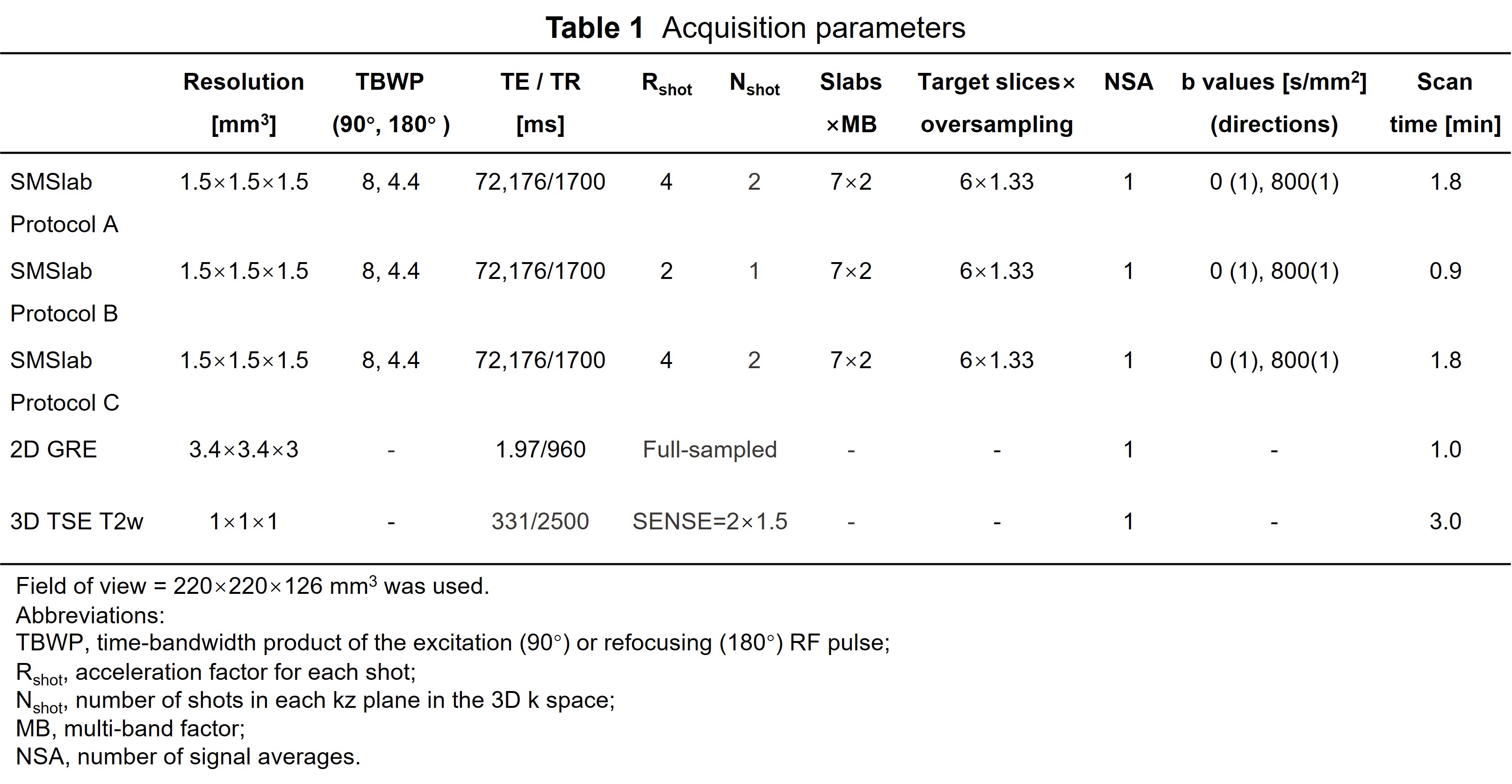
All images were acquired on a 3T scanner (Philips, Ingenia CX) using a 32-channel coil. The SMSlab acquisitions mainly followed 1,2, except that the PE gradient polarity was alternated for the blip-up/down acquisitions. The resolution was 1.5 mm isotropic. The detailed parameters are listed in Table 1. Two-dimensional navigators (multi-band factor [MB] = 2) were acquired and unfolded to separate bands via SENSE 21. Sensitivity maps were estimated from a low-resolution GRE sequence using ESPIRiT 24.
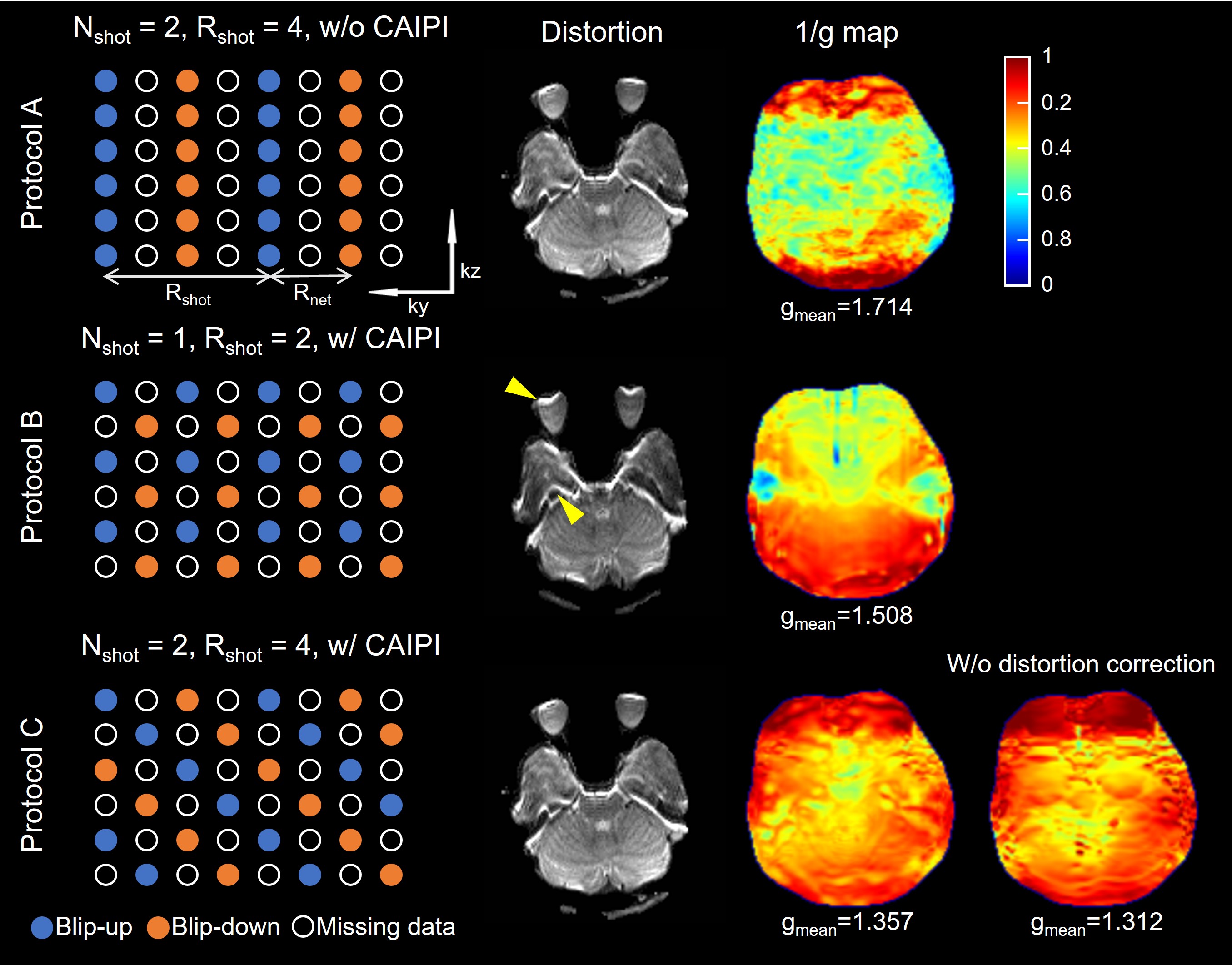
Three sampling patterns were compared (Fig. 2). Protocol A used a 4-shot acquisition with 2 shots undersampled (Nshot=2). Namely, for each shot, the undersampling factor Rshot=4. Then reversed PE directions were used in each kz plane without CAIPI sampling 25. Protocol B was a single-shot acquisition (Nshot=1) with Rshot=2 CAIPI sampling, and the PE gradient polarity was alternated for odd/even kz encodings. Protocol C repeated A but with CAIPI sampling. The g-factors of the joint reconstruction were calculated as follows 19,20.
$$g=\sqrt{X_{\text {cov}}^{\text {joint}} / X_{\text {cov}}^{\text {full}} / R_{\text {net}}}, (3)$$
where $$$X_{\text {cov}}^{\text {joint}} $$$ is the noise covariance matrix of the joint reconstruction matrix A. $$$X_{\text {cov}}^{\text {full}}$$$ is the noise covariance matrix for a full-sampled SENSE reconstruction without off-resonance effects. Rnet is the final effective undersampling factor, which is 2 for all three protocols after combining blip-up/down data.
Results & Discussion
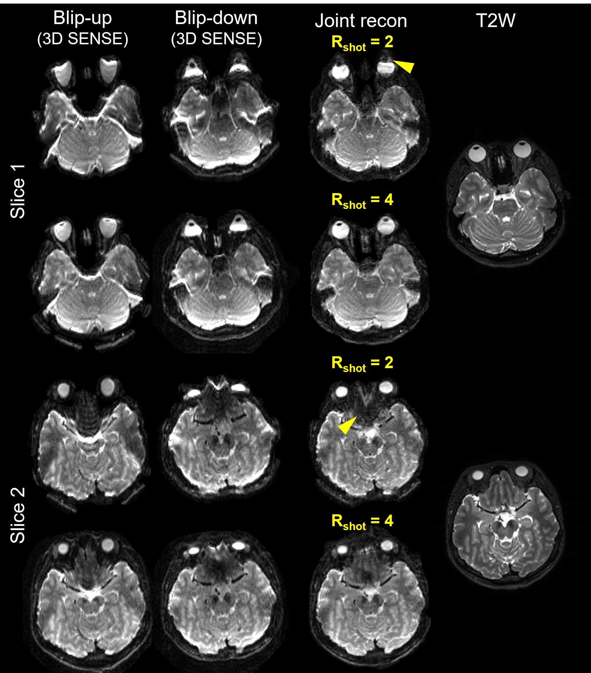
The levels of distortions and the 1/g maps of three sampling patterns are shown in Fig. 2. Protocol A shows higher g-factors compared with protocol B and C because CAIPI is not used. For protocol B, only Nshot=1 is used and the acquisition time is halved, but the reduced Rshot (=2) leads to stronger distortions (arrowheads), which introduces higher g-factors compared with protocol C. Protocol C uses Rshot=4, which reduces the distortion and thus reduces the g-factors. The 1/g map of protocol C but without off-resonance effects is shown in the lower right, which reflects the upper bound of 1/g because the phase accrual from off-resonance effects can affect the g-factors 18.The b=0 mm/s2 images of protocol B (Rshot=2) and C (Rshot=4) are shown in Fig. 3. Compared with protocol C, stronger distortions are found in protocol B, where more severe signal loss/pile-up exists (arrowheads). The correction performance at such regions is less satisfactory in protocol B compared with C. Therefore, for the proposed joint reconstruction method, multiple shots in each kz plane should be used to improve the Rshot and thus improve the performance of distortion correction.
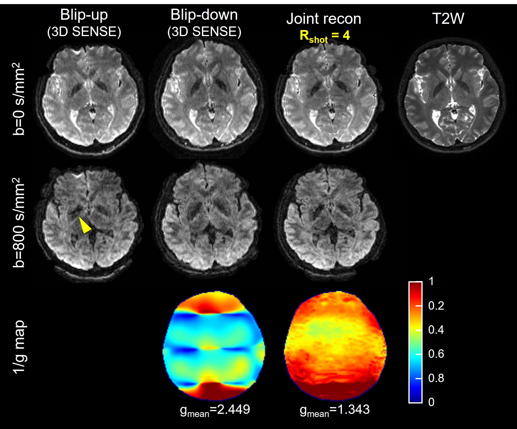
Fig. 4 shows the b=0 mm/s2 and b=800 mm/s2 images from protocol C. The 1/g maps of step 1 and joint reconstruction are also shown. Compared with step 1, joint reconstruction utilizes the acquired data more completely and distinctly reduces the g-factors. Residual aliasing exists (arrowhead) due to imperfect correction for inter-shot phase variations. Advanced MUSE-like reconstruction 14,26,27 can be implemented to solve this problem in the future.
Conclusion
In this study, we proposed a framework to jointly reconstruct blip-up/down SMSlab imaging with intrinsic distortion correction, which reduces the g-factors. We also used a large Rshot to improve the performance of distortion correction. Combined with the CAIPI sampling, it can further reduce the g-factors. In the future, receiving coil with more channels can be applied to increase the net acceleration factor.Acknowledgements
No acknowledgement found.References
1. Dai E, Liu S, Guo H. High-resolution whole-brain diffusion MRI at 3T using simultaneous multi-slab (SMSlab) acquisition. NeuroImage 2021;237:118099.2. Dai E, Wu Y, Wu W, et al. A 3D k-space Fourier encoding and reconstruction framework for simultaneous multi-slab acquisition. Magn Reson Med 2019;82(3):1012-1024.
3. Frost R, Jezzard P, Porter DA, Tijssen R, Miller K. Simultaneous multi-slab acquisition in 3D multi-slab diffusion-weighted readout-segmented echo-planar imaging. Proceedings of the 21st Annual Meeting of ISMRM. Salt Lake City, Utah, USA, 2013. p. 3176.
4. Barth M, Breuer F, Koopmans PJ, Norris DG, Poser BA. Simultaneous multislice (SMS) imaging techniques. Magn Reson Med 2016;75(1):63-81.
5. Chang HC, Guhaniyogi S, Chen Nk. Interleaved diffusion‐weighted improved by adaptive partial‐Fourier and multiband multiplexed sensitivity‐encoding reconstruction. Magn Reson Med 2015;73(5):1872-1884.
6. Dai E, Ma X, Zhang Z, Yuan C, Guo H. Simultaneous multislice accelerated interleaved EPI DWI using generalized blipped‐CAIPI acquisition and 3D K‐space reconstruction. Magn Reson Med 2017;77(4):1593-1605.
7. Feinberg DA, Reese TG, Wedeen VJ. Simultaneous echo refocusing in EPI. Magn Reson Med 2002;48(1):1-5.
8. Larkman DJ, Hajnal JV, Herlihy AH, Coutts GA, Young IR, Ehnholm G. Use of multicoil arrays for separation of signal from multiple slices simultaneously excited. J Magn Reson Imaging 2001;13(2):313-317.
9. Nunes R. G. HJV, Golay X., Larkman D. J. Simultaneous slice excitation and reconstruction for single shot EPI. Proceedings of the 14th Annual Meeting of ISMRM. Seattle, Washington, USA, 2006. p. 293.
10. Setsompop K, Gagoski BA, Polimeni JR, Witzel T, Wedeen VJ, Wald LL. Blipped-controlled aliasing in parallel imaging for simultaneous multislice echo planar imaging with reduced g-factor penalty. Magn Reson Med 2012;67(5):1210-1224.
11. Engstrom M, Skare S. Diffusion-weighted 3D multislab echo planar imaging for high signal-to-noise ratio efficiency and isotropic image resolution. Magn Reson Med 2013;70(6):1507-1514.
12. Frost R, Miller KL, Tijssen RHN, Porter DA, Jezzard P. 3D Multi-slab diffusion-weighted readout-segmented EPI with real-time cardiac-reordered k-space acquisition. Magn Reson Med 2014;72(6):1565-1579.
13. Moeller S, Ramanna S, Lenglet C, et al. Self‐navigation for 3D multishot EPI with data‐reference. Magn Reson Med 2020;84(4):1747-1762.
14. Bruce IP, Chang H-C, Petty C, Chen N-K, Song AW. 3D-MB-MUSE: A robust 3D multi-slab, multi-band and multi-shot reconstruction approach for ultrahigh resolution diffusion MRI. Neuroimage 2017;159:46-56.
15. Setsompop K, Fan Q, Stockmann J, et al. High‐resolution in vivo diffusion imaging of the human brain with generalized slice dithered enhanced resolution: Simultaneous multislice (gSlider-SMS). Magn Reson Med 2018;79(1):141-151.
16. Liu S, Xiong Y, Dai E, Zhang J, Guo H. Improving distortion correction for isotropic high‐resolution 3D diffusion MRI by optimizing Jacobian modulation. Magn Reson Med 2021;86(5):2780-2794.
17. Andersson JL, Skare S, Ashburner J. How to correct susceptibility distortions in spin-echo echo-planar images: application to diffusion tensor imaging. Neuroimage 2003;20(2):870-888.
18. Liao C, Bilgic B, Tian Q, et al. Distortion‐free, high‐isotropic‐resolution diffusion MRI with gSlider BUDA‐EPI and multicoil dynamic B0 shimming. Magn Reson Med 2021;86(2):791-803.
19. Zahneisen B, Aksoy M, Maclaren J, Wuerslin C, Bammer R. Extended hybrid-space SENSE for EPI: Off-resonance and eddy current corrected joint interleaved blip-up/down reconstruction. NeuroImage 2017;153:97-108.
20. Zhu K, Dougherty RF, Wu H, et al. Hybrid-space SENSE reconstruction for simultaneous multi-slice MRI. IEEE transactions on medical imaging 2016;35(8):1824-1836.
21. Pruessmann KP, Weiger M, Scheidegger MB, Boesiger P. SENSE: sensitivity encoding for fast MRI. Magn Reson Med 1999;42(5):952-962.
22. Jenkinson M, Beckmann CF, Behrens TEJ, Woolrich MW, Smith SM. FSL. NeuroImage 2012;62(2):782-790.
23. Chen N-k, Guidon A, Chang H-C, Song AW. A robust multi-shot scan strategy for high-resolution diffusion weighted MRI enabled by multiplexed sensitivity-encoding (MUSE). Neuroimage 2013;72:41-47.
24. Uecker M, Lai P, Murphy MJ, et al. ESPIRiT—an eigenvalue approach to autocalibrating parallel MRI: where SENSE meets GRAPPA. Magn Reson Med 2014;71(3):990-1001.
25. Breuer FA, Blaimer M, Heidemann RM, Mueller MF, Griswold MA, Jakob PM. Controlled aliasing in parallel imaging results in higher acceleration (CAIPIRINHA) for multi-slice imaging. Magn Reson Med 2005;53(3):684-691.
26. Zhang Z, Huang F, Ma X, Xie S, Guo H. Self-feeding MUSE: a robust method for high resolution diffusion imaging using interleaved EPI. NeuroImage 2015;105:552-560.
27. Chu M-L, Chang H-C, Chung H-W, Truong T-K, Bashir MR, Chen N-k. POCS-based reconstruction of multiplexed sensitivity encoded MRI (POCSMUSE): A general algorithm for reducing motion-related artifacts. Magn Reson Med 2015;74(5):1336-1348.
Figures
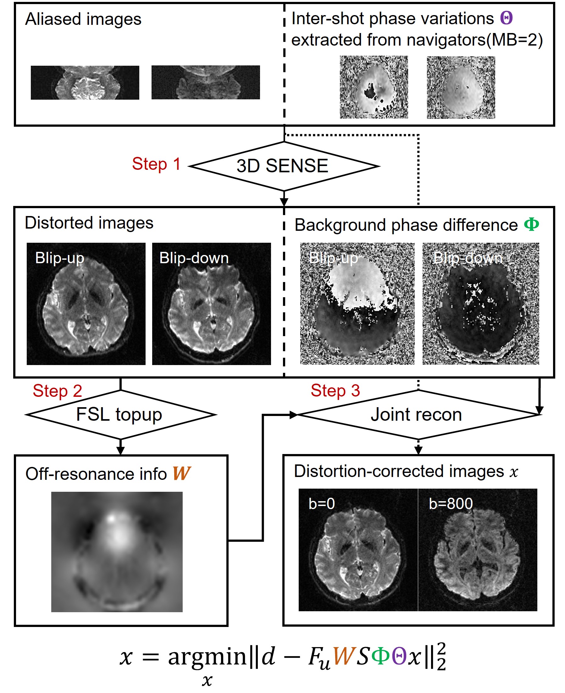
Figure 1. The flowchart of the proposed reconstruction framework. Abbreviations: MB, multi-band factor.