4685
Application of Compressed Sensing Technology in Rapid Lumbar Magnetic Resonance Imaging1Department of Radiology, the First Affiliated Hospital of Dalian Medical University, Dalian, Dalian, China, 2Philips Healthcare, Beijing, China, Beijing, China
Synopsis
Lumbar magnetic resonance imaging has a significant diagnostic value for spine lesions. Reducing scan time is critical to improve patient compliance and comfort. The purpose of this study aims to introduce a combination of compressed-sensing and parallel imaging for high acceleration and motion artifact reduction for the lumbar spine MRI, and to determine an optimal acceleration factor, meanwhile, provide high quality images .
Introduction
Lumbar magnetic resonance imaging has a wide range of clinical applications due to its good soft-tissue contrast, high spatial resolution, and no ionizing radiation1. During the scan examination, patients must remain still for a long time to prevent motion artifacts. However, it is difficult for the patients with severe lumbar disease2. Compressed sensing (CS) can significantly shorten the scan time, and ensure image quality via sparse sampling at the same time3,4. The purpose of this study was to explore the influence of CS on the lumbar image quality and to find an optimal acceleration factor.Materials and methods
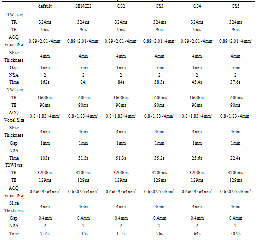
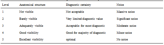
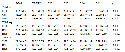
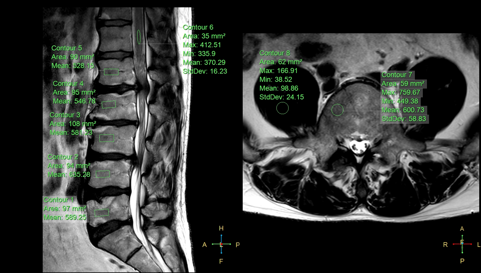
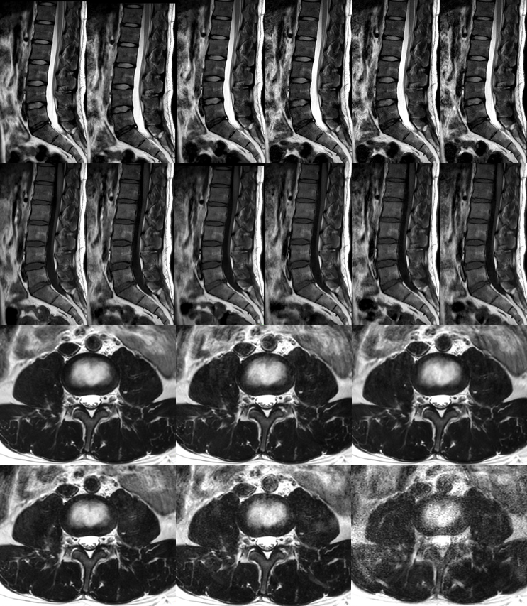
A total of twenty healthy volunteers (mean age: 37.2 ± 11.3, range: 21-57 years; 7 women) with informed consent were scanned using a 3.0 T MR scanner (Ingenia CX, Philips Healthcare, Best, the Netherlands). The performed MR protocols included sagittal T1WI, T2WI TSE and transverse T2WI TSE sequences without SENSE and CS-SENSE, with SENSE factor (SENSE=2), with CS-SENSE factors (CS=2, 3, 4, 5), respectively. Other scan parameters were shown in Table 1 as below. Regions of interest (ROIs) for signal intensity and standard deviation measurements were manually placed on the first to fifth lumbar spines, the psoas major and the middle spinal cord. Signal to noise ratio (SNR) and contrast to noise ratio (CNR) were calculated for all volunteers accordingly. Two radiologists scored the images independently based on the anatomical structures, diagnostic certainty and artifacts. Five-point scale criteria was used (the scoring system was listed in Table 2) to evaluate the image quality, and the score more than 3 was considered to meet clinical requirements. The Kappa test was adopted to evaluate the consistency of the scores from the two radiologists. If the consistency was in good agreement, the corresponding images would be adopted for the analysis in further by senior physicians. In the following analysis, the Kruskal-Wallis test was used to assess the difference of SNR, CNR and score between groups, and the LSD test was employed to make a pairwise comparison.Results
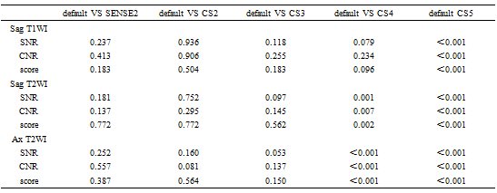
The scores by the two observers were in good agreement (=0.757-0.997). In Table 3, it was shown that there were statistically significant differences in SNR, CNR and score of sagittal T1WI, T2WI TSE and transverse T2WI TSE sequences. When CS=5, the SNR, CNR and scores of sagittal T1WI are significantly different from those of conventional sequences (p<0.05); when CS=4, the SNR, CNR and subjective scores of sagittal and axial T2WI were significantly different from conventional sequences (p<0.05).Discussion and Conclusions
Scan time for the lumbar spine decreased gradually with increase of the CS acceleration factor. CS factor of 4 was recommended for clinical sagittal T1WI, and CS factor of 3 was best for clinical sagittal and transverse T2WI to achieve an optimal balance between scan time and image quality.Acknowledgements
No acknowledgement found.References
1. Morita, K; Nakaura, T; Maruyama, N, et al. Hybrid of Compressed Sensing and Parallel Imaging Applied to Three-dimensional Isotropic T2-weighted Turbo Spin-echo MR Imaging of the Lumbar Spine, Magn Reson Med Sci.2020 Feb 10 ;19(1):48-55.
2. Bratke, G; Rau, R; Weiss, K. et al. Accelerated MRI of the Lumbar Spine Using Compressed Sensing: Quality and Efficiency. Journal of Magnetic Resonance Imaging, 2019;49(7):e164-175.
3. Seung Hyun Leea, Young Han Lee, Jin-Suck Suh. Accelerating knee MR imaging: Compressed sensing in isotropic three-dimensional fast spin-echo sequence. Magnetic Resonance Imaging, 2018;46:90-97.
4. Alessandro Furlana, Ersin Bayram, Senthur Thangasamy, Dale Barleya, Anil Dasyam. Application of compressed sensing to 3D magnetic resonance cholangiopancreatography for the evaluation of pancreatic cystic lesions. Magnetic Resonance Imaging, 2018;52:131-136.
Figures