4680
Regularized SUPER-CAIPIRINHA: accelerating 3D variable-flip-angle T1 mapping up to 16-fold with fast reconstruction1Institute of Medical Imaging Technology, School of Biomedical Engineering, Shanghai Jiao Tong University, Shanghai, China, 2United Imaging Healthcare Co., Ltd, Shanghai, China
Synopsis
Three-dimensional variable-flip-angle (VFA) T1 mapping is an accurate T1 quantification method suffering from long scan time. SUPER is a contrast-domain acceleration technique with strength in noise suppression and fast reconstruction. Here, we develop regularized SUPER-CAIPIRINHA to accelerate 3D VFA T1 mapping up to 16-fold with 10 flip angles, and perform fast reconstruction with the proposed proximal-Levenberg Marquardt algorithm. The novel method is validated with both retrospective and prospective experiments, compared to Locally Low Rank and Compressed Sensing. The results show that rSUPER-CAIPIRINHA is an accurate and computationally efficient technique, reducing the 3D scan time from 14:10 minutes to 0:59 minutes.
Introduction
Three-dimensional Variable-Flip-Angle (VFA) T1 mapping is an established method for accurate and volumetric T1 quantification1-4 with various clinical applications2,3,5-7. However, 3D VFA T1 mapping requires a long scan time, since multiple 3D volumes need to be acquired, especially for 3D isotropic high-resolution imaging. Most of existing acceleration methods, including Compressed Sensing8 (CS) and Locally Low-Rank9 (LLR), incur a long reconstruction time, which is a more serious concern for 3D parametric mapping since a large data size is present. An accurate, fast-imaging, and computationally-economic reconstruction is highly desired for 3D VFA T1 mapping.We have previously developed SUPER-CAIPIRINHA10—a combination of SUPER11 and CAIPIRINHA12—to accelerate 3D VFA T1 mapping with a validated 5-fold acceleration. At higher acceleration rates, however, increasing noise amplification poses a concern for the original SUPER-CAIPIRINHA method. Here we propose a combination of Total Variation13 (TV) and SUPER-CAIPIRINHA to suppress the noise at high acceleration rates. Furthermore, a computationally efficient algorithm is developed to rapidly minimize the regularized cost function. The proposed method, regularized SUPER-CAIPIRINHA (rSUPER-CAPIRINHA), is compared to CS and LLR at acceleration rates of 4-16 in 9 healthy subjects.
Methods
The cost function associated with rSUPER-CAIPIRINHA differs from SUPER-CAIPIRINHA10 by introducing the TV regularization term:||Y - WSΦ(U)||F2 + μ||U||TV (1)
where Y is the aliased 3D volume generated by zero-filling and 3D inverse DFT, W the modulation matrix containing aliasing coefficients for each voxel, S the sensitivity matrix of all coils, U the 3D M0 and T1 maps, ||·||TV the 3D total variation operator, and Φ the VFA signal model14 .
Introduction of the TV term prohibits a direct block-by-block solution of Eq 1, which underpins the fast reconstruction of SUPER11. To maintain the reconstruction efficiency, we propose a proximal-Levenberg Marquardt algorithm, which is inspired by the proximal-gradient algorithm15, by replacing the gradient descent step with Levenberg-Marquardt iterations. The proposed algorithm essentially toggles between SUPER reconstruction and the TV regularization, both of which can be rapidly solved on their own yet a combination would severely increase the computational burden.
Nine healthy subjects (5 male, age 24±2) were scanned after providing written informed consent, using a 3D FLASH sequence in a 3T scanner (uMR790, Shanghai United Imaging Healthcare, Shanghai, China) with a 24-channel head coil. Ten flip angles were used, namely 1◦, 3◦, 5◦, 7◦, 9◦, 11◦, 13◦, 15◦, 18◦ and 21◦. FOV was 300×300×220mm3, covering the entire cerebrum, and the image size was 192×192×44. Other sequence parameters were TR/TE/Bandwidth=10ms/4.48ms/135Hz/pixel. k-Space was retrospectively and prospectively undersampled by shift undersampling11 for SUPER-related methods and by pseudorandom sampling8,9 for CS and LLR. CS was performed with conjugate gradient algorithm8. LLR was performed using the code provided in [9]. In one healthy subject, imaging with an isotropic resolution of 1.6×1.6×1.6mm3 was performed, with prospective acceleration of 4-fold and 16-fold for CAIPIRINHA and rSUPER-CAIPIRINHA, respectively.
Results
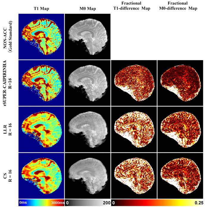
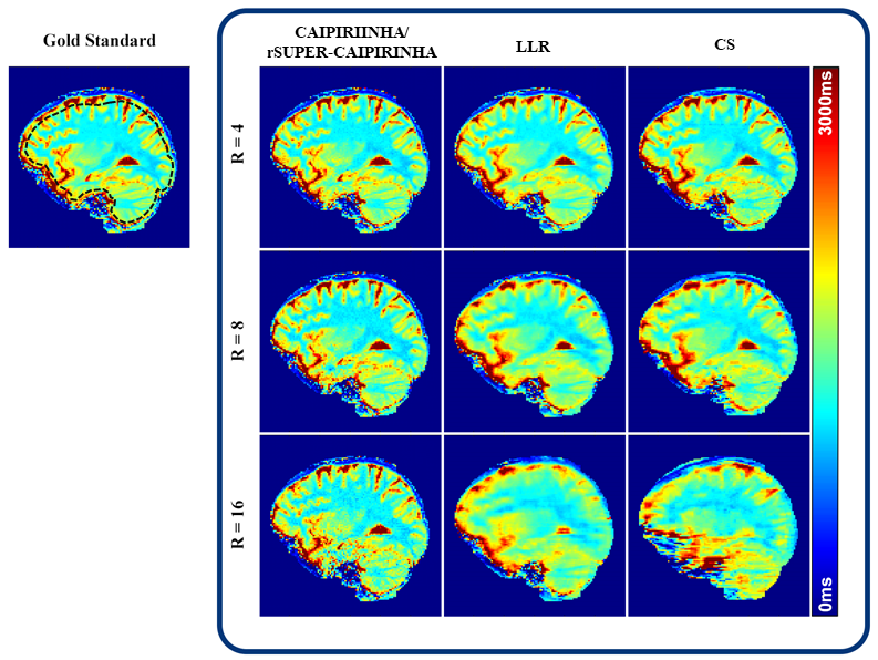
Figure 1 shows reconstruction results of a healthy subject with 16-fold retrospective undersampling. rSUPER-CAIPIRINHA achieved better performance than LLR and CS, with well-preserved image fine details. Both LLR and CS led to blur. Furthermore, LLR showed ringing artifacts in the M0 map.Figure 2 shows results of retrospectively undersampled T1 mapping at 4-, 8- and 16-fold accelerations. At 4-fold acceleration, all methods achieved consistent image quality compared with the gold standard. As the acceleration rate increased, rSUPER-CAIPIRINHA outperformed LLR and CS with better preservation of image details, despite a slightly and reasonably increased noise. rSUPER-CAIPIRINHA reduced the scan time from 14:10 minutes to 1:52 minutes (R=8) and 0:59 minutes (R=16). Prospective reconstruction led to consistent performance with retrospective reconstruction.
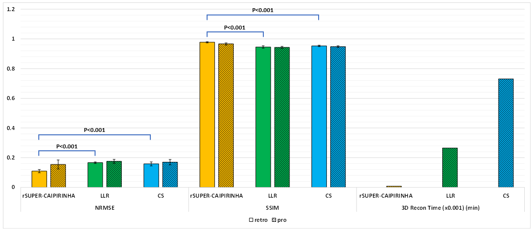
Figure 3 shows statistical comparison of NRMSE and SSIM between 16-fold rSUPER-CAIPIRINHA, LLR, and CS. Over 9 subjects, rSUPER-CAIPIRINHA obtained a lower NRMSE than LLR (0.11±0.01 vs 0.17±0.01, P<0.001) and CS (0.11±0.01 vs 0.16±0.01, P<0.001), and a higher SSIM than LLR (0.98±0.00 vs 0.95±0.01, P<0.001) and CS (0.98±0.00 vs 0.95±0.01, P<0.001) by paired t-test. The 3D reconstruction time of rSUPER-CAIPIRINHA, LLR and CS was 8, 265 and 730 minutes, respectively. rSUPER-CAIPIRINHA spent only 3% and 1% of the reconstruction time used by LLR and CS.
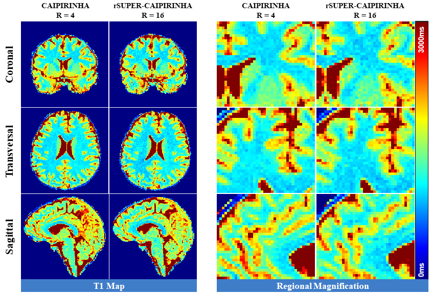
Figure 4 shows the prospective reconstruction of the isotropic-resolution T1 maps (1.6×1.6×1.6mm3) and their regional magnification in 3 orthogonal directions. The 16-fold accelerated rSUPER-CAIPIRINHA achieved similar image quality compared with 4-fold accelerated CAIPIRINHA. However, the imaging time was reduced from 11:42 minutes to 2:54 minutes by a 4-fold increase of the acceleration rate. A video of the 3D isotropic-resolution T1 maps of one subject in 98 slices over all 3 directions is shown in Figure 5.
Discussion and conclusions
The proposed method, rSUPER-CAIPIRINHA, successfully accelerates 3D VFA T1 mapping up to 16-fold, without conceivable blurring, whereas LLR and CS suffer from severe blurring artifacts. In the original work of LLR9, the author showed 4-fold acceleration and pointed out that there was blurring for higher acceleration rates, which is consistent with our work. CS shows overwhelming artificial noise at high acceleration rates. A satisfactory suppression warrants an exceedingly large regularization weight, which then introduces oversmoothing into the reconstruction. All of the results indicate that rSUPER-CAIPIRINHA is a feasible 3D acceleration technique for VFA T1 mapping.Acknowledgements
No acknowledgement found.References
1 Deoni, S. C. L., Peters, T. M. & Rutt, B. K. High-resolution T1 and T2 mapping of the brain in a clinically acceptable time with DESPOT1 and DESPOT2. Magnetic Resonance in Medicine 53, 237-241, doi:https://doi.org/10.1002/mrm.20314 (2005).
2 Vrenken, H. et al. Whole-Brain T1 Mapping in Multiple Sclerosis: Global Changes of Normal-appearing Gray and White Matter. Radiology 240, 811-820, doi:10.1148/radiol.2403050569 (2006).
3 Koh, T. S. et al. Hepatic Metastases: In Vivo Assessment of Perfusion Parameters at Dynamic Contrast-enhanced MR Imaging with Dual-Input Two-Compartment Tracer Kinetics Model. Radiology 249, 307-320, doi:10.1148/radiol.2483071958 (2008).
4 Li, Z. et al. Assessment of liver fibrosis by variable flip angle T1 mapping at 3.0T. Journal of Magnetic Resonance Imaging 43, 698-703, doi:10.1002/jmri.25030 (2016).
5 Vymazal, J. et al. T1 and T2 in the Brain of Healthy Subjects, Patients with Parkinson Disease, and Patients with Multiple System Atrophy: Relation to Iron Content. Radiology 211, 489-495, doi:10.1148/radiology.211.2.r99ma53489 (1999).
6 Zhu, Z. et al. Sparse precontrast T(1) mapping for high-resolution whole-brain DCE-MRI. Magnetic resonance in medicine 86, 2234-2249, doi:10.1002/mrm.28849 (2021).
7 Brookes, J. A., Redpath, T. W., Gilbert, F. J., Murray, A. D. & Staff, R. T. Accuracy of T1 measurement in dynamic contrast-enhanced breast MRI using two- and three-dimensional variable flip angle fast low-angle shot. Journal of Magnetic Resonance Imaging 9, 163-171, doi:https://doi.org/10.1002/(SICI)1522-2586(199902)9:2<163::AID-JMRI3>3.0.CO;2-L (1999).
8 Lustig, M., Donoho, D. & Pauly, J. M. Sparse MRI: The application of compressed sensing for rapid MR imaging. Magn Reson Med 58, 1182-1195, doi:10.1002/mrm.21391 (2007).
9 Zhang, T., Pauly, J. M. & Levesque, I. R. Accelerating parameter mapping with a locally low rank constraint. Magnetic Resonance in Medicine 73, 655-661, doi:https://doi.org/10.1002/mrm.25161 (2015).
10 Yang F, Zhang J, Li G, Zhu J, Tang X, Hu C. Accelerating 3D variable-flip-angle T1 mapping: a prospective study based on SUPER-CAIPIRINHA. ISMRM 2021.
11 Hu, C. & Peters, D. C. SUPER: A blockwise curve-fitting method for accelerating MR parametric mapping with fast reconstruction. Magnetic Resonance in Medicine 81, 3515-3529, doi:10.1002/mrm.27662 (2019).
12 Breuer, F. A. et al. Controlled aliasing in volumetric parallel imaging (2D CAIPIRINHA). Magn Reson Med 55, 549-556, doi:10.1002/mrm.20787 (2006).
13 Rudin, L. I., Osher, S. & Fatemi, E. Nonlinear total variation based noise removal algorithms. Physica D: Nonlinear Phenomena 60, 259-268, doi:https://doi.org/10.1016/0167-2789(92)90242-F (1992).
14 Deoni, S. C. L., Rutt, B. K. & Peters, T. M. Rapid combined T1 and T2 mapping using gradient recalled acquisition in the steady state. Magnetic Resonance in Medicine 49, 515-526, doi:https://doi.org/10.1002/mrm.10407 (2003).
15 Parikh, N. & Boyd, S. Proximal Algorithms. Found. Trends Optim. 1, 127–239, doi:10.1561/2400000003 (2014).
Figures