4672
High Temporal and Spatial Pulmonary Dynamic Ventilation Imaging Using Hyperpolarized 129Xe MRI1Key Laboratory of Magnetic Resonance in Biological Systems, State Key Laboratory of Magnetic Resonance and Atomic and Molecular Physics, National Center for Magnetic Resonance in Wuhan, Wuhan Institute of Physics and Mathematics, Innovation Academy for Precision Measurement Science and Technology, Chinese Academy of Sciences- Wuhan National Laboratory for Optoelectronics, Wuhan, China
Synopsis
The mutual restriction of temporal resolution and spatial resolution is a challenge for hyperpolarized gas dynamic MRI. Herein, we proposed a method to enhance the temporal resolution of pulmonary dynamic ventilation imaging without spatial resolution loss using hyperpolarized 129Xe MRI. Furthermore, compressed sensing undersampling technique was used to accelerate the MRI data acquisition.
Introduction
Hyperpolarized (HP) 129Xe gas MRI has been widely used in lung diseases evaluation because of its ability in quantifying the lung structure and function without invasion and ionizing radiation1-3. However, these pulmonary physiological parameters are generally obtained during the breath-hold after subjects inhaled HP xenon gas4,5. Dynamic ventilation MRI could image the processes of inspiration and expiration, and it has great potential in quantifying pulmonary pathophysiological abnormalities such as airflow restriction, obstruction, and air trapping6-8. Unfortunately, limited by the short breathing cycle, it is difficult to obtain high-temporal-resolution dynamic images. Moreover, HP gas MR signal is unrecoverable, and would be depolarized by RF pulses and the longitudinal relaxation, which makes it a challenge to obtain high quality dynamic images with high temporal and spatial resolution. In this study, we proposed a multi-respiration acquisition scheme to enhance the temporal resolution to 5.6 ms without reducing the SNR and spatial resolution.Methods
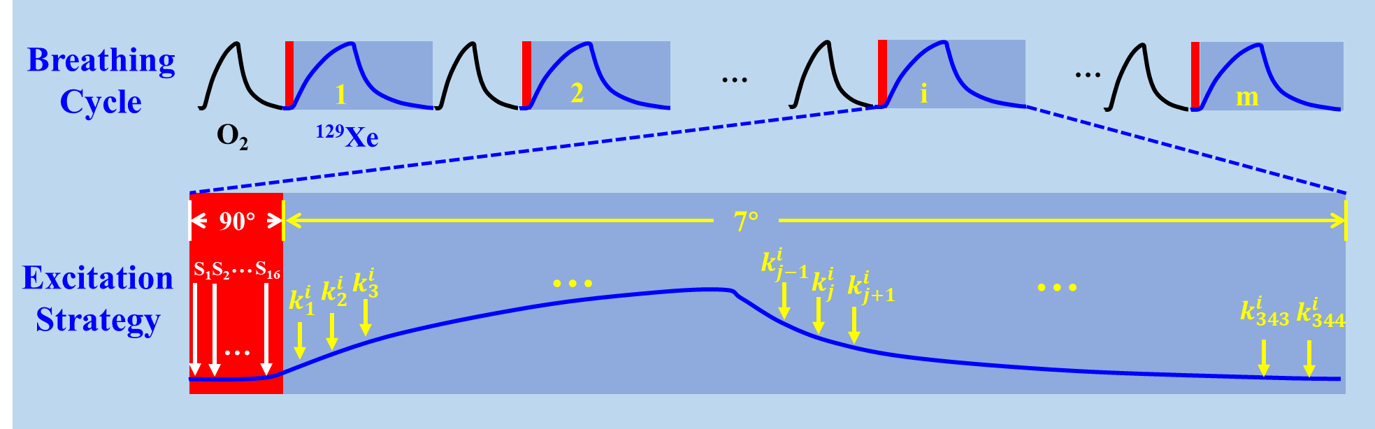
The ventilation and acquisition strategies were shown in Figure 1. The data acquisitions for all frame images are performed in multiple HP 129Xe breathes (breath number (m) equals to the phase encoding steps). To maintain normal physiology, one oxygen breath is used between every two 129Xe breathes. The biggest feature of our dynamic MRI acquisition strategy is that the k-space lines of all the frame images with the same phase encoding are acquired in the same respiratory cycle of xenon. Therefore, different k-space lines of the same will be distributed in different respiratory cycles. Before the data acquisition, sixteen 90° saturation RF pulses were applied to destroy the residual magnetization. Then, all the collected data were reorganized according to the time series in one respiratory cycle, and the dynamic images were obtained with high temporal resolution, which equals to one repetition time (TR). For the MRI experiments, the scanning parameters are as follows: TR/TE = 5.6 ms/1.7 ms; flip angle = 7°, FOV = 45×45 mm2; matrix = 96×96; and scanning parameters for the CS acquisition are the same (two folder undersampling factor were used for accelerating the acquisition).Results
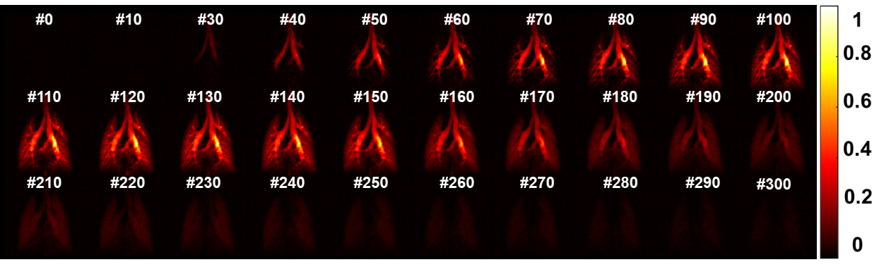
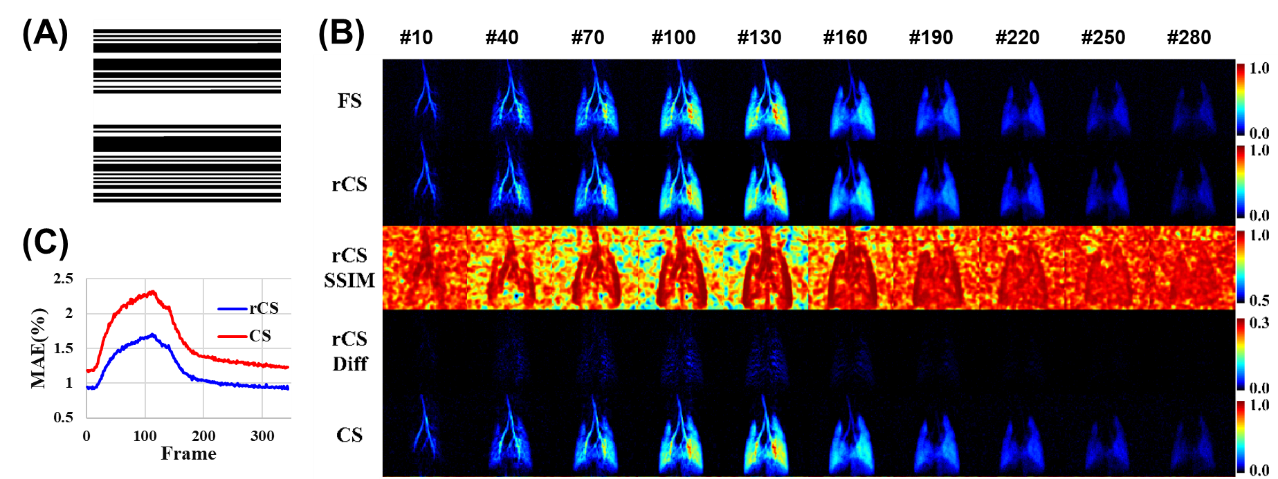
Figure 2 showed the representative dynamic ventilation images obtained with HP 129Xe gas dynamic MRI using the proposed method with flip angle of 7° from a healthy rat. The temporal and spatial resolution of the dynamic images were 5.6 ms and 0.5 mm, respectively. Signal in trachea and bronchi are much higher than that in lung parenchyma during the inspiration process, and opposite trends are found during the expiratory process. Figure 3 (B) showed 10 typical frames of the dynamic ventilation images using fully sampled (FS), retrospective compressed sensing (rCS) and compressed sensing (CS) acquisition strategies from the same healthy rat. The MAE of the images obtained using rCS and CS strategies are 0.016±0.004 and 0.023±0.003 when compared with FS strategy, respectively. Moreover, the details of the images using rCS and CS are comparable with images using FS, and the measured SSIM are 0.84±0.02 and 0.75±0.02 for images acquired with rCS and FS, respectively. The difference and SSIM maps showed the accelerated dynamic ventilation images have the comparable image quality and details compared with FS strategy.Discussion and Conclusion
Although numerous studies have been reported about the HP gas dynamic ventilation imaging, most of them are qualitative analysis due to the limited temporal and spatial resolution, and only the inspiration or expiration data was obtained6,9-12. An accelerated method for dynamic ventilation imaging using HP 129Xe MRI was proposed. Dynamic ventilation images with a high temporal resolution of 5.6 ms and high spatial resolution of 0.47 mm×0.47 mm were obtained. By using the technique of compressed sensing, the acquisition time could be reduced, and the images details and quality could be well preserved.In this study, we proposed a method to enhance the temporal resolution of pulmonary dynamic ventilation imaging using hyperpolarized 129Xe MRI without loss of spatial resolution. Our preliminary results demonstrated the feasibility of the proposed method for evaluating the dynamic ventilation function regionally in vivo, which has the potential in lung diseases diagnosis that related to the dynamic ventilation functional injuries.
Acknowledgements
This work is supported by National Natural Science Foundation of China (91859206, 21921004, 81825012), National key Research and Development Project of China (2018YFA0704000), Key Research Program of Frontier Sciences (ZDBS-LY-JSC004) and Scientific Instrument Developing Project of the Chinese Academy of Sciences (GJJSTD20200002, YJKYYQ20200067), CAS. Haidong Li acknowledges the support from Youth Innovation Promotion Association, CAS (2020330). Xin Zhou acknowledges the support from the Tencent Foundation through the XPLORER PRIZE.References
1. Zhang, M., et al. Quantitative evaluation of lung injury caused by PM2.5 using hyperpolarized gas magnetic resonance. Magnetic Resonance in Medicine 84, 569-578 (2020).
2. Li, H.D., et al. Quantitative evaluation of pulmonary gas-exchange function using hyperpolarized Xe-129 CEST MRS and MRI. Nmr in Biomedicine 31(2018).
3. Li, H.D., et al. Quantitative Evaluation of Radiation-Induced Lung Injury with Hyperpolarized Xenon Magnetic Resonance. Magnetic Resonance in Medicine 76, 408-416 (2016).
4. Duan, C.H., et al. Fast and accurate reconstruction of human lung gas MRI with deep learning. Magnetic Resonance in Medicine 82, 2273-2285 (2019).
5. Zhang, H.T., et al. Lung morphometry using hyperpolarized Xe-129 multi-b diffusion MRI with compressed sensing in healthy subjects and patients with COPD. Med. Phys. 45, 3097-3108 (2018).
6. Chen, M.H., et al. Delayed ventilation assessment using fast dynamic hyperpolarised Xenon-129 magnetic resonance imaging. European Radiology 30, 1145-1155 (2020).
7. Hamedani, H., et al. Regional Fractional Ventilation by Using Multibreath Wash-in He-3 MR Imaging. Radiology 279, 917-924 (2016).
8. Wild, J.M., Horn, F.C., Collier, G.J. & Marshall, H. Dynamic Imaging of Lung Ventilation and Gas Flow With Hyperpolarized Gas MRI. in Hyperpolarized and Inert Gas MRI 47-59 (2017).
9. Chen, B.T., Brau, A.C.S. & Johnson, G.A. Measurement of regional lung function in rats using hyperpolarized (3)helium dynamic MRI. Magnetic Resonance in Medicine 49, 78-88 (2003).
10. Collier, G.J. & Wild, J.M. In vivo measurement of gas flow in human airways with hyperpolarized gas MRI and compressed sensing. Magnetic Resonance in Medicine 73, 2255-2261 (2015).
11. Doganay, O., et al. Fast dynamic ventilation MRI of hyperpolarized Xe-129 using spiral imaging. Magnetic Resonance in Medicine 79, 2597-2606 (2018).
12. Deppe, M.H., Parra-Robles, J., Ajraoui, S. & Wild, J.M. Combined measurement of pulmonary inert gas washout and regional ventilation heterogeneity by MR of a single dose of hyperpolarized 3He. Magn Reson Med 65, 1075-1083 (2011).
Figures