4669
Feasibility of non-invasive IDEAL-IQ MR imaging in assessment the correlation of fat fraction and hepatosomatic index in two in vivo fish species1Department of Radiology, Affiliated Tongji Hospital, Tongji Medical College, Huazhong University of Science and Technology, wuhan, China, 2Faculty of Resources and Environmental Science, Hubei University, wuhan, China, 3GE Healthcare, beijing, China, 4Affiliated Tongji Hospital, Tongji Medical College, Huazhong University of Science and Technology, Wuhan, China
Synopsis
Fish is widely consumed as one of the main sources of animal food and plays a key role in the ecological processes in aquatic ecosystems. Of particular importance to assess the effects of aquatic pollution on the development and health of fish are organ somatic indices (HSI) and physiological indices (e.g. lipid deposition). Traditional assessment of somatic indices and lipid deposition is invasive and required to sacrifice examined animals. The present study was designed to assess HSI and lipid deposition in intraperitoneal tissues and livers of two fish species using a rapid, accurate and noninvasive magnetic resonance imaging method.
Introduction
Fish is widely consumed as one of the main animal foods and plays a key role in the ecological processes in aquatic ecosystems. Adverse effects on fish caused by aquatic pollutants have been reported. Of particular importance to assess the effects of aquatic pollution on the development and health of fish are organ somatic indices (e.g. hepatosomatic index, HSI) and physiological indices (e.g. lipid deposition). Traditional assessment of somatic indices and lipid deposition is invasive and required to sacrifice examined animals, and cannot perform longitudinal and fish-relocated follow-ups. The present study was designed to assess HSI and lipid deposition in intraperitoneal tissues and livers of two fish species, snakehead Channa argus and big-eyed mandarin fish Siniperca knerii, using a rapid, accurate and noninvasive magnetic resonance imaging (MRI) method.Materials and methods
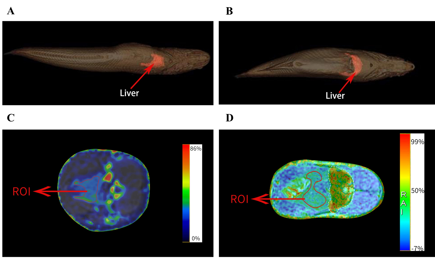
A total of 93 fresh fish, including 43 C. argus and 50 S. knerii were randomly purchased. Fish were anesthetized with MS-222 before MRI examination to ensure immobilization. All fish survived after the MRI scans on a 3.0 T clinical MRI scanner (Signa Pioneer; GE Healthcare) with a 48-channel head coil with no negative effects on health. IDEAL-IQ and 3D-FSE-Cube-Flex T2 sequence was used to measure fish whole-body volume due to its better edge discrimination of tissues and organs (Figure 1). 3D reconstruction of liver and adipose deposition in both species were done on a workstation (Advantage Windows Workstation 4.7; GE Healthcare). The percentage volume of liver (%) was determined from the measured volume of liver and that of the entire body of the fish; fat fraction maps were automatically computed. Two doctors with more than 3-year experience in clinical MR and 4-year experience in research MR selected the representative axial slice across the dome of each fish liver and obtained signal to noise ratio (SNR) of each sketched ROI on the largest liver cross-sectional area. After MRI examinations, each fish was weighed and dissected to obtain the hepatosomatic index (HSI) as liver weight over whole-fish weight. All statistical analyses were performed using R software. All figures were conducted using PRISM Statistical Software Package. Pearson’s correlation analysis was used to assess the relationships between MRI measurements and HSI, IPF, chemical approaches, respectively.Results
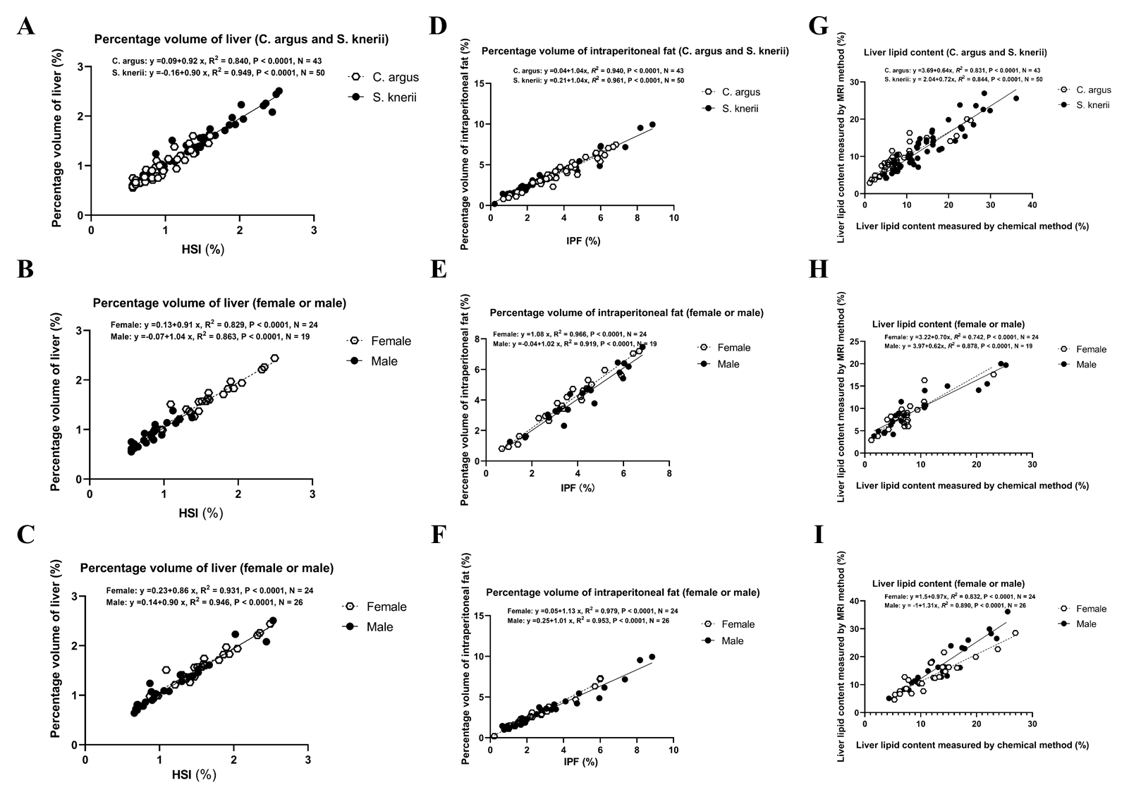
The positive correlation between HSI and the percentage volume of liver was excellent for C. argus andS. knerii. There was no significant difference between these two linear regressions (F value = 1.58; P value = 0.21). After controlling for gender, the positive correlation between HSI and percentage volume of liver was excellent for female and male in C. argus and S. knerii, respectively. There was no significant gender difference of the correlations between HSI and percentage volume of liver. (Figure 2 A, B, C)The positive correlation between mean IPF and percentage volume of intraperitoneal fat was excellent for C. argus and S. knerii. There was no significant difference between these two linear regressions (F value = 1.85; P = 0.16). After controlling for gender, the positive correlation between IPF and percentage volume of intraperitoneal fat was excellent for female and male in C. argus and S. knerii, respectively. There was no significant gender difference of the correlations between IPF and percentage volume of intraperitoneal fat (C. argus: F value = 2.41; P = 0.10; S. knerii: F value = 2.21; P = 0.12). (Figure 2 D, E, F) The positive correlation of liver lipid content measured by the chemical method and MRI was excellent for C. argus and S. knerii There was a significant difference between these two linear regressions (F value = 25.82; P < 0.001). After controlling for gender, the positive correlation of liver lipid content measured by the chemical method and MRI method was good for female and male in C. argus and S. knerii, respectively. There was no significant gender difference of liver lipid content between two methods for C. argus (F value = 0.32; P = 0.73) but for in S. knerii (F value = 6.90; P = 0.00). (Figure 2 G, H, I)Discussion
This study showed MRI was a fast, accurate and noninvasive method to assess HSI and lipid deposition (intraperitoneal fat and liver) in two live fish species (C. argus and S. knerii). Parameters of HSI and lipid deposition were widely reckoned as key biomarkers in reflection of aquatic pollutants. As the fat fraction using IDEAL-IQ was significantly correlated with hepatosomatic index using chemical approaches, MRI might have potential to not only detect but also long-term track excess lipid deposition that was caused by aquatic pollutants in the fish throughout the river and ocean ecosystem as well as toxicity mechanism especially in these two fish species with advent of no sacrifice and adverse effects. Moreover, as HSI and lipid deposition, have been shown to be dynamic, time-dependent process values, it is necessary to repeatedly observe the same fish via a non-invasive approach. Therefore, this approach could help us to investigate toxic mechanism of aquatic pollutants in the longitudinal studies (e.g. juvenile to brood stock) and reduce the number of sacrificed animals.Conclusions
IDEAL-IQ is a promising, rapid, accurate and noninvasive MR imaging method in assessment of HSI and lipid depositions in ecotoxicology.Acknowledgements
No acknowledgement found.References
1.Birgisdottir BE et al. (2012) Fish liver and seagull eggs, vitamin D-rich foods with a shadow: results from the Norwegian Fish and Game Study. Mol Nutr Food Res 56:388-398
2. Bock C, Wermter FC, Mintenbeck KJMRI (2017) MRI and MRS on preserved samples as a tool in fish ecology.
3.Brinkmann M, Rizzo LY, Lammers T, Gremse F, Schiwy S, Kiessling F, Hollert H (2016) Micro-computed tomography (muCT) as a novel method in ecotoxicology--determination of morphometric and somatic data in rainbow trout (Oncorhynchus mykiss). Sci Total Environ 543:135-139
4.Cardoso C, Afonso C, Lourenço HM, Nunes ML (2015) Assessing risks and benefits of consuming fish muscle and liver: Novel statistical tools. Journal of Food Composition and Analysis 38:112-120
5. Du ZY (2014) Causes of fatty liver in farmed fish: a review and new perspectives. Journal of Fisheries of China 38:1628-1638
6.Fu J, Guo Y, Wang M, Yang L, Han J, Lee JS, Zhou B (2020) Bioconcentration of 2,4,6-tribromophenol (TBP) and thyroid endocrine disruption in zebrafish larvae. Ecotoxicol Environ Saf 206:111207
7. Hong MY, Lumibao J, Mistry P, Saleh R, Hoh E (2015) Fish Oil Contaminated with Persistent Organic Pollutants Reduces Antioxidant Capacity and Induces Oxidative Stress without Affecting Its Capacity to Lower Lipid Concentrations and Systemic Inflammation in Rats. J Nutr 145:939-944
8. Idilman IS, Keskin O, Celik A, Savas B, Elhan AH, Idilman R, Karcaaltincaba M (2016) A comparison of liver fat content as determined by magnetic resonance imaging-proton density fat fraction and MRS versus liver histology in non-alcoholic fatty liver disease. Acta Radiol 57:271-278
9. Johnson MS, Watts RJ, Hammer HS, Nagy TR, Watts SA (2017) Validation of Dual-energy X-ray Absorptiometry to Predict Body Composition of Channel Catfish,Ictalurus punctatus. Journal of the World Aquaculture Society 48:122-131
10.Khieokhajonkhet A, Klongchai S, Maphum O, Kaneko G (2019) Lipid distribution patterns of nine commercial fish in Thailand. Aquaculture Research 50:1348-1360
11.Landgraf K et al. (2017) Short-term overfeeding of zebrafish with normal or high-fat diet as a model for the development of metabolically healthy versus unhealthy obesity. BMC Physiol 17:4
12. Li DL, Huang YJ, Gao S, Chen LQ, Zhang ML, Du ZY (2019) Sex-specific alterations of lipid metabolism in zebrafish exposed to polychlorinated biphenyls. Chemosphere 221:768-777
13. Liu P, Wang Q, Peng C, Luo B, Zhang J (2018) Combined application of isotropic three-dimensional fast spin echo (3D-FSE-Cube) with 2-point Dixon fat/water separation (FLEX) and 3D-FSE-cube in MR dacryocystography. The British Journal of Radiology 92:20180157
14. Lu L, Zhao J, Li C (2020) High-Quality Genome Assembly and Annotation of the Big-Eye Mandarin Fish (Siniperca knerii). G3 (Bethesda) 10:877-880
15. Montano M et al. (2011) Effects of mixtures of persistent organic pollutants (POPs) derived from cod liver oil on H295R steroidogenesis. Food Chem Toxicol 49:2328-2335
16. Nian R et al. (2020) Online fat detection and evaluation in modelling digital physiological fish. Aquaculture Research 51:3175-3190
17. Pan L, Hu J, Peng C, Liu H, Zhang Z, Xie J, Qin J (2016) Use of magnetic resonance imaging to assess ovarian maturation in live Rhinogobio ventralis (Sauvage & Dabry de Thiersant, 1874). Theriogenology 86:1969-1974
Figures