4635
Evaluation of condylar osseous changes using wireless detector combined with PDWI sequence1Guizhou Provincial People's Hospital, Guiyang,Guizhou, China, 2Zunyi Medical University, Guiyang,Guizhou, China, 3Michigan State University, East Lansing, MI, USA, MI, United States, 4GE Healthcare, MR Research China, Beijing, Beijing,China, China
Synopsis
MRI is considered to be the reference method for the imaging and understanding of temporomandibular disease (TMD) , but it provides little or no detectable signal from cortical bone. A disc-shaped wireless detector (WD), which is more suitable for body superficial tissue or organ examination, is used to overcome the sensitivity limitation of conventional MRI in condyle imaging. This study aims to observe the capability of WD in showing the bone changes of TMD condyle on PDWI. Compared with the traditional MRI, WD is suitable for imaging the condyle bone changes of TMD patients, and significantly improves the image quality.
Synopsis
MRI is considered to be the reference method for the imaging and understanding of temporomandibular disease (TMD) , but it provides little or no detectable signal from cortical bone. A disc-shaped wireless detector (WD), which is more suitable for body superficial tissue or organ examination, is used to overcome the sensitivity limitation of conventional MRI in condyle imaging. This study aims to observe the capability of WD-coupled head neck coupling coiling (WD-HNCC) in showing the bone changes of TMD condyle on proton density-weighted imaging (PDWI). Compared with the traditional MRI, WD-HNCC in PDWI sequence is suitable for imaging the condyle bone changes of TMD patients, and significantly improves the image quality.Introduction
Condylar osseous change is one of the most common causes and pathological manifestations of TMD, which is characterized by erosion and wearing of the articular surface cartilage and remodeling of subchondral cartilage due to overload. MRI is considered to be the reference method for imaging the soft tissue structures of the temporomandibular joint (TMJ). However, due to its low sensitivity in condyle bone imaging, MRI provides little or no detectable signal from cortical bone[1]. Recently, animal-based experiments have shown that the resolution and visibility of tiny tissues and organs can be enhanced greatly using a WD. This proof-of-concept study aims to qualitatively and quantitatively evaluate the feasibility and advantages of using WD with PDWI sequences to image condyle changes in patients with TMD.Materials and Methods
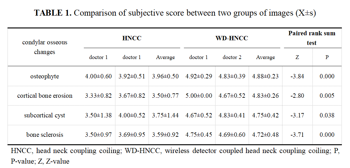
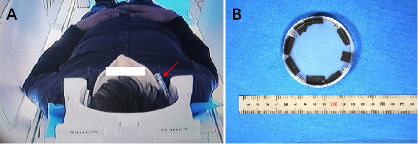
This study was approved by the institutional review board, and written informed consent was obtained from all patients. This study involved twenty patients (male=8, female=12, average age of 31.65 ±12.68 years old) with TMD. All subjects underwent a closed oblique sagittal PDWI scanning with HNCC and WD-HNCC on a 3.0 T MRI scanner (Discovery MR 750W, GE Healthcare, Waukesha, USA). The WD coil was fixed to the center of the bilateral TMJ using medical adhesive tape and sponge, and the scanning range included the entire TMJ region. Subsequently, the changes in the condyle bones in the scanned images for the two groups were scored subjectively and compared qualitatively. The contrast noise ratio (CNR) of the scanned images for the two groups was compared quantitatively. The comparison of CNR differences between the two types of images was performed using the paired t-test. The kappa statistic was used to test the consistency of quantitative analyses of MRI images between observers. The subjective scores of condylar osseous changes in the two types of images were compared by paired rank-sum test. A P<0.05 was considered statistically significant.Results
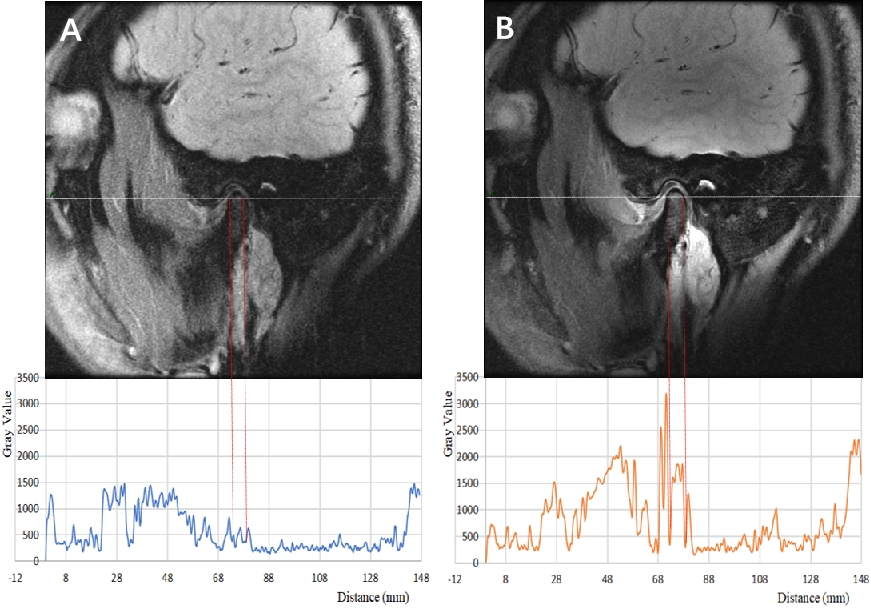
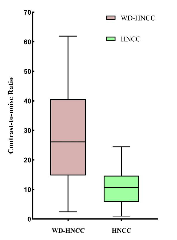
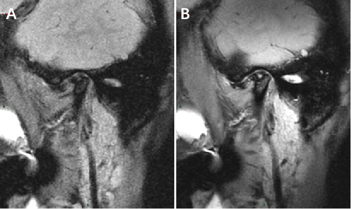
A total of 40 condyles from 20 patients were scanned. 8 condyles showed no bone changes, and the other 32 condyles demonstrated condyles osseous changes of varying degrees and nature. These 32 condyles were used in the subsequent analysis. As compared to images acquired by HNCC in the PDWI sequence, the WD-HNCC images showed mandibular osteophyte, cortical bone erosion, subcortical cystic focus, and cortical bone hyperplasia and thickening more clearly. In addition, the WD-HNCC was demonstrated to improve image CNR significantly (28.17 ±16.01 vs 10.88 ±6.53, t = 8.63, P = 0.001).Discussion and Conclusions
WD is a kind of local miniature signal receiving coil, which directly converts the energy collected wirelessly into amplified MR signal by nonlinear circuit. High-resolution scanning with WD can significantly improve tissue resolution. Compared with conventional external receivers, WD implantation can increase detection sensitivity by more than 5 times[2]. WD implanted in the corresponding position of the esophagus can accurately display the distal end of the common carotid artery and the vascular wall of the bifurcation of the internal and external carotid arteries after further scanning[3], while WD was used as an implantable detector mounted on the surface of the kidney,it can improve the high resolution imaging of renal tubules[4]. The main purpose of this study is to extend the use of WD from animal research to non-invasive imaging of human TMJ for the first time. The results showed that WD coupled with head and neck combined coil has improved the image quality of TMD, highlighting condylar osseous changes on the conventional PDWI sequences.So in the routine scanning sequence, the WD, which simultaneously images the TMJ disc and condyle with high resolution, provided the basis for a comprehensive clinical evaluation of TMD and demonstrated a high potential for clinical application.
Acknowledgements
No acknowledgement found.References
[1] Xiong X, Ye Z, Tang H, et al. MRI of Temporomandibular Joint Disorders: Recent Advances and Future Directions. J Magn Reson Imaging. 2020.
[2] Qian C, Yu X, Chen DY, et al. Wireless amplified nuclear MR detector (WAND) for high-spatial-resolution MR imaging of internal organs: preclinical demonstration in a rodent model. Radiology. 2013;268(1):228-36.[3] Zeng X, Barbic M, Chen L, Qian C. Sensitive enhancement of vessel wall imaging with an endoesophageal Wireless Amplified NMR Detector (WAND). Magn Reson Med. 2017;78(5):2048-54.[4] Zeng X, Ma S, Kruger JM, et al. High-resolution MRI of kidney microstructures at 7.05T with an endo-colonic Wireless Amplified NMR detector. J Magn Reson. 2019;303:121-7.
Figures