4634
Study on the relationship between accurate quantitative fat fraction of quadriceps femoris and patellofemoral arthritis by IDEAL-IQ1Honghui Hospital,Xi'an jiaotong University, Xi'an, China, 2GE Healthcare, Beijing, China
Synopsis
we aim to to explore the relationship between fat infiltration in quadriceps femoris with patellofemoral arthritis,It was concluded that IDEAL-IQ canbe a tool for quantification and prediction of patellar cartilage defect progression
INTRODUCTION:
patellofemoral OA is very common desease, with a prevalence of 25% of people in population-based cohorts[1]. It is the leading cause of pain and disability among adults,and caused huge economic burden to patients and society. [2]..Patellofemoral arthritis is closely related to the stability of knee joint, and the strength distribution of quadriceps femoris is an important factor to stabilize patellofemoral joint.Patella malacia is the early manifestation of the lesion[3]. Several radiological techniques are currently available for lipid quantification. Recently presented MR technique, termed as Iterative Decomposition of water and fat with Echo Asymmetry and Least square estimation (IDEAL-IQ), can obtain tissues proton density fat fraction (PDFF) with high spatial resolution. It has been successfully applied in several parts of the body,[4-5] In recent years, some studies have reported that IDEAL-IQ can indirectly reflect muscle strength. The increased fat infiltration in muscle reduce its functionality, which leads to the decrease of muscle strength[6-7]. The purpose of the study was to investigate the potential clinical value of IDEAL-IQ for quantification fat filtration in early-stage patellofemoral arthritis.Materials and methods
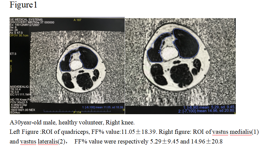
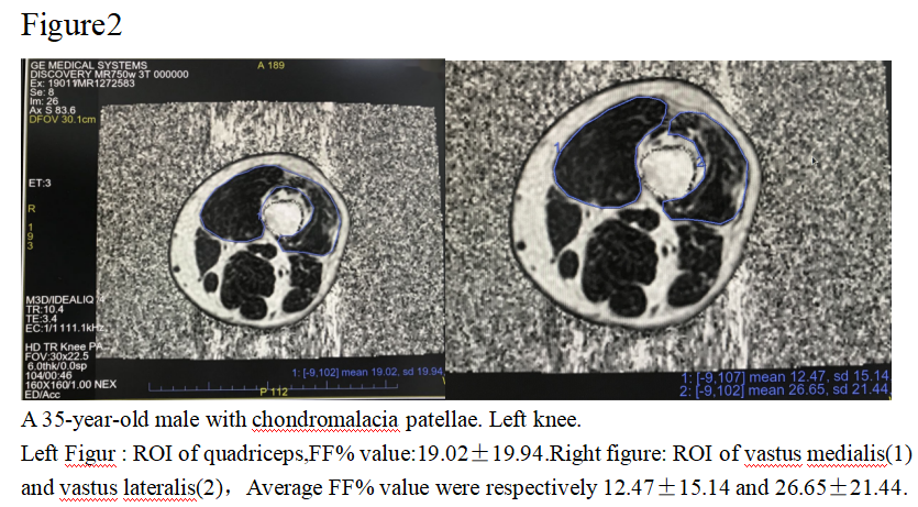
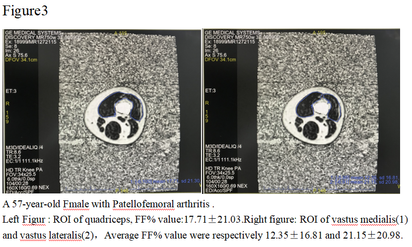
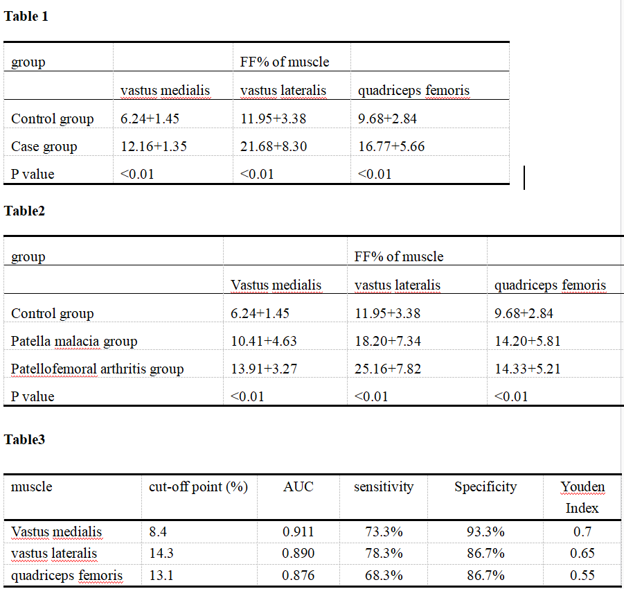
Our Institutional Review Board approved the protocol and written informed consent was obtained from each subject. This prospective study included 30 patients with patellar malacia, 30 patients with patellofemoral arthritis and 30 healthy volunteers from July 2018 to August 2021,All lesions were diagnosed by two musculoskeletal radiologists. The subjects underwent MR examination on a 3.0 T scanner (Discovery 750w, GE Healthcare)with 8-channel knee coil, IDEAL-IQ were selected for the examinations. IDEALIQ imaging was performed with detailed parameters list as below: TR=10.4ms, two echo times (TE, 1.1and5.8 ms),, Bandwidth=111.1kHz, FOV=30cm×22.5cm, slice thickness=6/0mm, 31 axial slices, echo train length=3, matrix=160×160, and scanning time = 1.32mins,Vendor provided inline post processing of IDEAL-IQ images was done automatically to generate PDFF maps of quadriceps femoris muscle. The fat fraction of vastus medial(FFm%), vastus lateral (FFl%) and total quadriceps(FFt%) were measured on the PDFF maps (Figure 1-3). Mann-Whitney U test(Table1) was used to compare the FFm%, FFl% and FFt% between case group (patellar malacia group + patellofemoral arthritis group) and healthy control group. Diagnosis efficacy including area under the receiver operating characteristic curve (AUC)(Table2), sensitivity and specificity were calculated at maximal Youden’s index. In addition, Kruskal-Wallis test(Table3) was separately used to compare fat fraction values in all three groups.Result
All FFm%, FFl% and FFt% were significantly higher in case group than that in the healthy control group(P <0.01)Table1. There were significant differences in FF% of quadriceps femoris among normal group、patellar malacia group and patellofemoral arthritis group(P<0.01)Table2. FFt% did not demonstrate significant difference between patellar malacia group and patellofemoral arthritis group. The median FF% showed an increasing trend with the progression of the disease. The ROC analysis showed an AUC of 0.911(95%CI 0.855-0.968)、0.890(95%CI0.824-0.956)、0.876(95%CI0.786-0.937)for FFm%, FFl% and FFt% respectively. Table3.CONCLUSION
Our study demonstrated that IDEAL-IQ can quantify fat fraction in vastus muscle, potentially can be used as a tool for patellofemoral arthritis early diagnosis. The fat fraction was found to be significant higher in patella malacia group than control group, which imply that fat infiltration in quadriceps femoris muscle occur in the early stage of patellofemoral arthritis . However, further evaluations are needed in order to determine ‘cut-off values’ for the reproducible assessment of Disease state that could be used to define the appropriate time points for intervention, as proposed by Surowiec R.K. et al[8]. In addition,we found that the fat infiltration in vastus medialis and vastus lateralis are different. Our result also shown compared to FFt%, FFm% and FFl% are seems more sensitive in detecting early fat infiltration. To our knowledge, this is the first study in patellar arthritis quantitatively use PDFF to assess the Fat infiltration. One of the major limitations in our study is the relatively small number of subjects. To conclude, this study provides evidence regarding the possible potential of IDEAL-IQ as a tool for quantification and prediction of patellar cartilage defect progression.This might further provide the basis for an MRI-guided tool , which could be an effective means of monitoring the subsequent treatment options of early OA.Acknowledgements
Thanks to all the leaders and colleagues who have helped my article.References
[1]Kobayashi S , Pappas E , Fransen M , et al. The prevalence of patellofemoral osteoarthritis: A systematic review[J]. Osteoarthritis & Cartilage, 2016:1697-1707.
[2]Hunter D J , D Schofield, Callander E . The individual and socioeconomic impact of osteoarthritis[J]. Nature Reviews Rheumatology, 2014, 10(7):437-441.
[3]SRA †, MMS †, PS‡ §, et al. Potential predictive value of axial T2 mapping at 3 Tesla MRI in patients with untreated patellar cartilage defects over a mean follow-up of four years[J]. Osteoarthritis and Cartilage, 2020, 28(2):215-222.
[4]Bailey J F , Fields A J , Ballatori A , et al. The Relationship Between Endplate Pathology and Patient-reported Symptoms for Chronic Low Back Pain Depends on Lumbar Paraspinal Muscle Quality[J]. Spine, 2019, 44.
[5]Lansdown, Drew A , Morrison, et al. Preoperative IDEAL (Iterative Decomposition of Echoes of Asymmetrical Length) magnetic resonance imaging rotator cuff muscle fat fractions are associated with rotator cuff repair outcomes[J]. Journal of Shoulder and Elbow Surgery, 2019, 28( 10):1936-1941.[6]Heskamp L , Okkersen K , Nimwegen M V , et al. Quantitative Muscle MRI Depicts Increased Muscle Mass after a Behavioral Change in Myotonic Dystrophy Type 1[J]. Radiology, 2020, 297(1):192518.[7]Stouge A , Khan K S , Kristensen A G , et al. MRI of Skeletal Muscles in Participants with Type 2 Diabetes with or without Diabetic Polyneuropathy[J]. Radiology, 2020, 297(3):608-619.
[8] Surowiec R K , Lucas E P , Ho C P . Quantitative MRI in the evaluation of articular cartilage health: reproducibility and variability with a focus on T2 mapping[J]. Knee Surgery Sports Traumatology Arthroscopy, 2014, 22(6).
Figures