4628
Periodic assessment of four horns of knee meniscus using MR T2mapping images in volunteers before and after amateur marathons1The Third Hospital of Hebei Medical University, Shijiazhuang, China, 2Siemens Healthineers, Ltd., Shanghai, 201318, China., Shanghai, China
Synopsis
T2 mapping can evaluate the dynamic changes of the meniscal microstructure. The study was designed to evaluate the T2 values of the menisci of amateur marathon volunteers at three different timings. Compared with the T2 values of menisci one week before running, the values 12 h after running increased significantly (P <0.05), and the values 2 months after running did not change significantly (P > 0.05). The result suggests that marathons do not necessarily cause irreversible knee damage.
Introduction
Marathon running may affect the condition of the meniscus given the sport’s repetitive loading of the knee joint 1. The meniscus is an integral part of the knee and is considered the second stabilizer of the knee joint 2, playing an important role in shock absorption, joint stability, joint lubrication, load transmission, and maintenance of articular cartilage integrity 3. The meniscus is also one of the most frequently injured parts of the knee joint 2. Whether marathons can induce the irreversible changes of the meniscus is a pertinent issue. MRI has become integral for the diagnosis and treatment of knee pathology 4. Among various quantitative methods, T2 mapping has been shown to provide enhanced morphologic and biochemical assessments of soft tissue structures, including the menisci 5. In the present study, the T2 values of the menisci were obtained at three distinct time points in volunteers who are marathon runners; these values were then analyzed to detect meniscal changes and assess the effects of marathon running on the meniscal microstructure.Methods
Twelve healthy marathon volunteers, including 5 males and 7 females, were prospectively recruited. The 24 knee joints were scanned using a 3T MR scanner (MAGNOTOM Verio, Siemens Healthcare, Erlangen, Germany) at 1 week before the event, an at 12 hours and 2 months (within 60 - 65 days) after the event. The T2 mapping parameters were as follows: TR, 3410.0 ms; TE, 13.8, 27.6, 41.4, 55.2, and 69.0 ms; FOV 160.0 mm x 160.0 mm2; flip angle, 180°; and slice thickness, 2.7 mm. Subsequent procedures were as follows:1. All the raw data were transferred to a Siemens workstation after the images were collected. The menisci were divided into four subregions, corresponding to the anterior horn of the medial meniscus (MMAH), posterior horn of the medial meniscus (MMPH), anterior horn of the lateral meniscus (LMAH), and posterior horn of the lateral meniscus (LMPH).
2. The images were analyzed independently by two diagnostic radiologists who each had 5 or more years of experience in diagnostic musculoskeletal imaging. T2 values of the four horns were measured by drawing the regions of interest (ROI) on the T2 map images. The intraclass correlation coefficient (ICC) was used to evaluate the consistency of the data.
3. The Wilcoxon sign rank test was used to compare the T2 values between 1 week before and 12 hours after the event, and between 1 week before and 2 months after the event in each anatomical region, respectively.
Results
The measured consistency range was 0.764 to 0.956, indicating good consistency and reproducibility. (Table 1)The T2 values of the menisci at 12 hours after the event (timing 2) were significantly higher (P <0.05) than those at 1 week before the event (timing 1). No statistically significant differences in the T2 values of the menisci were found between 2 months after (timing 3) and 1 week before the event (P > 0.05). (Table 2)
Discussion
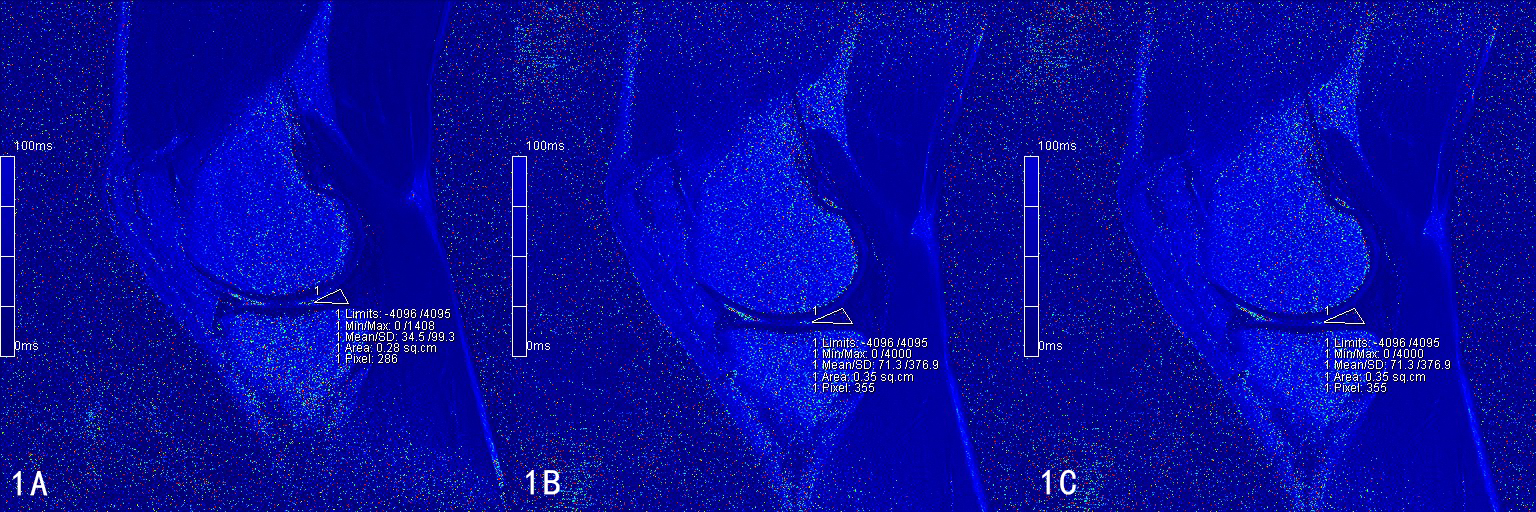
MRI may be more suitable for prospective and longitudinal imaging studies for large populations. T2 mapping MRI can detect meniscus degeneration and injury quantitatively 6 and may be more appropriate for measuring dynamic changes in the meniscus, which can be implemented in a clinical environment without requiring hardware modifications 7.Our results revealed that the T2 values of menisci were significantly higher at 12 hours after a marathon compared to values at 1 week before the run (Fig. 1A, 1B), suggesting an acute change in the collagen and water content. Interestingly, the condition of the menisci had recovered by 2 months after the event, returning to T2 values that did not differ significantly from those obtained at baseline (1 week before the run) (Fig. 1A, 1C). Our results also suggested that marathon exercise does not cause unrecoverable damage to the meniscus. Although the meniscal condition of amateur marathoners might fluctuate in the short term after running, it would return to pre-run levels over a longer period, a change that could be assessed by the measurements of T2 values.
Conclusions
The meniscus microstructures of marathon runners undergo dynamic changes before and after running, and these can be detected by T2 mapping. The menisci may recover to pre-run levels in the long-term, although the T2 values rise in the early hours after a run. Therefore, marathons do not necessarily cause irreversible meniscus damage.Acknowledgements
NoneReferences
1. Wang, Z. et al. Higher Body Mass Index Is Associated With Biochemical Changes in Knee Articular Cartilage After Marathon Running: A Quantitative T2-Relaxation MRI Study. Orthop J Sports Med 8, 2325967120943874, doi:10.1177/2325967120943874 (2020).
2. Hornakova, L. et al. In vivo assessment of time dependent changes of T2* in medial meniscus under loading at 3T: A preliminary study. J Appl Biomed 16, 138-144 (2018).
3. Nebelung, S. et al. Functional MRI Mapping of Human Meniscus Functionality and its Relation to Degeneration. Sci Rep 10, 2499, doi:10.1038/s41598-020-59573-4 (2020).
4. Nacey, N. C., Geeslin, M. G., Miller, G. W. & Pierce, J. L. Magnetic resonance imaging of the knee: An overview and update of conventional and state of the art imaging. J Magn Reson Imaging 45, 1257-1275, doi:10.1002/jmri.25620 (2017).
5. Paproki, A. et al. Automated T2-mapping of the Menisci From Magnetic Resonance Images in Patients with Acute Knee Injury. Acad Radiol 24, 1295-1304, doi:10.1016/j.acra.2017.03.025 (2017).
6. Le, J., Peng, Q. & Sperling, K. Biochemical magnetic resonance imaging of knee articular cartilage: T1rho and T2 mapping as cartilage degeneration biomarkers. Ann N Y Acad Sci 1383, 34-42, doi:10.1111/nyas.13189 (2016).
7. Meng, X. H. et al. Quantitative evaluation of knee cartilage and meniscus destruction in patients with rheumatoid arthritis using T1rho and T2 mapping. Eur J Radiol 96, 91-97, doi:10.1016/j.ejrad.2017.09.018 (2017).
Figures

Table 1. The ICC of T2 values in bilateral menisci as independently measured by two radiologists
ICC, intraclass correlation coefficient

Table 2. Comparison of T2 values in each subregion at different timings [M (P25,P75) ] (ms)
* Statistically significant difference (P <0.05).