4623
In vivo preliminary assessment of knee cartilage degeneration by Quantitative Ultrashort Echo Time MRI1Department of Radiology, Shanghai tenth People’s Hospital, Tongji University School of Medicine, Shanghai, China, shanghai, China, 2GE Healthcare, Beijing, China, Beijing, China
Synopsis
Recent advances in UTE-MRI techniques development allow for quantitative measurements of tissues with very short T2. However, in vivo experiments are needed to extend its application in clinical practice. In this in vivo study, we found that both UTE-MTR and UTE-adiabatic-T1rho values indicated a moderate correlation with MOAKS Grading. Meanwhile, both of them had moderate diagnostic efficacy for the mild cartilage degeneration (MOAKS = 1). These findings suggested that quantitative UTE-MRI techniques have a great potential as imaging biomarkers for early OA diagnosis in clinic.
Introduction
In recent years, with the rapid development of magnetic resonance imaging (MRI) techniques, ultrashort echo time (UTE) sequence has been proposed to capture signal of short T2 components in tissues, such as cartilage (1). Specifically, by using half pulse variable-rate selective excitation (VERSE) and radial ramp sampling, TE can be reduced to a micro second range in order to seize the signal of tissues with very short T2, which are not well-appreciated by conventional morphologic MRI or arthroscopy (2,3). In combination with UTE technique, several quantitative UTE-MRI sequences such as UTE-adiabatic-T1rho, UTE-T2*mapping, and UTE-MT have emerged, bringing new perspectives and possibilities to diagnose early cartilage degeneration, and has been proved by lots of in vitro experiments (4-6). However, their clinical application remains to be evaluated by extensive in vivo studies. This study aimed to investigate the feasibility of quantitative UTE-MRI sequences, including UTE-MT, UTE-adiabatic-T1rho, and UTE-T2*mapping, for in vivo assessment of early cartilage degeneration in clinic.Methods
Forty-six volunteers with knee pain as the main complaint were recruited in this study. All MRI examinations were performed on a clinical 3.0T scanner (MR750; GE Healthcare, Milwaukee, WI). Sequence including UTE-MT, UTE-adiabatic-T1rho, UTE-T2*mapping and PDWI/Sag/FS were acquired from all participants with following parameters: A)3D UTE-MT:off-resonance frequency=2kHz, saturation power=750 kHz, TR/TE=100/0.032ms, scan time=6:24min. B)3D UTE-adiabatic-T1rho: spin-locking time =0, 4, 8, 16ms, scan time=16:32min. C) UTE-T2*mapping:TEs=0.032, 4.9, 9.8, 14.7ms, TR=30.9ms, scan time=5:12min. D) PDWI/Sag/FS: TR=3000ms, TE=32ms, scan time=3:22min. Other imaging parameters are consistent: field of view (FOV)=16cm, acquisition matrix=256×256 pixels, scanning thickness =3mm. UTE-MTR, UTE-adiabatic-T1rho, and UTE-T2* values were calculated using Advantage Workstation 4.6 (GE Healthcare). Three regions of interest (ROIs) were manually delineated on the medial and lateral femoral condyles and the corresponding medial and lateral tibial plateaus, resulting in a total of 12 ROIs for a knee. A total of 561 ROIs were finally included and divided into three groups according to the MRI Osteoarthritis Knee Score (MOAKS): normal cartilage (n = 175, MOAKS = 0), mild cartilage degeneration (n = 283, MOAKS = 1), and moderate cartilage degeneration (n = 103, MOAKS = 2). The differences among different groups based on MOAKS were evaluated and compared using one-way analysis of variance (ANOVA) with post-hoc Tamhane-T2 test, and Spearman correlation analysis. Receiver-operating characteristic (ROC) curve was conducted to assess the diagnostic efficacy of three quantitative UTE-MRI techniques in detecting mild cartilage degeneration. For all tests, a difference of P < 0.05 was considered statistically significant.Results
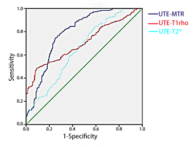
Both UTE-MTR and UTE-adiabatic-T1rho values indicated a moderate correlation with MOAKS Grading (r = -0.523, P < 0.001; r = 0.531, P < 0.001, respectively), while UTE-T2* showed a relatively weak correlation with MOAKS Grading (r = -0.396, P < 0.001). UTE-MTR and UTE-T2* values in the normal group were significantly higher than those in the mild group (both P < 0.001) and moderate group (both P < 0.001). There were significantly differences in values between the normal group and the mild group (P < 0.001), and between the mild group and moderate group (P < 0.001). AUC of UTE-MTR, UTE-adiabatic-T1rho, and UTE T2* mapping value in discriminating the normal group and mild group were 0.794,0.732, and 0.651, respectively (Fig. 1).Discussion
This is the first in vivo study using three different UTE-MRI sequences, including UTE-MT, UTE-adiabatic-T1rho, and UTE-T2*mapping, in detecting the early cartilage degeneration of human. Previous in vitro experiments have shown the advantages of UTE-MRI sequences for the imaging of human cartilage. In the present study, we showed that UTE-MTR, and UTE-adiabatic-T1rho values had moderate correlation with MOAKS Grading. Meanwhile, both of them had a higher diagnostic efficacy than UTE-T2*mapping, suggesting that both UTE-MT and UTE-adiabatic-T1rho methods had a higher potential for clinical applications in vivo. The variability in diagnostic efficacy of different UTE-MRI sequences may be due to the different quantitative biochemical basis of cartilage. In early cartilage degeneration, changes in proteoglycan (PG) and collagen content precede changes in collagen fiber and water content (7). Loss of collagen and PG leads to a decrease in locally bound water, resulting in decreased T2* values. However, at the same time, collagen fiber destruction increases the exposed surface area of collagen, which may also lead to an increase in locally bound water (8). These two pathophysiological processes simultaneously affect the diagnosis efficacy of UTE-T2* values. In general, quantitative UTE-MRI sequences (UTE-MT, UTE-adiabatic-T1rho, and UTE-T2*mapping) have the potential to serve as reliable tools to identify early cartilage degeneration and provide novelty perspectives in clinic utilization.Acknowledgements
This work was supported by grants from the National Natural Science Foundation of China (No.81801656), Science and technology innovation action project of STCS [20Y11911800].
