4622
Application of MR diffusion tensor imaging (DTI) in acute anterior cruciate ligament (ACL) injury1Department of Magnetic Resonance Imaging, Hong Hui Hospital of Xi’an Jiaotong University, Xi’an, China, 2GE Healthcare, Beijing, China
Synopsis
In this study, we aim to investigate whether DTI is superior to conventional MRI in evaluating acute ACL injury. It was concluded that the mean FA values is lower and the mean ADC values is higher in the ACL injury group than control group. The sensitivity, specificity, accuracy and Kappa value of DTI were higher than those of conventional MRI.
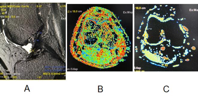
Material and Methods Our Institutional Review Board approved the protocol and written informed consent was obtained from each subject. From June 2019 to June 2021, 40 patients ranging from 23 to 61 years old (41.9±8.0 years) were included in our study. All subjects were confirmed by arthroscopy to have ACL injury. As a control group, 35 age-matched healthy volunteers from the community were recruited. All subjects underwent MR exams on a 3.0 T whole body scanner (GE Discovery 750W) with an 8-channel knee coil. Conventional MRI and DTI scans were performed. The detailed DTI parameters were as follows: number of gradient direction=25, b=400s/mm2, TE=90 ms, TR=2500 ms, slice thickness=2.9 mm,slice gap= 0 mm,FOV=180 mm×180 mm, matrix=128 ×128, NEX=4. For DTI images, post processing was done on vender provided workstation (AW 4.6, GE Healthcare) to generate FA and MD maps. Measurements were done with manually drawn region of interest (ROI) on femoral end, middle segment and tibial end of ACL respectively. For comparison purpose, one experienced radiologist who was blinded to arthroscopy results reviewed conventional MRI images for diagnosis of ACL injury. Differences in the FA and MD between acute ACL injury group and control group were compared with an independent Student’s t-test. Diagnostic efficacies of DTI and conventional MRI approach in ACL injury were also compared. For all test, P<0.05 was considered statistically significant.
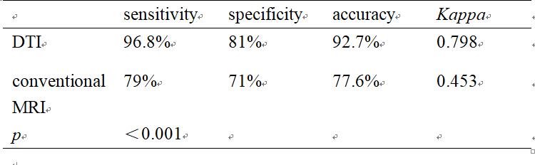
Results The mean FA values of ACL injury group and control group were 0.279±0.041 and 0.495±0.038 respectively, and the MD values were 2.884±0.163 (×10-3 mm2/s) and 1.582±0.320 (×10-3mm2/s) respectively, all of which were statistically significant (P<0.001). Based on conventional MR images, the radiologist achieved the sensitivity, specificity, accuracy and Kappa value of 79%, 71%, 77.6% and 0.453 respectively. On the other hand, the sensitivity, specificity, accuracy and Kappa value of DTI to diagnose ACL injury were 96.8%, 81%, 92.7% and 0.798 respectively, which were significantly higher than those of conventional MRI (P=0.012). (Table 1.2)
Discussion and conclusion The FA value of ACL injury area decreased obviously, while ADC value increased, and the difference between them was significant. The reason was attributed to the fracture of ligament fiber and the disorder of broken fiber bundle, which directly led to the loss of diffusion anisotropy of water molecules in ACL injury, which led to the decrease of FA value. Also, disruption of tissue fiber alignment freer motion of water. The aggregation of water molecules in ligament increases its diffusion degree, resulting an elevated MD in injured area. To conclude, diffusion tensor imaging can provide quantitative evaluation of acute ACL injury. The diagnosis approach based on DTI outperformed the approach based on conventional MR images.
Acknowledgements
No acknowledgement found.References
1. Lepley Adam S, Ly Monica T, Grooms Dustin R, etc. Corticospinal tract structure and excitability in patients with anterior cruciate ligament reconstruction: A DTI and TMS study. Neuroimage Clin 2020;25:102-157.
2. Zhao Q, Ridout RP, Shen J, etc. Effects of Angular Resolution and b Value on Diffusion Tensor Imaging in Knee Joint. Cartilage 2021;Apr 10.
3. Van Dyck P, Billiet T, Desbuquoit D, etc. Diffusion tensor imaging of the anterior cruciate ligament graft following reconstruction: a longitudinal study. Eur Radiol 2020;30(12).
4. Van Dyck P, Froeling M, Heusdens CHW, etc. Diffusion tensor imaging of the anterior cruciate ligament following primary repair with internal bracing: A longitudinal study. J Orthop Res 2021; 39(6):1318-1330.
Figures