4620
In vivo Feasibility of Three-Dimensional Simultaneous Quantitative T1-T2-T2* Mapping (SQUMA) for Knee Cartilage1Department of Radiology, Beijing Tsinghua Changgung Hospital, School of Medicine, Tsinghua University, Beijing 102218, China, Beijing, China, 2The Center for Biomedical Imaging Research, Department of Biomedical Engineering, School of Medicine, Tsinghua University, Beijing 100084, China, Beijing, China
Synopsis
T1, T2, and T2* mapping provide valuable information for evaluating joint cartilage. However, few studies simultaneously quantified T1, T2, and T2* map to evaluate knee cartilage. In this study, a three-dimensional (3D) simultaneous quantitative T1-T2-T2* mapping (SQUMA) sequence is utilized in knee cartilage. Excellent correlation and good consistency in measuring T1, T2 and T2* values were found between 3D SQUMA sequence and referenced sequences in phantom studies. The T1, T2 and T2* values of knee cartilage were 949.90±82.59 ms, 36.85±2.25 ms and 27.89±5.74 ms, respectively, with good repeatability. 3D SQUMA sequence is feasible for in vivo evaluation of knee cartilage.
Introduction
Magnetic resonance quantitative T11, T22, and T2*3 mapping can provide information for human tissue characterization and clinical diagnosis such as tumor, multiple sclerosis and neurodegenerative diseases. Increasing evidence has shown that changes in T1 and T2 values reflect the biochemical composition and microstructure variations in the cartilage at early stages of knee cartilage injury. Meanwhile, T2* mapping is more sensitive to detecting cartilage degenerative lesions. In practice, however, T1, T2, and T2* mapping often require a separate scan and may be subject to some uncertainties, such as complex imaging workflows and misalignment of parameter maps. 4 The purpose of this study is to determine the feasibility of a proposed sequence of three-dimensional (3D) simultaneous quantitative T1-T2-T2* mapping (SQUMA) for characterizing human knee joint cartilage.Methods
MRI Sequence: The 3D SQUMA sequence proposed by our group consists of six dynamic scans, using variable flip angle, variable T2 preparation time and multi echo acquisition similar to the previous method. 5 The 3D SQUMA sequence was acquired within 10 minutes on a 3.0 Tesla MR scanner (Ingenia, CX, Philips Healthcare, Best, The Netherlands) with a dedicated quadrature head coil and 8 channel knee joint special coil.Phantom experiment: The phantom was produced using previously described methods. 6 For phantom scan, the T1, T2, and T2* maps were acquired using the golden standard referenced sequences including spin echo imaging with different inversion recovery time (TR/TE = 10000/9.6 ms, TI = 100, 200, 300, 400, 500, 600, 700, 800, 900, 1000, 1500, 2000, 2500, 3000 ms), spin echo imaging with multiple echo times (TEs) (TR/TE = 10000/8*9.5 ms) and gradient echo imaging with 25 echoes (TR/TE = 10000/3.1/Δ5 ms). Clinical standard referenced sequences, such as T1 Native (TR/TE =3.1/1.4 ms), turbo spin echo with multiple TEs (TR/TE = 2000/6*13 ms) and turbo field echo with 15 echoes (TR/TE = 30/1.83/Δ1.8 ms), were also performed to acquire T1, T2 and T2* values, respectively. The 3D SQUMA sequence was also scanned in phantom and the imaging parameters are presented in Table 1.
Volunteer experiment: In vivo scans were performed on nine young health volunteers using the clinical standard referenced sequences and 3D SQUMA sequence. Five of health subjects were scanned to acquire 3D SQUMA images twice within seven days.
Data analysis: The T1, T2 and T2* maps of phantom and in-vivo subjects were fitted using MATLAB 2020a (MathWorks, Inc. Natick, Massachusetts, USA). The T1, T2 and T2* values of phantom and human knee patella cartilage were recorded.
Statistical analysis: Linear correlation analysis and Bland-Altman analysis were used to evaluate the correlation and agreement between the 3D SQUMA sequence and the referenced sequences in measuring T1, T2 and T2* values. The repeatability of T1, T2, and T2* values in phantom and in vivo were evaluated using intraclass correlation coefficient (ICC). Wilcoxon rank-sum test was used to compare the 3D SQUMA sequence with the clinical standard referenced sequences in quantitatively evaluating T1, T2 and T2* maps of the phantom and human knee cartilage.
Results
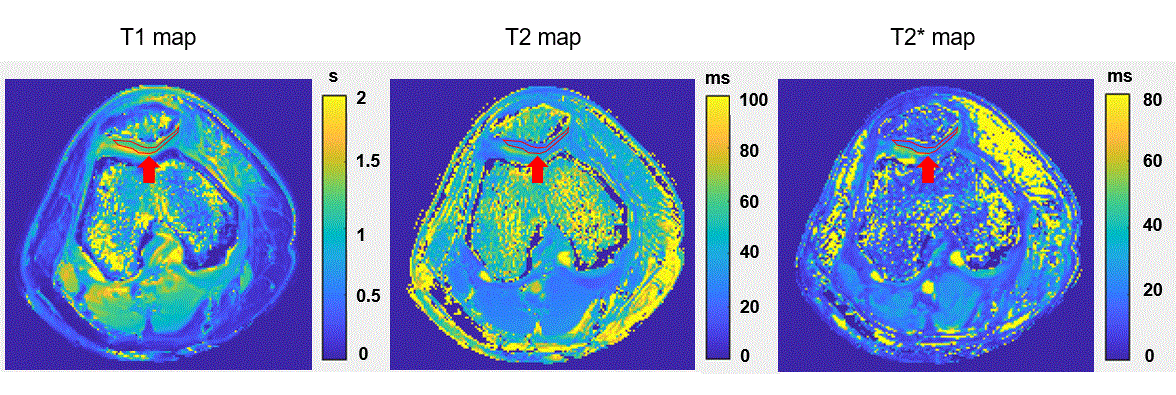
Phantom measurements: Figure 1 shows representative images of the phantom. In estimation of T1, T2, T2* values, strong correlations were found between 3D SQUMA sequence and golden standard referenced sequences (r=0.99, 0.87, 0.93 for T1, T2 and T2* values, respectively), between 3D SQUMA sequence and clinical standard referenced sequences (r=0.98, 0.87, 0.92 for T1, T2 and T2* values, respectively), and between golden standard referenced sequences and clinical standard referenced sequences (r=0.98, 0.99, 0.99 for T1, T2 and T2* values, respectively), respectively (Figure 2). The Bland-Altman plots showed a good consistency among the 3D SQUMA sequence, golden standard referenced sequences, and clinical standard referenced sequences (Figure 3). In addition, excellent repeatability was found between two scans of the phantom in measuring T1 (ICC= 0.999), T2 (ICC=0.996), and T2* (ICC=0.962) values, and no significant differences were observed in T1 (P=0.43), T2 (P=0.18), T2* (P=0.11) values between two scans of the phantom, respectively.Volunteer measurements: Figure 4 shows representative images in a healthy volunteer. The T1, T2, T2* values of knee patella cartilage by 3D SQUMA sequence (T1 =949.90±82.59 ms, T2=36.85±2.25 ms, T2*=27.89±5.74 ms) and clinical standard referenced sequences (T1 =882.96±89.95 ms, T2 =42.27±5.91 ms, T2*=30.20±5.13 ms) were successfully achieved and no significant differences were found in T1 (P=0.07), T2 (P=0.06), and T2* (P=0.11) values. For the repeated scans of knee patella cartilage, T1 (ICC=0.982), T2 (ICC=0.972), and T2* (ICC=0.978) values showed excellent repeatability, and the difference between two scans was not significant in T1 (P=0.50), T2 (P=0.23), T2* (P=0.35) values.
Discussion and conclusion
In this study, the proposed 3D simultaneous quantitative T1-T2-T2-mapping (SQUMA) sequence was proved to be a promising technique for quantitative evaluation of knee patella cartilage. The usefulness of SQUMA in diagnosing knee cartilage diseases needs to be further investigated in future studies.Acknowledgements
NoneReferences
1. Chen M, Qiu L, Shen S, et al. The influences of walking, running and stair activity on knee articular cartilage: Quantitative MRI using T1 rho and T2 mapping. PLoS One. 2017;12(11): e0187008.2. Mamisch TC, Trattnig S, Quirbach S, et al. Quantitative T2 mapping of knee cartilage: differentiation of healthy control cartilage and cartilage repair tissue in the knee with unloading--initial results. Radiology. 2010;254(3): 818-826.
3. Qian Y, Willianms AA, Chu CR, et al. Multicomponent T2* mapping of knee cartilage: technical feasibility ex vivo. Magn Reson Med. 2010;64(5): 1426-1431.
4. Cao T, Ma S, Wang N, et al. Three-dimensional simultaneous brain mapping of T1, T2, T2* and magnetic susceptibility with MR Multitasking. Magn Reson Med. 2021.
5. Qiao H, Chen S, Ning Z, et al. Three-Dimensional Simultaneous Quantitative T1-T2-T2* Mapping of Carotid Vessel Wall: Sequence Design and In-vivo Feasibility. In the proceeding of 32nd SMRA Annual International Conference #43.
6. Ikemoto Y, Takao W, Yoshitomi K, et al. Development of a human-tissue-like phantom for 3.0-T MRI. Med Phys. 2011;38(11): 6336-6342.
Figures
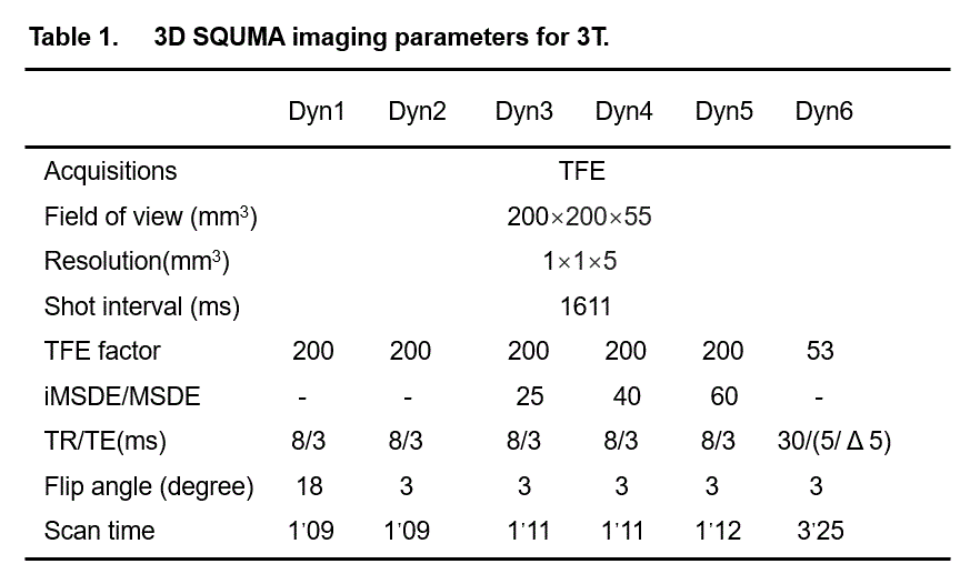

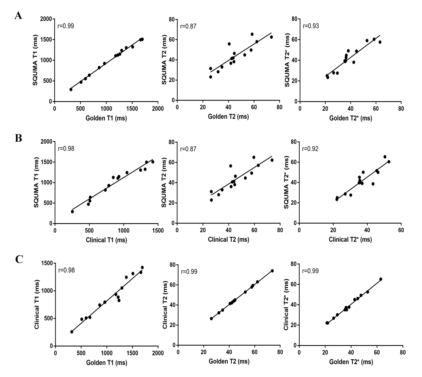
Figure 2. Correlation of T1, T2 and T2* measurements of phantoms between T1-T2-T2* mapping (SQUMA) imaging and standard reference methods (A: golden standard referenced sequences vs. 3D SQUMA, B: clinical standard referenced sequences vs. 3D SQUMA). Correlation of T1, T2 and T2* measurements of phantoms between the two reference imaging methods (C: golden standard referenced sequences vs. clinical standard referenced sequences).