4618
Quantitative evaluation of cartilage damage in hemophiliac arthropathy using automatic segmentation of 3D high resolution MRI images1Fuwai Central China Cardiovascular Hospital, Zhengzhou, China, 2MR Collaboration, Siemens Healthineers Ltd, Beijing, China, 3Siemens Shenzhen Magnetic Resonance Ltd, Shenzhen, China, 4Siemens Healthcare GmbH, Erlangen, Germany
Synopsis
Hemophilic arthropathy (HA) is a serious complication of haemophilia, characterized by bone and cartilage damage due to recurrent joint bleeding. Existing evidence showed HA has a fast disease progression. MRI-based evaluation plays an important role in the detection, categorization and staging of soft tissue and osteochondral changes in HA, which is important for assessing and monitoring treatment outcomes. However, currently semi-quantitative scoring system based on conventional MRI images cannot give an objective assessment of knee cartilage damage. To this end, this study for the first time used an automatic segmentation algorithm to quantitatively evaluate the morphological change of knee cartilage in HA.
Introduction
Haemophilia is an X-linked recessive disease that manifests in males and results in a blood coagulation defect. In patients with haemophilia, recurrent joint bleeding triggers a cascade of pathological changes that eventually cause progressive arthropathy with cartilage and bone damage [1]. Hemophilic arthropathy (HA) significantly contributes to the morbidity of the disease. Currently MRI is considered as the reference modality in detection and staging of soft tissue and osteochondral changes in hemophilic HA. Morphological quantification of articular cartilage using automatic segmentation techniques has been widely investigated in osteoarthritis and shown promise for detecting early cartilage changes. However, it has been seldomly used in HA. The purpose of this study was to: 1) to determine the reproducibility of an automatic cartilage quantification method for segmenting three-dimensional double-echo steady state (3D-DESS) images in HA; 2) to investigate the knee cartilage morphological characteristics in HA by a comparison between HA patients with different degree of cartilage injury and healthy controls.Methods
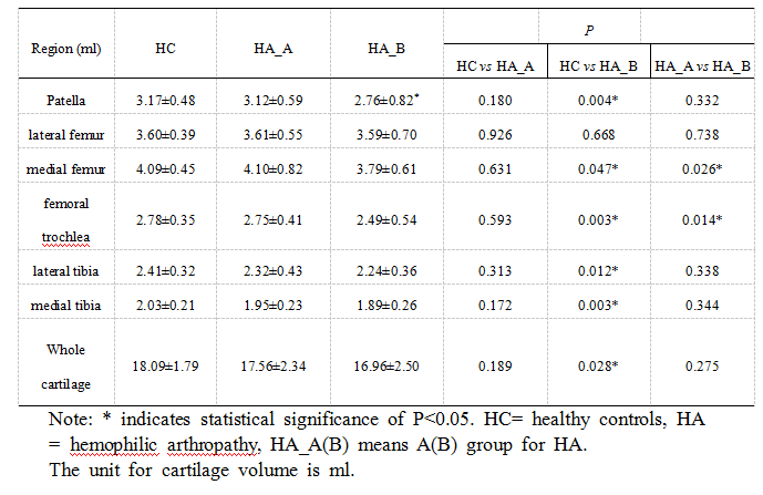
This study prospectively recruited 39 male patients (age: 16.87 ± 5.75 years old, 68 knees) with confirmed hemophilia joint disease from the Hemophilia Diagnosis and Management Center in Henan Province, China. 39 age, gender matched volunteers (age: 16.85±5.72 years old, 68 knees) were recruited as healthy control (HC) from local community. All the participants were scanned on 3T MAGNETOM Prisma scanner (Siemens Healthcare, Erlangen, Germany) equipped with a 15-channel knee coil with routine MRI and 3D DESS sequences. The parameters for the 3D DESS sequence: TR=14.1ms; TE=5ms ; FOV=160x160 mm2; acquisition time = 4:05mins.The HA knees were rated according to the criteria of International Cartilage Repair Society (ICRS) for cartilage damage and grouped into HA group A (score 0-I, with mild damage) and B (score II-IV, with severe damage). Finally, HA-A group enrolled 45 knees, HA-B group enrolled 23 knees, and HC enrolled 68 knees.A prototype post-processing software MR ChondralHealth V2.0 (Siemens Healthcare, Erlangen, Germany) was used to automatically segment the knee cartilage into 6 sub-regions on 3D DESS images using a scheme based on 3D shape models and statistical modeling [2]. More recent versions of this software provide 21 subregions and we may try it in the future. The segmentation result was checked visually in the tool after the automatic segmentation was completed. If the segmentation was accurate, the result was accepted. Otherwise, a manual correction was performed. Two experienced radiologists independently completed the segmentation.Because of the involvement of manual correction, intraclass correlation coefficient (ICC) was performed to evaluate the segmentation consistency between the two radiologists. One way analysis of variance (ANOVA) was used to explore whether the differences in sub-regional and whole cartilage volume between the three study groups were significant. P<0.05 was considered as statistically significantly different.Results
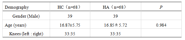
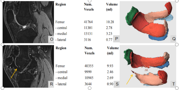
Subjects’ demography was listed in Figure 1. Age and gender were strictly controlled between HA groups and HC (P=0.984). Segmentation consistency between two radiologists was high for each subregion with minimum ICC value of 0.890. The knee cartilage volume comparison (Figure 2) found HA_B group have a significant lower cartilage volume than HC in patella, medial femur, femoral trochlea, lateral tibia and medial tibia. HA_B have a significant lower cartilage volume than HA_A in two bearing zones of knee cartilage of medial femur and femoral trochlea. No significant difference was found between HA_A and HC group.Knee cartilage was automatically segmented into 6 sub-regions. Representative segmentation results for both knees from one HA patient were shown in Figure 3.Discussion
This study for the first time used an automatic segmentation algorithm to quantitatively evaluate the morphological change of knee cartilage in HA. The 3D view of the segmentation results provided more intuitive data to observe the location and scope of cartilage defect, providing more accurate clinical evaluation of articular cartilage damage in HA patients. The segmentation consistency between two radiologists was high for each cartilage subregion (all > 0.890), demonstrating the automatic segmentation has excellent reproducibility and was not affected by inter-observer variation. Cartilage volume analysis found HA-B group have a lower cartilage volume than HC group in almost all the subregions except lateral femur, while HA-A group has no significance with HC, suggesting the morphology and volume of knee cartilage in early HA patients were intact and normal. The history of hemophilia may have no significant influence on the development of articular cartilage in patients, and the change of cartilage volume is mainly caused by repeated joint bleeding. Furthermore, it’s found that cartilage volume of patients with severe HA is significantly lower than that in the early stage (HA_B < HA_A) in the main weight-bearing area (medial femoral area). Therefore, the close tracking and monitoring of the volume of knee cartilage in the main weight-bearing area of HA patients and timely detection of the changes are conducive to early clinical intervention and treatment, which is of great value in slowing down the course of the disease.Conclusion
Automatic cartilage segmentation technology based on high resolution MRI images can reliably and efficiently quantify the morphological change of knee joint in HA, which may serve as an objective quantitative biomarker for dynamic monitoring HA disease progression.Acknowledgements
No acknowledgement found.References
[1] Doria, A. S. “State‐of‐the‐art Imaging Techniques for the Evaluation of Haemophilic Arthropathy: Present and Future.” Haemophilia, vol. 16, 2010, pp. 107–114.
[2] Fripp J, Crozier S,Warfield SK, Ourselin S (2010) Automatic segmentation and quantitative analysis of the articular cartilages from magnetic resonance images of the knee. IEE Trans Med Imaging 29(1):55–64. https://doi.org/10.1109/TMI.2009.2024743
Figures