4612
Improved Dynamic contrast in Motion Corrected Dynamic Contrast Enhancement MRI Using Low Rank with Soft Weighting and Joint sparsity1SPMIC, The University of Nottingham Ningbo China, Ningbo, China, 2COMSATS University Islamabad, Islamabad, Pakistan, 3SPMIC, The University of Nottingham, Nottingham, United Kingdom
Synopsis
This work presents a motion corrected free breathing Dynamic Contrast Enhanced MRI (DCE-MRI) with improved dynamic contrast reconstruction method called L+S with soft weighting and joint sparsity. Soft weighting achieves accurate motion correction at expense of dynamic contrast. An additional temporal Fast Fourier Transform (FFT) constraint is used to recover the dynamic contrast. A simulated phantom dataset was created with ground truth in this work, enabling quantification of the motion correction and dynamic contrast performance. The proposed method achieved improved dynamic contrast, better motion correction and high reconstruction efficiency simultaneously.
Introduction
Golden Angle Radial Sparse Parallel (GRASP) MRI1 and L+S decomposition2 are two increasingly popular reconstruction frameworks for free breathing DCE-MRI. By exploring temporal sparsity among image frames, prior spatio-temporal resolution is achieved in these two frameworks. L+S further subdivides the image series into low rank background components L and sparse dynamic components S, enabling efficient reconstruction of highly under-sampled datasets with Iterative Soft Thresholding Algorithm (ISTA). However, periodic respiratory motion still degrades reconstruction quality in GRASP and L+S decomposition. Extra dimension (XD) GRASP3 and motion weighted (RACER) GRASP4,5 involves further motion states subdivision to compress motion blurring at expense of reconstruction efficiency. Grappa operator gridding (GROG) has been introduced to accelerate the reconstruction in RACER-GRASP4. The motion compression can also be achieved by employing a soft weighting function to control the contribution of spokes acquired at different motion phases6.However, both motion subdivision and soft weighting methods theoretically amplify the contribution of temporal Total Variation (TV) constraint during the iterative reconstruction. The temporal blurring effect is increased which leads to further degradation of dynamic contrast. The dynamic contrast can be recovered by employing an additional sparsity constraint temporal FFT7. Here we introduce both soft weighting and joint sparsity constraints into L+S decomposition model, compressing motion blurring and recovering the dynamic contrast simultaneously for improved DCE-MRI reconstruction.
Method
The proposed L+S with soft weighting and joint sparsity scheme is mathematically formulated as: $$ argmin_{L,S}=\frac{1}{2}\left \| W\{E(L+S)-d\}\right \|_{2}^{2}+\lambda_{L}\left \| L \right \|_{*}+\lambda_{T}\left \| TS \right \|_{1}+\lambda_{F}\left \| FS \right \|_{1} \qquad \qquad [1]$$ where $$$ E $$$ is the multi-coil encoding operator and $$$ d $$$ is the acquired data. $$$ W $$$ is a soft weighting matrix. $$$ T $$$ and $$$ F $$$ present the temporal TV and temporal FFT sparsity transforms respectively. Penalty factors $$$ \lambda_{L} $$$ , $$$ \lambda_{T} $$$ and $$$ \lambda_{F} $$$ trade off data consistency versus complexity of solution given by the nuclear norm $$$ \left \| \cdot \right \|_{*} $$$ and $$$ L_{1} $$$ norm $$$ \left \| \cdot \right \|_{1} $$$. In this work, Fast Composite Splitting Algorithm (FCSA) was employed to solve the reconstruction scheme with multiple constraints efficiently8,9.A simulated phantom dataset was produced to quantify the dynamic degradation caused by motion correction and dynamic recovery from joint sparsity. A modified Shepp-logan computer model was created with a total of 512*512 voxels, six dynamic regions and 3 motion regions. The signal intensity of dynamic regions is varied with designed dynamic curve. The position of motion regions was rotated periodically at a maximum angle of 12 degrees, simulating respiratory motion. The simulated dataset contains 8 virtual channels, 1100 golden angle spokes with 768 readout points in each. A free breathing liver DCE-MRI dataset offered by Li.et al3 was adopted to evaluate the proposed method further. The liver dataset contains 8 channels, 1100 golden angle spokes with 512 readout points in each. Both two datasets were subdivided into 11 time frames with the temporal resolution of 100 spokes/frame. The time series were reconstructed by GRASP, L+S decomposition, XD-GRASP, RACER-GRASP, L+S with soft weighting and proposed method with a matrix size 512*512*11 in phantom dataset and 384*384*11 in liver dataset. All the reconstructions were performed using MATLAB 2020b (MathWorks, Natick, MA) on an Intel Core i7-10700 PC with a 2.9 GHz processor.
Results & Discussion
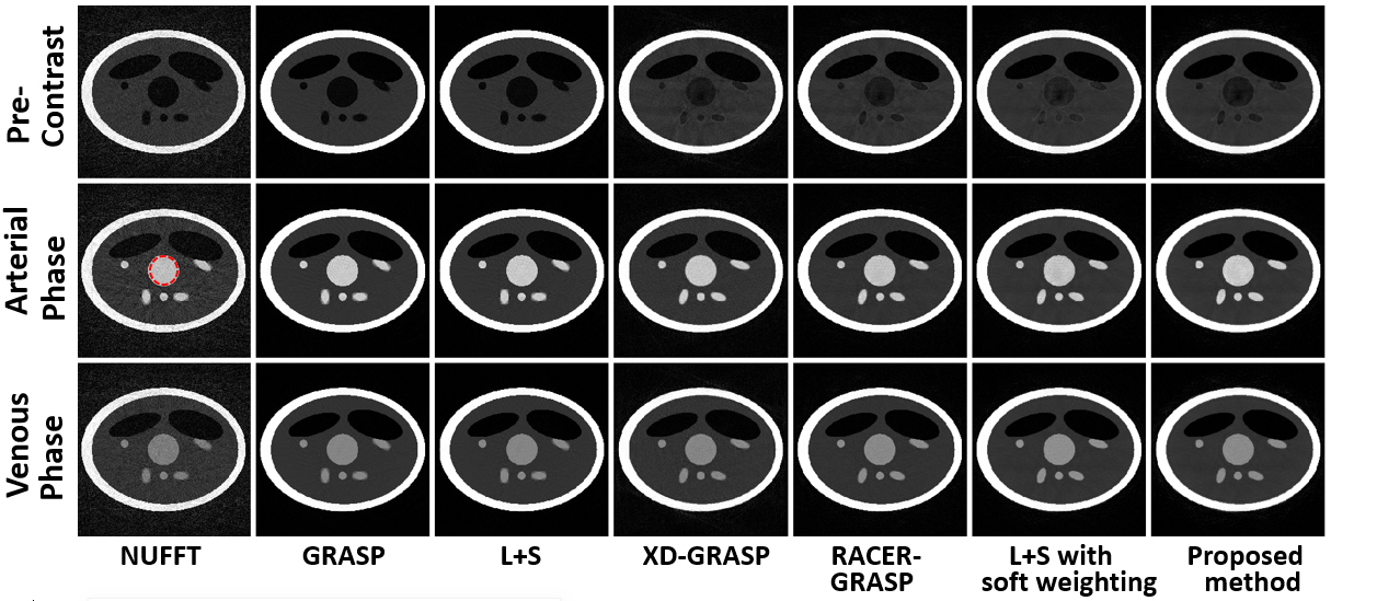
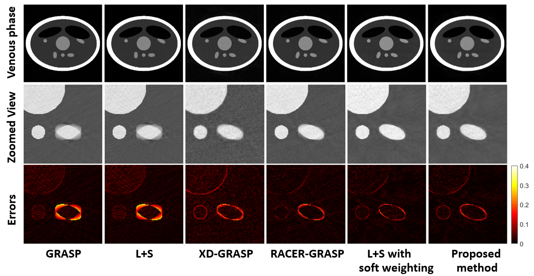
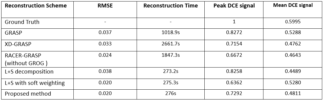
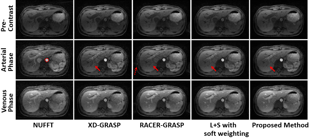
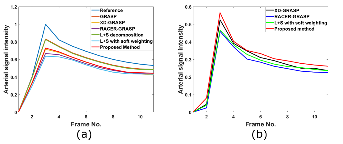
Figure 1 and Figure 2 show a comparison of six reconstruction schemes in simulated phantom dataset. Without motion correction, obvious motion blurring was observed in GRASP and L+S decomposition. XD-GRASP, RACER-GRASP, L+S with soft weighting and proposed method compressed motion blurring effectively. Figure 3 summarizes the reconstruction period and motion correction errors of different schemes. Using the selected motion section in reference as the benchmark, the minimum RMSE was achieved in L+S with soft weighting and the proposed method, showing the robustness of soft weighting. The performance of soft weighting is not degraded by joint sparsity. Our proposed method and standard L+S achieved similar reconstruction efficiency. Figure 4 shows a comparison of 4 motion corrected reconstruction schemes in liver DCE-MRI dataset. Residual streaking artefacts and convolutional artefacts were obtained in XD-GRASP and RACER-GRASP respectively. Better motion correction and dynamic contrast were observed in the proposed method. Figure 5 demonstrates dynamic performance of the different reconstruction schemes in two datasets. GRASP and L+S decomposition showed similar dynamic degradation compared with reference. Further dynamic degradation was observed in motion corrected schemes as -28.5%, -33.3% and -36.4% in XD-GRASP, RACER-GRASP and L+S with soft weighting respectively. The proposed method demonstrated an increase in peak DCE signal by 14.6% than that of L+S with soft weighting (Figure 5a). In Figure 5b, the proposed method demonstrated an increase in peak DCE signal by 7.8%, 23.1% and 20% than that of XD-GRASP, RACER-GRASP and L+S with soft weighting in liver DCE-MRI dataset.Conclusion
A low rank with soft weighting and joint sparsity framework is proposed and evaluated for improved motion corrected DCE-MRI reconstruction. Soft weighting leads to better motion correction without additional computation cost. Joint sparsity constraints recover the dynamic contrast degraded by motion correction effectively while the computation cost from additional sparsity constraint is negligible with the support of FCSA.Acknowledgements
No acknowledgement found.References
1. Feng L, Grimm R, Block KT, Chandarana H, Kim S, Xu J, Axel L, Sodickson DK, Otazo R. Golden-angle radial sparse parallel MRI: combination of compressed sensing, parallel imaging, and golden-angle radial sampling for fast and flexible dynamic volumetric MRI. Magn Reson Med. 2014;72(3):707-717.
2. Otazo R, Cande`s E, Sodickson DK. Low-Rank Plus Sparse Matrix Decomposition for Accelerated Dynamic MRI with Separation of Background and Dynamic Components. Magn Reson Med. 2015;73(3):1125–1136.
3. Feng L, Axel L, Chandarana H, Block KT, Sodickson DK, Otazo R. XD-GRASP: golden-angle radial MRI with reconstruction of extra motion-state dimensions using compressed sensing. Magn Reson Med. 2016;75:775–788.
4. Feng L, Huang C, Shanbhogue K, Sodickson DK, Chandarana H, Otazo R. RACER-GRASP: Respiratory-Weighted, Aortic Contrast Enhancement-Guided and Coil-Unstreaking Golden-Angle Radial Sparse MRI. Magn Reson Med 2018;80:77–89.
5. Chen L, Zeng X, Ji B, Liu D, Wang J, Zhang J, Feng L. Improving dynamic contrast-enhanced MRI of the lung using motion-weighted sparse reconstruction: Initial experiences in patients. Magnetic resonance imaging. 2020; 68:36-44.
6. Najeeb F, Zhang J, Wang X, Wang C, Omer H, Gowland P, Francis S, Glover P. Efficient DCE-MRI image reconstruction with feasible temporal resolution in L+S Decomposition model. Proceedings of the ISMRM 30th Annual Meeting & Exhibition 2021. International Society for Magnetic Resonance in Medicine.
7. Zhang J, Najeeb F, Wang X, Xu P, Omer H, Gowland P, Francis S, Glover P, Bowtell R, Wang C. Improved Dynamic Contrast Enhanced MRI Using Low Rank with Joint Sparsity Reconstruction. Proceedings of the ISMRM 30th Annual Meeting & Exhibition 2021. International Society for Magnetic Resonance in Medicine.
8. Huang J, Zhang S, Metaxas D. Efficient MR image reconstruction for compressed MR imaging. Medical Image Analysis.2011; 15(5):670-679.
9. Huang J, Wang L, Zhu Y. Compressed Sensing MRI Reconstruction with Multiple Sparsity Constraints on Radial Sampling. Mathematical Problems in Engineering. 2019; 2019: 1–14.
Figures