4595
A 72-channel coil system for MR vessel wall imaging of intracranial and carotid arteries at 3 T1Paul C. Lauterbur Research Center for Biomedical Imaging, Shenzhen Institutes of Advanced Technology, Chinese Academy of Sciences, Shenzhen, China, 2Key Laboratory for Magnetic Resonance and Multimodality Imaging of Guangdong Province, Shenzhen, China, 3Shanghai United Imaging Healthcare, Shanghai, China
Synopsis
MR vessel wall imaging of intracranial and carotid arteries is still challenging because of the small cross-sectional size of the vessel wall and the cord, and susceptibility effects, especially in the carotid and the spinal cord. In this study, a 72-channel coil system that consists of a 64-channel head coil combined with an 8-channel carotid coil was proposed and characterized in its performance by comparison with a 40-channel coil system. As a result, the proposed 72-channel coil system provides improved performance in SNR, parallel imaging capability, and image quality.
Introduction
Atherosclerotic plaque is a primary cause of ischemic stroke and is prevalent in the aorta and carotid vessels [1]. Simultaneous intracranial and extracranial arterial wall imaging would greatly enhance the convenience of clinical diagnosis. However, the normal middle cerebral artery and basilar artery wall thickness is 0.2–0.3 mm, which needs a high spatial resolution imaging. Previous studies showed that a high isotropic spatial resolution of 0.6 mm can be achieved by using a 40-channel coil system including a 32-channel head coil and an 8-channel carotid coil [2]. In this work, we have designed and built a 72-channel coil system with a 64-channel head coil and an 8-channel carotid coil for MR vessel wall imaging at a 3 T MRI system (uMR 880, Shanghai United Imaging Healthcare, Shanghai, China). The coil performance was characterized with improved SNR, enhanced parallel imaging capability, and superior image quality in human studies, compared to a 40-channel coil system with a 32-channel head coil and an 8-channel carotid coil.Methods
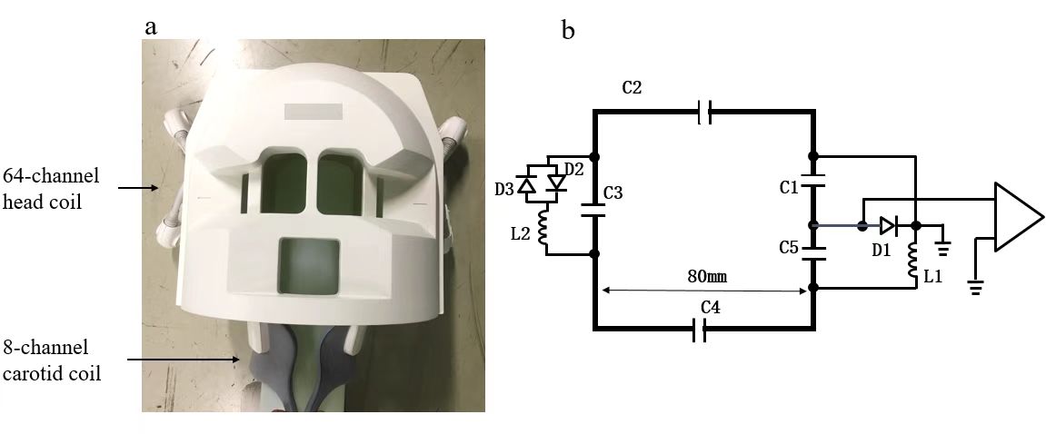
Figure 1 shows the photograph and the circuit schematic of the 72-channel coil system. The head coil elements with an 80 mm diameter were arranged in six rows in H-F direction. The inner dimensions of the head coil shell were: 230 mm in the Anterior-Posterior (AP) direction, 205 mm in the Left-Right (LR) direction, and 255 mm in the Inferior-Superior (IS) direction. Conventional front-end circuits generally include matching and tuning circuits, detuning circuits and preamplifier. Each element was tuned to 128.23 MHz and 50 ohm impedance-matched to minimize the noise of the preamp.A 2D density-weighted gradient echo (GRE) sequence was applied for signal acquisitions with a phantom. The parameters were as followings: TR/TE =400/20 ms, flip angle=30o, slice thickness=5mm, matrix=256×256, FOV=250mm×250mm. The noise images were obtained by setting the flip angle to zero. For SNR comparisons, SNR maps were calculated using the sum-of-squares method [3]. For parallel imaging capability evaluation, the inverse g-factor maps were analyzed by using sensitivity encoding (SENSE) reconstructions [4].
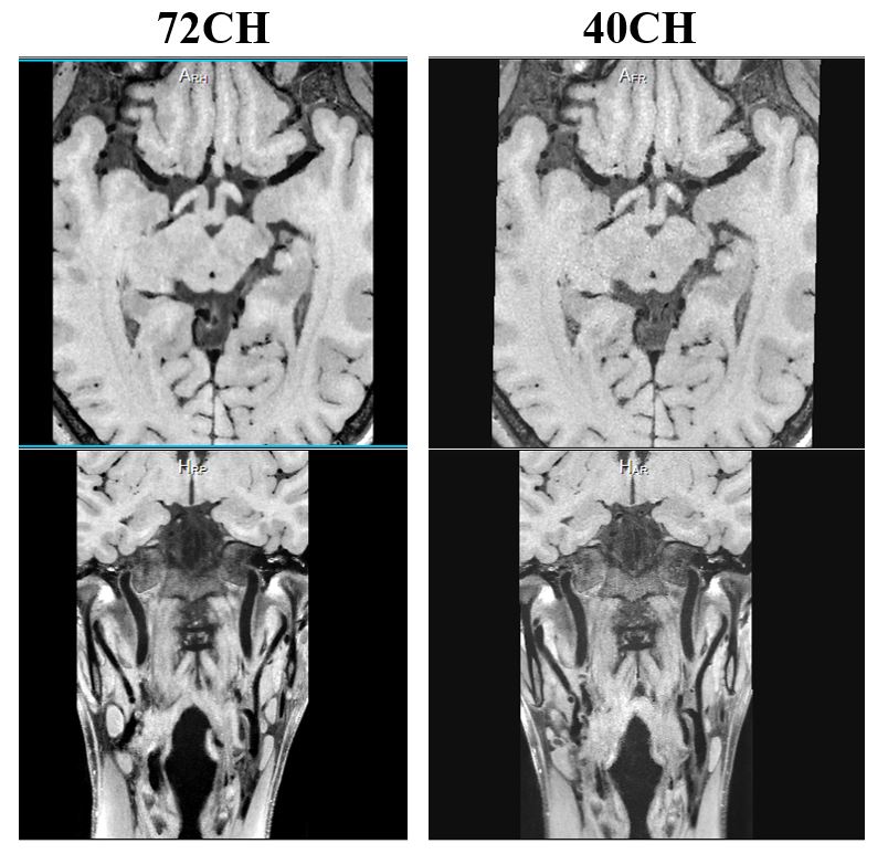
Human anatomy images were acquired using a 3D GRE sequence with with following parameters: TR/TE=8.4/3.4 ms, flip angle=9o, FOV=220mm×256mm, matrix=316×368, reconstructed resolution=0.7×0.7×0.7mm3, bandwidth=250 Hz/pixel, compression channel = 36, uCS (united compressed sensing) acceleration factor=5. Vessel wall imaging of intracranial and carotid arteries were acquired using a T1-weighted fast spin echo (FSE) MATRIX (Modulated flip angle technique in refocused imaging with extended echo train) sequence with following parameters: TR/TE=800/14.82 ms, flip angle=75o, FOV=180mm×220mm, matrix=380×464, reconstructed resolution=0.47×0.47×0.47mm3, bandwidth=440 Hz/pixel, compression channel = 36, uCS acceleration factor=5.5.
Results
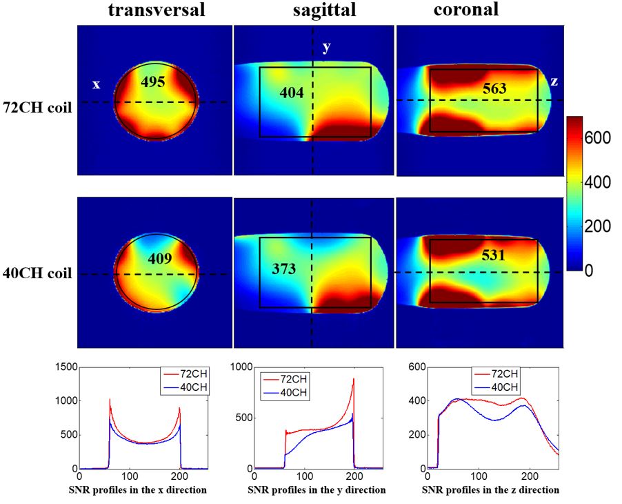
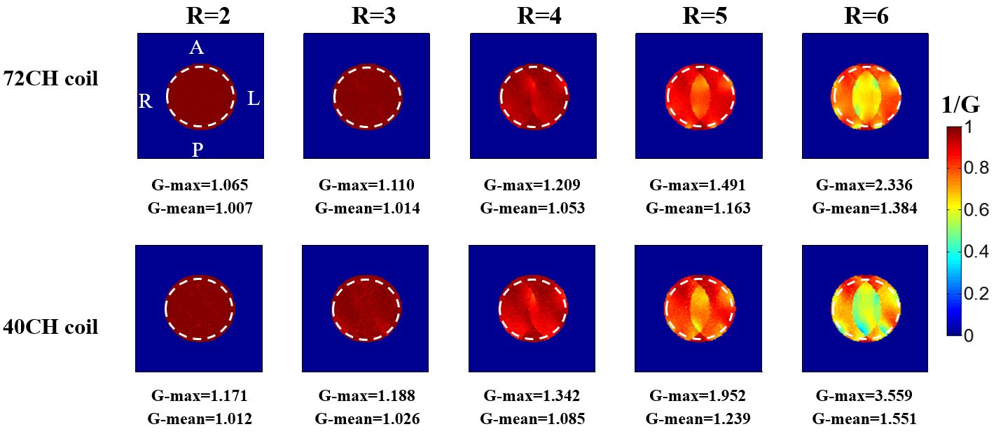
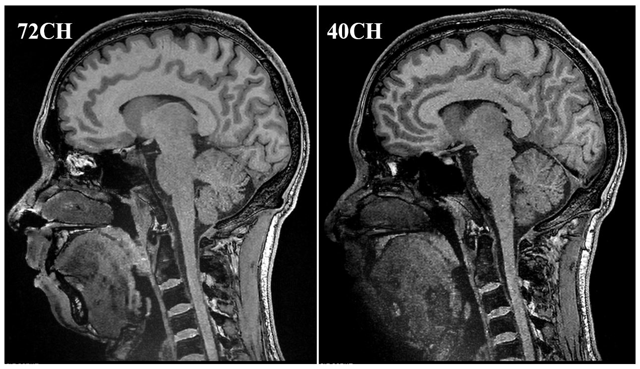
Figure 2 shows phantom SNR maps in the transverse, sagittal and coronal planes. Additionally, SNR profiles in x, y and z directions were also depicted in Figure 2. The mean SNR values in the ROIs depicted as the circles or rectangles and SNR profiles both demonstrated that the 72-channel coil system had higher SNR than the 40-channel coil system. Figure 3 depicts the inverse g-factor maps in the transverse plane with acceleration factors R from 2 to 6. The results indicate that the parallel imaging capability of the 72-channel coil system was better than that of the 40-channel coil system, particularly at high acceleration factors. Figure 4 displays the anatomy images of human brain with a 0.7 mm resolution and an acceleration factor of 5 using the 72-channel and 40-channel coil systems. Figure 5 displays the MR vessel wall images using the 72-channel and the 32-channel coil systems. Both these images using 72-channel coil system showed a higher quality than those using 40-channel coil system.Discussions and Conclusions
A 72-channel array coil system for MR vessel wall imaging of intracranial and carotid arteries was designed, constructed and evaluated by imaging experiments in phantom and human studies. Compared to the 40-channel coil system, the 72-channel coil system achieves better performance in MR SNR, acceleration capacity and quality of MR vessel wall images.Acknowledgements
This work is supported by the Strategic Priority Research Program of Chinese Academy of Sciences, XDB25000000; National Key R&D Program of China, 2021YFE0204400; Shenzhen city grant, RCYX20200714114735123, ZDKJ20190204003, ZDKJ20190204004.References
[1] Mandell DM, Mossa-Basha M, Qiao Y, Hess CP, Hui F, Matouk C, Johnson MH, Daemen MJ, Vossough A, Edjlali M, Saloner D, Ansari SA, Wasserman BA, Mikulis DJ; Vessel Wall Imaging Study Group of the American Society of Neuroradiology. Intracranial Vessel Wall MRI: Principles and Expert Consensus Recommendations of the American Society of Neuroradiology. AJNR Am J Neuroradiol. 2017, 38(2):218-229.
[2] Q. Chen, Z. Wei, L. Zhang, C. Tie, Q. He, X. Zhang, X. Liu, H. Zheng, and Y. Li. “MR vessel wall imaging of intracranial and carotid arteries with a 40-channel coil system at 3 T,” ISMRM, 2019, p1488.
[3] Roemer PB, Edelstein WA, Hayes CE, Souza SP, Mueller OM. The NMR phased array. Magn Reson Med. 1990, 16(2):192-225.
[4] Pruessmann KP, Weiger M, Scheidegger MB, Boesiger P. SENSE: sensitivity encoding for fast MRI. Magn Reson Med. 1999, 42(5):952-62.
Figures