4566
Clinical assessment of high spatial resolution myocardial T2-weighted dark blood imaging based on deep learning1Department of Radiology, Tongji Hospital, Tongji Medical College, Huazhong University of Science and Technology, Wuhan, China, 2United Imaging Healthcare, Shanghai, China, 3UIH America, Inc., Houston, TX, United States
Synopsis
High resolution T2-weighted dark blood (HR-T2W-DB) imaging is not always robust for clinical use because of low SNR and long scan time. The purpose of this study was to evaluate a novel deep learning (DL) based reconstruction method in T2W-DB sequence that achieves higher spatial resolution and same scan duration compared with traditional reconstruction method. Quantitative and qualitative image assessment demonstrated that DL based HR-T2W-DB sequence showed better CNR in region of edema and LV free wall visibility, which might help detecting myocardial edema.
Introduction
T2-weighted dark blood (T2W-DB) imaging is an important technique to evaluate myocardial edema. Constrained by long breath-hold time, 2D dual inversion breath-hold fast spin echo sequence for cardiac T2W-DB imaging requires high acceleration factors to achieve high spatial resolution, which usually causes a compromise in image quality due to low signal-to-noise ratio (SNR).Deep learning has been used frequently in MRI reconstruction, especially in de-noising, and restoration, et al. It has been proven to have the potential to shorten MR scan time or to improve image quality.
The purpose of this study was to implement the combination of deep learning based acceleration1 and reconstruction2 method on a high resolution T2W-DB sequence, and to compare the image quality, diagnostic value with the conventional T2W-DB with routine reconstruction method and same scan duration.
Methods
Deep learning reconstruction:DL-based HR-T2W-DB imaging acquisition was under the scheme of a novel deep learning based acceleration method (AI-assisted Compressed Sensing, ACS) 1. Reconstructed multi-channel data was then transferred to a novel deep learning based image reconstruction neural network 2 as input.
MR Scan:
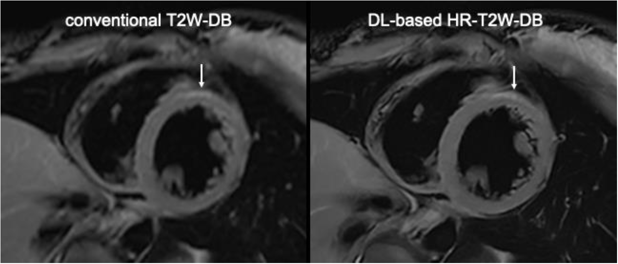
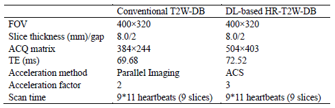
This clinical study was approved by the local Institution Review Board. Five healthy volunteers (age 29±4, five male) and twenty eight patients (age 43±15, twenty one male) were prospectively recruited to undergo cardiac magnetic resonance imaging (CMR). Clinical indications included ischemic and non-ischemic cardiomyopathy, cardiac valve disease, myocarditis, and other diseases that were not classified. CMR was performed on a 3T scanner (uMR 790, United Imaging Healthcare, Shanghai, China) with a 24-channel dedicated cardiac coil. Conventional T2W-DB imaging was achieved with parallel imaging. Both conventional T2W-DB and DL-based HR-T2W-DB imaging were applied in short-axial view at the same position (Figure 1). Detailed scan parameters were shown in Table1.
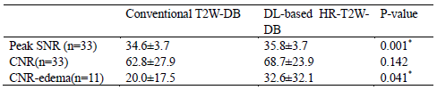
Image Analysis: Two radiographers (X.Y., Y.L.) contoured regions of interest on the ventricular septal myocardium and the blood pool on mid-ventricular short-axis images independently. Peak SNR 3 and CNR which was defined as ($$\frac{SI_{myocardium-mean}-SI_{blood-mean}}{SI_{blood-SD}}$$) were calculated as quantitative measurements.
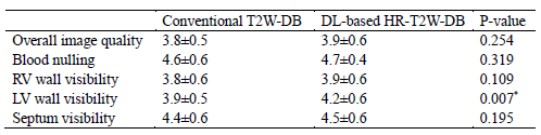
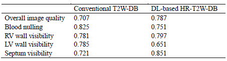
Images were blindly reviewed by two radiologists (L.H., L.R.), both with more than 5 years of experience in cardiovascular imaging. Overall image quality, blood nulling, right ventricular (RV) free wall visibility, left ventricular (LV) free wall visibility, and septum visibility were scored individually on mid-ventricular short-axis images with a 5-point Likert scale as: 1-non-diagnostic; 2-poor; 3-fair; 4-good; 5-excellent 4. In eleven patients with myocardial edema 5, CNR-edema, which was defined as ($$\frac{SI_{edema-mean}-SI_{muscle-mean}}{SI_{blood-SD}}$$), was also calculated.
Statistical Analysis:
All data were described as mean ± standard deviation. Statistical analysis was performed using SPSS (version 23.0, Chicago, IL). Ordinal Likert scores from both observers were averaged prior to analysis, and differences in Conventional T2W-DB and DL-based HR-T2W-DB scores were assessed using paired t-test and paired Wilcoxon signed-rank test, respectively. Kendall's W was used to assess inter-observer agreement of scoring data. ICC was used to assess inter-observer agreement of CNR. P<0.05 was considered as statistically significant.
Results
Peak SNR and CNR-edema of DL-based HR-T2W-DB sequence were both significantly higher than conventional T2W-DB (Table 2). Image quality scores of LV free wall visibility in DL-based HR-T2W-DB sequence were also significantly higher than conventional T2W-DB (Table 3). Overall image quality, blood nulling, RV free wall visibility and septum visibility shows no statistical difference between the two sequences. The results of CNR measured by the two radiographers in DL-based HR-T2W-DB and conventional T2W-DB were in good agreement (ICC 0.669 and ICC=0.677, respectively), and scoring data also had good inter-observer agreement (Table 4).Discussion
The DL-based HR-T2W-DB sequence doubles the resolution without increasing scan duration or compromising image quality compared with conventional T2W-DB sequence. In our study, deep learning based HR-T2W-DB sequence could achieve better peak SNR, better CNR in region of edema and LV free wall visibility, which might be benefit for detecting myocardial edema, especially in cases of myocarditis to reveal LV lateral wall edema.Acknowledgements
No acknowledgement found.References
1. Renkuan Zhai, et al. Intelligent Incorporation of AI with Model Constraints for MRI Acceleration. Proc. Intl. Soc. Mag. Reson. Med. 27 (2021): 1760.
2. Aowen Liu, et al. A lightweight and efficient convolutional neural network for MR image restoration Proc. Intl. Soc. Mag. Reson. Med. 27 (2021): 1952.
3. Wang Z, et al. Image quality assessment: from error visibility to structural similarity[J].IEEE transactions on image processing : a publication of the IEEE Signal Processing Society,2004,13(4):600-612.
4. Edelman RR, Botelho M, Pursnani A, Giri S, Koktzoglou I. Improved dark blood imaging of the heart using radial balanced steady-state free precession. J Cardiovasc Magn Reson. 2016;18(1):69.
5. Friedrich MG, Sechtem U, Schulz-Menger J, et al. Cardiovascular magnetic resonance in myocarditis: A JACC White Paper. J Am Coll Cardiol. 2009;53(17):1475-1487.
Figures