4546
Acceleration Motion Compensation Diffusion-weighted Imaging for Large Vessel Vasculitis: Phantom Model and Initial Clinical Experience
Tomoko Hyodo1, Daisuke Morimoto-Ishikawa2,3, Hayato Kaida1, Yu Ueda4, Daisuke Tomita5, Atsuhiro Yamamoto5, Makoto Itoh2, Nao Yasuda2, Hiroyuki Fukushima2, Yuji Nozaki5, Itaru Matsumura5, and Kazunari Ishii1
1Radiology, Kindai University Faculty of Medicine, Osaka-Sayama, Japan, 2Radiology Center, Kindai University Hospital, Osaka-Sayama, Japan, 3Medical Physics and Engineering, Osaka University Graduate School of Medicine, Suita, Japan, 4Philips Japan, Tokyo, Japan, 5Hematology and Rheumatology, Kindai University Faculty of Medicine, Osaka-Sayama, Japan
1Radiology, Kindai University Faculty of Medicine, Osaka-Sayama, Japan, 2Radiology Center, Kindai University Hospital, Osaka-Sayama, Japan, 3Medical Physics and Engineering, Osaka University Graduate School of Medicine, Suita, Japan, 4Philips Japan, Tokyo, Japan, 5Hematology and Rheumatology, Kindai University Faculty of Medicine, Osaka-Sayama, Japan
Synopsis
The ascending aorta is poorly delineated on DWI due to the heartbeat, making it difficult to assess the activity of large vessel vasculitis using MRI. We investigated the utility of acceleration motion compensation diffusion-weighted imaging (aMC-DWI) compared with conventional DWI (cDWI) to detect active inflammation of the accenting aorta. The phantom experiment revealed that aMC-DWI showed a significantly higher median visual score than cDWI in both experimenters (P =.048 for both). The clinical study on 11 patients showed that aMC-DWI had higher sensitivity, specificity, and positive predictive value (with FDG-PET/CT as the reference method) than cDWI.
Introduction
The activity of large vessel vasculitis is usually assessed by a segmental approach by using FDG-PET/CT1. The advantage of FDG-PET/CT is its well-documented clinical usefulness, but its disadvantages include limited applicability to diseases and conditions, high cost, and radiation exposure. The possible advantages of DWI over FDG-PET are that the signal and morphology of the arterial wall can be assessed unencumbered by the signal of the vessel lumen, the combination with anatomical MRI such as T2-weighted images or balanced turbo field-echo can provide a lot of histological information, and the cost is lower. However, one of the problems with DWI is that the depiction of the ascending aorta is reduced by the effect of the heartbeat. The acceleration motion compensation diffusion-weighted imaging (aMC-DWI) may visualize the ascending aorta and allow clinicians to use MRI for diagnosis and monitoring of disease activity of large vessel vasculitis. The purpose of this study was to investigate whether aMC-DWI could be an alternative to FDG-PET/CT for the detection of ascending aortitis.Methods
A phantom experiment and a clinical study were conducted on a 1.5 T MRI scanner. Qualitative comparisons were made between the aMC-DWI and conventional DWI (cDWI) (b factor=800 s/mm2 for both) since the apparent diffusion coefficient value of aMC-DWI could not be acquired in principle. The phantom was comprised of a syringe (internal diameter 9 mm), which was filled with agar, attached to a balloon. Two experimenters independently manipulated the balloon to move the syringe up and down in 5 mm range at 60 times per minute. Twenty image slices obtained over four sessions (two experimenters/two DWI techniques) were graded on a 5-point scale (1=poor to 5=as good as a static image by using cDWI). Mann-Whitney U tests with Bonferroni correction were used to assess differences among the sessions. The prospective clinical study was performed on patients with suspected aortitis undergoing DWI using both two techniques. cDWI and aMC-DWI were visually scored for active inflammation of the ascending aorta (0=negative or 1=positive), and the difference value (aMC-DWI - cDWI) was calculated for each patient. We compared the detectability of inflammation in the ascending, arch, and descending portions of the thoracic aorta between the two DWI techniques using FDG-PET/CT as the reference method. Statistical analyses included calculation of sensitivity, specificity, and positive and negative predictive value (PPV and NPV, respectively), as well as a two-sided one-sample t-test (assuming mean difference = 0).Results
In the phantom experiment, aMC-DWI showed a significantly higher median score than cDWI in both experimenter 1 (4 vs 3; P =.048) and experimenter 2 (3 vs 1; P =.048). In the clinical evaluation, 11 patients (median age, 37 years [range, 19-84 years]; 10 women) were assessed. Five patients were diagnosed as aortitis (two with giant cell arteritis and the other with Takayasu arteritis) and FDG-PET/CT indicated active inflammation in the ascending aorta in four patients (maximum standardized uptake value range, 3.2 and 4.4). The sensitivity, specificity, and PPV and NPV, respectively, for detecting active inflammation in the ascending aorta (with FDG-PET/CT as the reference method) were 0/4 (0 %), 7/7 (100 %), 0/0 (Inf), and 7/11(64 %) for cDWI; and 4/4 (100 %), 6/7 (86 %), 4/5 (80 %) and 6/6 (100%) for aMC-DWI. The one sample t-test revealed a significant difference between two DWI techniques (t = 2.887; P = .016; 95% CI = 0.10 – 0.81).Conclusion
The acceleration motion compensation DWI improves aortic delineation over conventional DWI and promises to increase the utility of MRI in assessing activity and extent of large vessel vasculitis.Acknowledgements
Not applicableReferences
1. Slart R, Writing g, Reviewer g, et al. FDG-PET/CT(A) imaging in large vessel vasculitis and polymyalgia rheumatica: joint procedural recommendation of the EANM, SNMMI, and the PET Interest Group (PIG), and endorsed by the ASNC. Eur J Nucl Med Mol Imaging. 2018;45:1250-1269.Figures
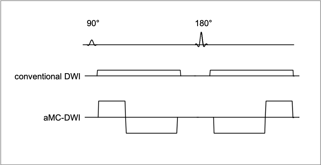
Conventional DWI is the diffusion sequence by using a mono-polar diffusion gradient. Acceleration motion compensation DWI (aMC-DWI) is the diffusion sequence by using the series of gradient which first and second moments are nulled. The spin phase error due to velocity and acceleration motion can be compensated.
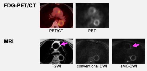
A 68 yo woman with giant cell arteritis. PET and MR T2-weighted images show wall thickening with inflammation in the left wall of the ascending aorta (arrow) and the entire circumference of the descending aorta. Acceleration motion compensation DWI (aMC-DWI) depicts high signal intensity of the ascending aorta (arrow) better than cDWI.
DOI: https://doi.org/10.58530/2022/4546