4541
Demonstrating the Facial Nerve in the Parotid Gland using 3 Dimension Fast Field Echo imaging: a Pilot Study1Department of Radiology, the First Affiliated Hospital of Dalian Medical University, Dalian, China, 2Philips Healthcare, Beijing, China, Beijing, China
Synopsis
Facial nerve MRI is a clinical challenge that it is difficult to differentiate parotid gland tumors from facial nerve on conventional imaging. It is great demand to develop new diagnostic technology to accurately display peripheral nerve and tumor for avoiding intraoperative injury. In this study, 3 dimension fast field echo imaging(T2WI-3D-FFE) is potentially a valuable sequence in displaying the intraparotid facial nerve and localizing the tumor.
Introduction
Facial nerve which is the VII of cranial nerves and part of the facial nerve transverses the parotid gland where the parotid gland tumors maybe ocurr. Thus,intraoperative injury may cause irreversible consequence that affect the quality of life seriously. However, the conventional MRI is unable to display the facial nerve in the parotid gland accurately1, 2. It is important to find an effective method to display the facial nerve. T2WI-3D-FFE sequence is a three-dimensional high-resolution T2-weighted sequence, which can inhibit the fat background and blood flow signals, and has significant advantages in peripheral nerve. In this study, we use T2WI-3D-FFE sequence to display the facial nerve and explore the localization with parotid tumors.Methods
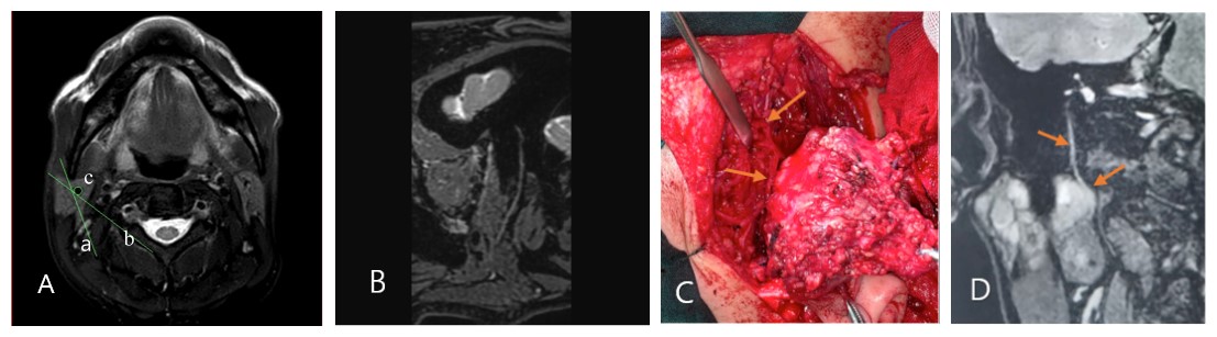
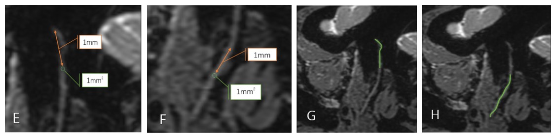
Thirty patients with solitary tumor of parotid glands (27 benign tumors and 3 malignant tumors) were recruited from the First Affiliated Hospital of Dalian Medical University. All patients were scanned using a 3.0 T MR scanner (Ingenia CX, Philips Healthcare, the Netherlands) with a 32 channel phase-array head coil. The MR protocol is T2WI-3D-FFE sequence. TR/TE = 8.3/4.1 ms, flip angle=30, FOV=220×220×65 mm3, Voxel=0.65mm×0.65mm×1.00mm, matrix=340×339×130, slice thickness=1.0mm, Gap=-0.5. The serial images were reconstructed in multiplanar reconstruction (MPR) and curvilinear planar reconstruction (CPR). Altogether there were 30 facial nerves on the tumor side and the contralateral sides were measured as the control group. Two radiologists (with 3 and 5 years radiology experiences respectively) independently scored the display of the facial nerve in T2WI-3D-FFE images according to Previous work 3, 4. ROI was placed on the nerve which is 1 mm far from the stylomastoid foramen and the place where the facial nerve enters the parotid gland. At the same time, the signal value of parotid parenchyma and image background were measured in the same slice. The signal intensity ratio (SIRN) is the comparison between the signal of facial nerve and the parotid gland in the same slice5. The nerve length of the mastoid and parotid gland is measured along the course of the reconstructed images. The tumor locations were categorized as deep or superficial lobes on the basis of observing the facial nerve on MRI and tumor directly. The other 3 indirect methods: the facial nerve line (FNL), retromandibular vein (RMV), and Utrecht line (UL) were also be recorded. The true positions of parotid tumors were confirmed by surgery. All the measurement data are expressed by mean ± standard deviation. The diagnostic accuracy, sensitivity and specificity for localizing parotid lesions using each method were calculated and compared using the McNemar tests. The scores of the image quality were evaluated on paired (mastoid and parotid gland) data by using a Wilcoxon test. For SNR and CNR, paired t testing was used to assess facial nerve (mastoid and parotid gland) differences. The interobserver reliability on qualitative evaluation was assessed via intraclass correlation coefficient (excellent agreement if ICC > 0.8; good agreement if ICC > 0.6).Results
Measurement consistency between the two observers was good (ICC>0.6, p<0.05). The average image quality scores are satisfied, SNR and CNR of the intraparotid facial nerve are better than the mastoid segment (Table 1). The SIRN and length of the mastoid and parotid gland segments are shown in Table 2. The direct method means judging the deep or superficial lobe by manifestation the facial nerve directly. It showed the highest sensitivity and specificity for localization of deep lobe parotid gland tumors (85.7%,100% respectively) (Table 3).Discussion
In this study, T2-3D-FFE sequence can show the facial nerve of the parotid gland segments with high signal and quality. Moreover, compared with other indirect methods, the direct method showed the highest sensitivity and specificity for localization of deep lobe parotid gland tumors. Although the direct method has no statistical differences, which may be due to the small sample size, direct display of the course of the facial nerve can help surgeons make appropriate surgery plan pre-operation. T2-3D-FFE is a sequence of stimulation echoes that outline the surrounding nerve structure through the water in the nerve, and combined with the fat inhibition sequence, the nerves are high signal, clearly showing the neural section in the soft tissue, thus, the parotid segments of the facial nerve can be significantly contrasted with the surrounding parotid tissue6.Conclusion
T2-3D-FFE is able to visualize the intraparotid facial nerves which can not be demonstrated on conventional MR imaging. It also provides detailed morphological information on the nerve relative to adjacent structures preoperatively.Acknowledgements
No acknowledgement found.References
1. Fujii H, Fujita A, Kanazawa H, Sung E, Sakai O and Sugimoto H. Localization of Parotid Gland Tumors in Relation to the Intraparotid Facial Nerve on 3D Double-Echo Steady-State with Water Excitation Sequence. AJNR Am J Neuroradiol. 2019; 40:1037-1042.
2. Zhao Y and Yang B. Value of Visualization of the Intraparotid Facial Nerve and Parotid Duct Using a Micro Surface Coil and Three-Dimensional Reversed Fast Imaging With Steady-State Precession and Diffusion-Weighted Imaging Sequence. J Craniofac Surg. 2018; 29:e754-e757.
3. Ciftci E, Anik Y, Arslan A, Akansel G, Sarisoy T and Demirci A. Driven equilibrium (drive) MR imaging of the cranial nerves V-VIII: comparison with the T2-weighted 3D TSE sequence. Eur J Radiol. 2004; 51:234-240.
4. Guenette JP, Seethamraju RT, Jayender J, Corrales CE and Lee TC. MR Imaging of the Facial Nerve through the Temporal Bone at 3T with a Noncontrast Ultrashort Echo Time Sequence. AJNR Am J Neuroradiol. 2018; 39:1903-1906.
5. Jiang YW, Sun C, Sun J, et al. The value of 3D-iMSDE MR neurography in the determination of the anatomical relationship between intraparotid facial nerve and parotid ducts and parotid tumors. Chin J Radiol 2019: 755-60.
6. Takahashi N, Okamoto K, Ohkubo M and Kawana M. High-resolution magnetic resonance of the extracranial facial nerve and parotid duct: demonstration of the branches of the intraparotid facial nerve and its relation to parotid tumours by MRI with a surface coil. Clin Radiol. 2005; 60:349-354.
Figures

Table 1. The image quality scores, SNR and CNR of the bilateral parotid gland and mastoid segment.
SNR: signal-intensity-to noise ratio.
CNR: contrast-to-noise ratio.
Score: the image quality of the facial nerve.
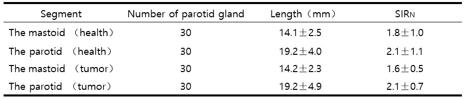
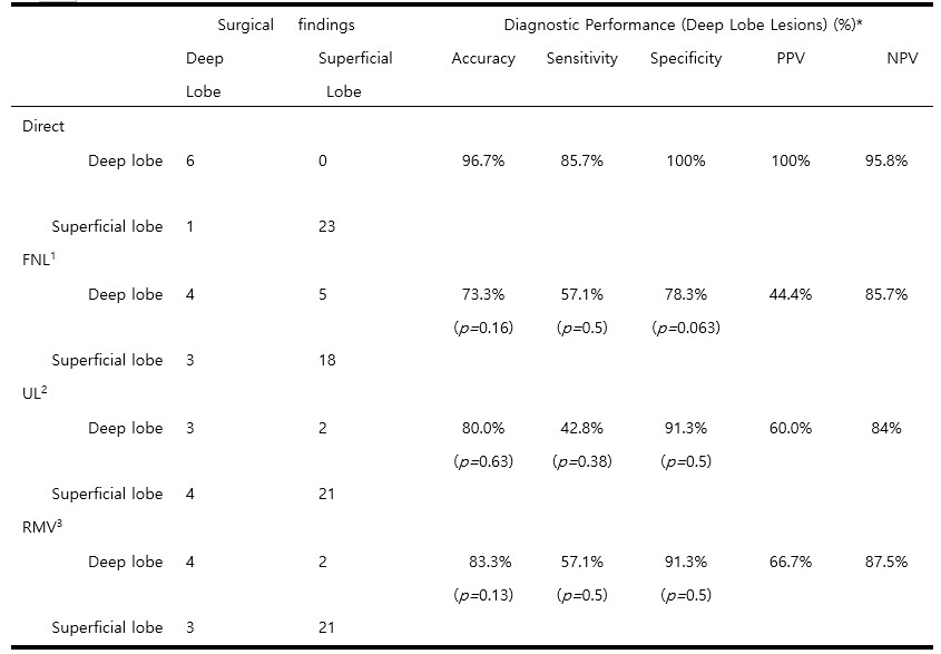
Table 3. Localization of parotid lesions with imaging and surgical findings.
PPV indicates positive predictive value; NPV, negative predictive value.
1FNL:the lateral surface of the posterior belly of the digastric muscle to the lateral surface of the ascending ramus of the mandible.
2UL:the most dorsal point observed on the ipsilateral half of a vertebra to the most dorsal point of the retromandibular vein (RMV).
3RMV: the retromandibular vein.
*Statistical analyses (P values) were performed comparing the direct method with each indirect method.