4533
Accelerated brachial Plexus MRI using deep learning constrained compressed SENSE reconstruction1Radiology, West China Hospital Of Sichuan University, Chengdu, China, 2Clinical Science, Philips Healthcare, Chengdu, China, 3Application, Philips Healthcare, Chongqing, China
Synopsis
This study presents an accelerated brachial Plexus MR imaging technique using deep learning constrained compressed SENSE reconstruction (CS-AI-SENSE). The results showed that CS-AI-SENSE could enable comparable diagnostic image quality to the conventional SENSE-accelerated 3D T2-TSE approaches while significantly reduced the data acquisition time, which has potential to enhance the workflow of brachial Plexus MR examinations in clinics.
Introduction
Currently, three-dimensional T2-weighted Turbo Spin Echo (3D T2-TSE) MRI is a non-invasive modality commonly used in clinical practice to evaluate the brachial plexus nerve. However, this technique is limited to long scan time due to the required high resolution, which is susceptible to motion artifacts and can cause a great interference to the physician's observation of the nerve during diagnosis in patients1. Compressed-SENSE (CS-SENSE), using signal processing optimization algorithms to obtain MRI images in the presence of K-space undersampling, has ben proposed to reduce MRI examination time and applied in various aspects2-3. Nevertheless, excessive shortening of acquisition time using high acceleration factors could also inevitably lead to image quality degradation due to insufficient noise removal , and hence how to further reduce the scan time while maintain diagnostic image quality is still a great challenge. Recently, a novel technique, integrating artificial intelligence (AI) into compressed SENSE reconstruction (CS-AI-SENSE), has been proposed to further accelerate MR scans and has shown excellent performance in highly undersampled knee imaging4. It is hypothesized that CS-AI-SENSE could significantly reduce the scan time of brachial plexus MRI while maintain diagnostic image quality. This study aims to achieve rapid brachial plexus MRI using CS-AI-SENSE, and compare its image quality with that using CS-SENSE and conventional SENSE-accelerated 3D T2-TSE approaches.Materials and Methods
This study was approved by the institutional ethics committee approved. Five patients (mean age 50.4, age-range 31-74 years, 3 men and 2 women) were included and underwent brachial plexus nerve MR examination on a 3T MRI scanner (Ingenia Elition, Philips Healthcare) using a 32-channel body matrix coil. The scan parameters of conventional 3D T2-TSE were as follows: FOV = 300 × 419 × 60mm3, slices = 68, voxel size = 1.2 × 1.35 × 1.8mm3, TR/TE = 2200/170 ms, NSA = 2. The imaging parameters of CS-SENSE and CS-AI-SENSE were nearly consistent to conventional 3D T2-TSE, but with an CS acceleration factor=8. In CS-AI approach, the CS reconstruction chain is merely replaced by a convolution neural network (CNN) reconstruction. Compared to conventional method, the acquisition time of CS & CS-AI decreased (2:34 vs. 4:46 min).The image quality was rated by two radiologists with more than 3 years of neuroimaging experience using a 4-point scale (4 indicates that the brachial plexus nerve and its major branches are complete, continuous, and clear with no venous interference. 3 indicates that the brachial plexus nerve and its branches are mostly continuous, with minor venous interference, which does not affect the evaluation. 2 indicates that most of the brachial plexus nerve and its branches are shown significant moderate venous interference. 1 score indicates that most brachial plexus structures are not shown with severe venous interference). The quantitative contrast-to-noise ratio (CNR) and signal-to-noise ratio (SNR) between the nerve and the surrounding tissue were calculated for all subjects and compared among SENSE, CS-SENSE and CS-AI-SENSE. The ANOVA was used to compare the subjective image quality scores and objective image quality scores, and the difference was statistically significant with a P value less than 0.05.
Results and Discussions
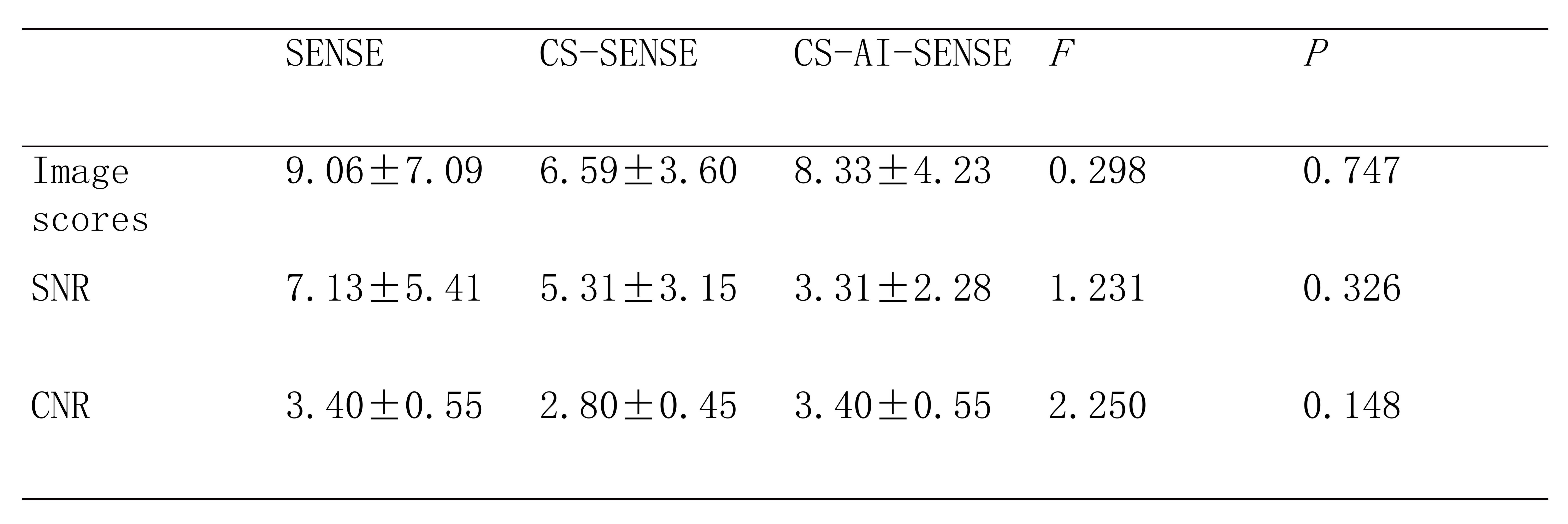
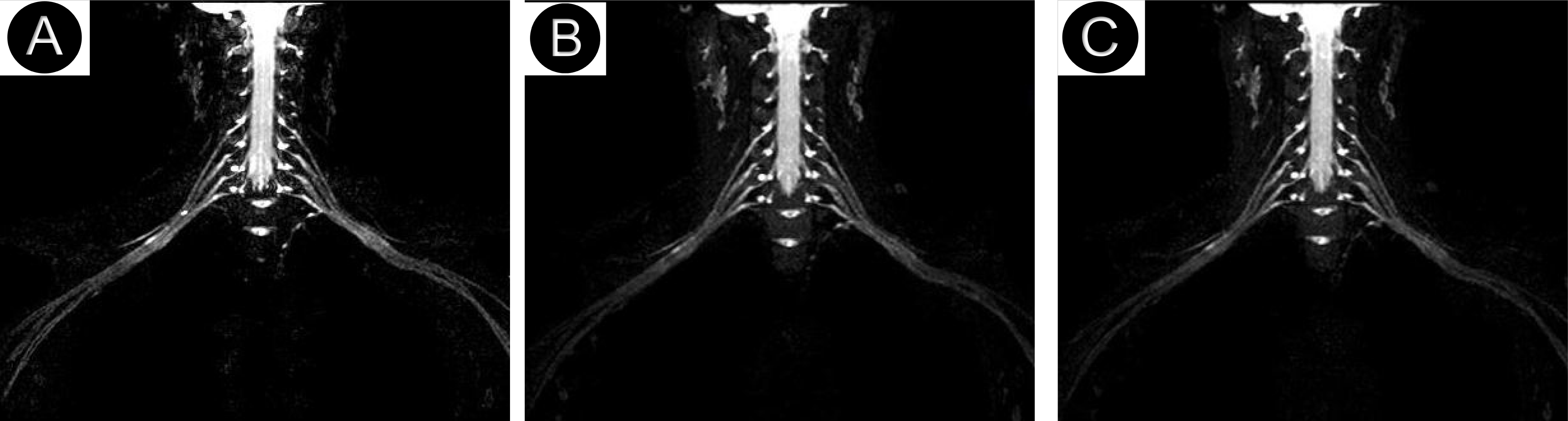
The image scores, SNRs and CNRs were shown in Table 1. The CS-AI-SENSE shows higher image quality to CS-SENSE (3.40±0.55 vs. 2.80±0.45) and comparable image quality to conventional SENSE with nearly half reduced san time (3.40±0.55 vs. 3.40±0.55), and the differences were not statistically significant (p = 0.148). Compared to CS-SENSE, CS-AI-SENSE has significantly higher SNR (8.33±4.23 vs 6.59±3.60, p = 0.747), and slightly CNR ( 3.31±2.28 vs. 5.31±3.15, p = 0.326). Pairwise comparisons between the three methods were not statistically significant, maybe in part due to current less subjects included in this study.The brachial plexus example acquired on a patient was shown in Figure 2, which indicated AI-CS-SENSE can enable comparable image quality with conventional SENSE accelerated method while significantly shortening the scan time.Conclusion
This study qualitatively and quantitatively compares the brachial plexus image quality among CS-AI-SENSE,CS-SENSE and conventional SENSE-accelerated approaches. The results showed that CS-AI-SENSE could enable comparable diagnostic image quality to the conventional method while significantly reduced the data acquisition time, which could avoid the effects of motion artifacts and reduce patient discomfort during the examination. A large clinical population is underway to validate the diagnostic value of this technique.Acknowledgements
No acknowledgement found.References
1. Chhabra A, Thawait GK, Soldatos T, et al. High-Resolution 3T MR Neurography of the Brachial Plexus and Its Branches, with Emphasis on 3D Imaging. AJNR Am J Neuroradiol. 2013;34(3):486-97.
2. Cho SJ, Choi YJ, Chung SR, et al. High-resolution MRI using compressed sensing-sensitivity encoding (CS-SENSE) for patients with suspected neurovascular compression syndrome: comparison with the conventional SENSE parallel acquisition technique. Clin Radiol. 2019;74(10):817.e9-817.e14.
3. Chen Z, Sun B, Xue Y, et al. Comparing compressed sensing breath-hold 3D MR cholangiopancreatography with two parallel imaging MRCP strategies in main pancreatic duct and common bile duct. Eur J Radiol. 2021;142:109833.
4. Knoll F, Murrell T, Sriram A, et al. Advancing machine learning for MR image reconstruction with an open competition: Overview of the 2019 fastMRI challenge. Magn Reson Med. 2020;84(6):3054-3070.
Figures