4530
Feasibility of inhomogeneous magnetization transfer for the peripheral nerve of the brachial plexus1Department of Radiology, Chiba university hospital, Chiba, Japan, 2Diagnostic Radiology and Radiation Oncology, Graduate School of Medicine, Chiba University, Chiba, Japan, 3Philips Japan, Tokyo, Japan, 4Philips Canada, Mississauga, ON, Canada
Synopsis
Magnetization transfer (MT) has been conventionally used for the determination of myelin. However, the problem with the MT is that it reflects bound water that is not related to myelin. Inhomogeneous Magnetization Transfer (ihMT) can act specifically on myelin, it has been reported to be a useful method for quantifying myelin in brain and spinal cord regions. In this study, we compared conventional MT and ihMT for the brachial plexus, and ihMT might be able to reflect more accurate information about myelin than MT.
Introduction
Magnetization transfer (MT) is a method that utilizes the interaction between bound water and water.1,2 From the two images (one with an MT pulse and one without it), the ratio of bound water to the sum of bound and free water (MTR) can be defined. In the central nervous system, bound water is abundant in myelin and nerve cell membranes, and therefore, myelin can be determined indirectly by calculating MTR. The MT method has been conventionally used for the determination of myelin but the MT has the problem that it reflects bound water that is not related to myelin. The development of myelin-specific MR imaging techniques remains an active and highly focused area of research in which novel approaches are being sought. One promising example is the recently described inhomogeneous magnetization transfer (ihMT) technique. 3 It can be envisioned as a method to image, in vivo, the dipolar order underlying broad tissue macromolecular lines, thereby providing higher specificity for myelinated structures. Although ihMT has been reported to be useful in the brain and spinal cord regions, the feasibility for peripheral nerves has not been studied.4,5,6,7 In this study, we compared conventional MT and ihMT for the brachial plexus and investigated the feasibility of ihMT in the peripheral nerve.Methods
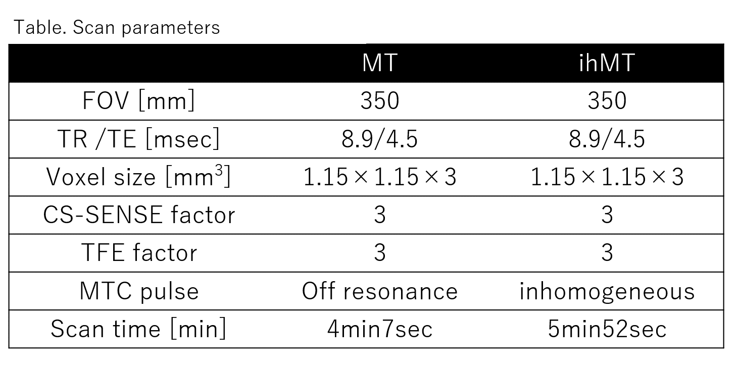
Nine volunteers with electrophysiological studies demonstrating the absence of peripheral neuropathy were examined on 3T MR unit (Ingenia 3T, Philips Healthcare). Scan parameters were shown in Figure 1. MTRs of MT and ihMT were calculated for brachial plexus. MTRs of MT were calculated as follows :MTR =100×( MT[OFF] - MT[ON] ) / MT[OFF] .
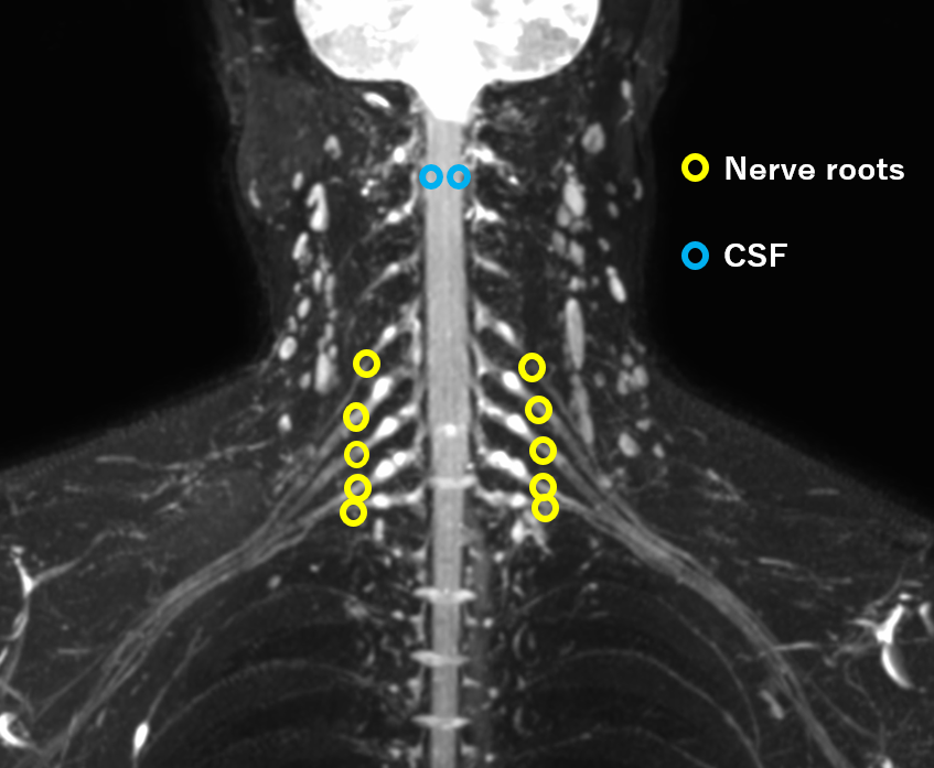
MTRs of ihMT were calculated by turning on and off the ihMT pulse as in MT. The measurement points were the nerve roots of the five nerves that make up the brachial plexus and MTRs of the cerebrospinal fluid was also measured to evaluate free water (Figure 2).The evaluation method was as follows.
1) The MTRs of MT and ihMT were compared.
2) Correlation between MTRs in MT and ihMT was evaluated.
3) The coefficients of variation (CV) of MT and ihMT were calculated and their reproducibility was evaluated.
Results
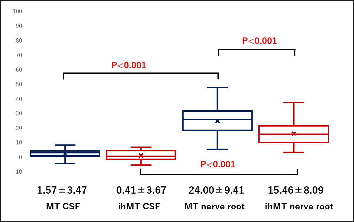
1) MTR-ihMT were lower than in MTR-MT (P<0.001). Cerebrospinal fluid was unchanged in both groups (Figure 3).2) There were no correlation between MTR-MT and MTR-ihMT (r2=0.215)(Figure 4).
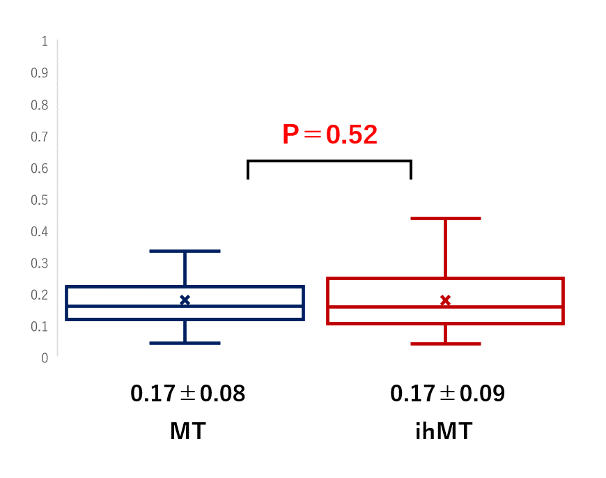
3) There were no significant difference in CV between MTR-MT and MTR-ihMT (P=0.52)(Figure 5).
Discussion
Myelin in peripheral nerves is affected in various diseases, and its damage is recognized as demyelinating disorders. Since MTR by MT does not accurately reflect the amount of myelin, a sequence that more accurately reflects the pathological state of the peripheral nerve has been desired. ihMT is a sequence with such potential. The MTRs of cerebrospinal fluid were unchanged in both groups. MTR-MT and MTR-ihMT might act only on bound water and not on free water. The MTRs were lower in ihMT than in MT. MT acts on myelin and other bound waters, whereas ihMT acts specifically on myelin. Therefore, ihMT might be able to reflect more accurate information about myelin. No correlation was found between MTR-MT and MTR-ihMT. The values of MTR-MT and MTR-ihMT were considered to reflect different information. There was no significant difference in CV between MT and ihMT. This suggests that the reproducibility of ihMT was comparable to that of the conventional MT method. This result might indicate the possibility of quantitative evaluation of myelin in the peripheral nerve.Conclusion
In the brachial plexus, ihMT was feasible as a quantitative assessment of myelin in the peripheral nerve.Acknowledgements
No acknowledgment was found.References
1. Wolff, S et al, Magnetization transfer contrast (MTC) and tissue water proton relaxation in vivo , Magn Reson Med. 1989 Apr;10(1):135-44. doi: 10.1002/mrm.1910100113. PMID: 2547135
2. E. Alonso-Ortiz et al, MRI-based myelin water imaging: A technical review, Magn Reson Med. 2015 Jan;73(1):70-81. doi: 10.1002/mrm.25198. Epub 2014 Mar 6. PMID: 24604728
3. Varma, G. et al, Magnetization transfer from inhomogeneously broadened lines: a potential marker for myelin , Magn Reson Med. 2015 Feb;73(2):614-22. doi: 10.1002/mrm.25174. Epub 2014 Mar 6.
4. Zhang L et al. A comparison study of inhomogeneous magnetization transfer (ihMT) and magnetization transfer (MT) in multiple sclerosis based on whole-brain acquisition at 3.0 T , Magn Reson Imaging. 2020 Jul;70:43-49. doi: 10.1016/j.mri.2020.03.010. Epub 2020 Mar 26.
5. Mancini et al, An interactive meta-analysis of MRI biomarkers of myelin, Elife. 2020 Oct 21;9:e61523. doi: 10.7554/eLife.61523.
6.Van Obberghen E, et al, Evaluation of the sensitivity of inhomogeneous magnetization transfer (ihMT) MRI for multiple sclerosis. J Magn Reson . 2018 Nov;296:60-71.AJNR Am J Neuroradiol. 2018 Apr;39(4):634-641. doi: 10.3174/ajnr.A5563. Epub 2018 Feb 22.
7. Varma, G. et al, In vivo measurement of a new source of contrast, the dipolar relaxation time, T1D, using a modified inhomogeneous magnetization transfer (ihMT) sequence, Magn Reson Med. 2017 Oct;78(4):1362-1372. doi: 10.1002/mrm.26523. Epub 2016 Nov 17.
Figures