4528
Improved visualization of MR Neurography in the lumbosacral and sciatic nerves using double-diffusion sensitized driven-equilibrium (D2SDE)1Department of Radiology, Eastern Chiba Medical Center, Chiba, Japan, 2Philips Japan, Tokyo, Japan, 3Diagnostic Radiology and Radiation Oncology, Graduate School of Medicine, Chiba University, Chiba, Japan, 4Orthopaedic Surgery, Eastern Chiba Medical Center, Chiba, Japan, 5General Medical Services, Graduate School of Medicine, Chiba University, Chiba, Japan
Synopsis
We developed a new prepulse scheme, D2SDE, inspired by filter-probe double diffusion scheme for improved visualization of MR Neurography in the lumbosacral and sciatic nerves. This technique may be useful for arriving at an accurate diagnosis of lumbosacral plexus pathology.
Purpose
MR neurography plays a major role in the diagnostic work-up of peripheral nerve pathologies, substantially improving early diagnosis and patient management1. SHINKEI, which consists of a fat suppression pre-pulse and a motion-sensitized driven-equilibrium (MSDE) blood suppression pre-pulse, followed by a 3D T2W TSE sequence, is one of the commonly used MR neurography sequences2. Although high-quality MR neurography of the brachial plexus and the lumbosacral plexus could be obtained using SHINKEI, it is still challenging to completely suppress signals from the cerebrospinal fluid (CSF), venous blood, and synovial fluid, which presented difficulties in distinguishing nerves from surrounding tissue signals in the lumbosacral plexus3,4. Recently, HIRE-SHINKEI4 (SHINKEI with high-intensity reduction) has been proposed, which improves visualization of the detailed anatomical structure of lumbosacral plexus nerves due to efficient background signal suppression using dual-echo TSE subtraction. On the other hand, called DSDE (diffusion-sensitized driven-equilibrium) prepared 3D gradient echo (TFE) is also used for peripheral nerve visualization5-8 thanks to its stronger motion (and diffusion) sensitized gradients and it is promising to enhance the nerve signal while suppressing fluid and venous signals sufficiently. However, these techniques may potentially suffer from unwanted signal loss of the nerves due to subtraction or applying strong diffusion gradients.Recently, double diffusion encoding (DDE) scheme has been introduced and it provides novel contrast based on microscopic diffusion anisotropy9. Filter-probe diffusion imaging, which is one of the unique DDE clinical applications, which provides diffusion maps of axial diffusivity devoid of CSF partial volume effects10-13. We hypothesized that such a filter-probe double diffusion scheme may provide improved nerve visualization while suppressing venous and fluid signals effectively without penalty for signal loss. In this study we developed a new prepulse scheme, called double-diffusion-sensitized driven equilibrium (D2SDE) for improved visualization of MR Neurography in the lumbosacral and sciatic nerves.
Methods
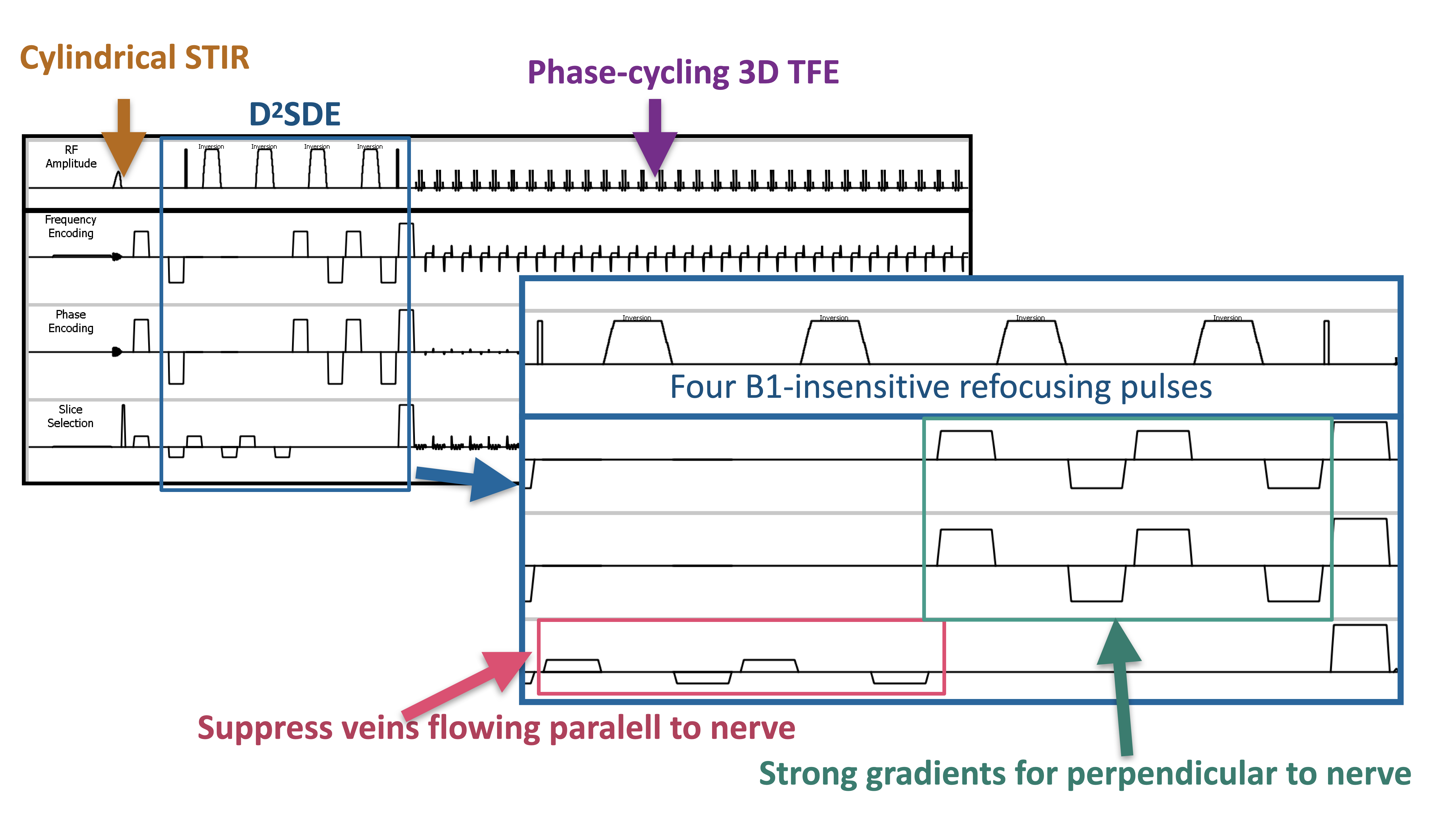
For visualizing peripheral nerves while suppressing vessel and fluid signals sufficiently, there are some unique MPG combination schemes has been proposed5-7. Because of the diffusion anisotropy of the human nervous system, peripheral nerves are best visualized when applying motion-sensitized gradients (MPGs) perpendicular to the course of the nerves, while nerves are not well visualized when applying MPGs parallel to the course of the nerves.In this study, we developed the D2DSE prepulse, which has four refocusing pulses and two MPG blocks with different timings. First, moderate bipolar MPGs were applied for the direction parallel to the nerve (slice direction) to reduce vessel signal while simultaneously having a small b-value contribution and thus negligible diffusion attenuation to the nerve signal. Subsequently, two directions of strong bipolar MPGs were placed in both the anterior-posterior and right-left directions, which are most perpendicular to the trajectories of the nerves in the lumbosacral nerves. Hence, the D2SDE pre-prepulse, in separate working regimes of gradient strength and with gradient-axis selection accommodating nerve orientation, would provide improved vessel reduction while preserving nerve signals. In addition, to eliminate the T1-effects, which are given by readout 3D spoiled gradient echo sequence (T1-turbo field echo: T1TFE) and deteriorate diffusion-weighted contrast, RF phase-cycling scheme is applied. Furthermore, bowel signals on the DWI sometimes obscure the nerve visualization. STIR may be useful in suppressing bowel signal14, which may have a short T1 value, similar to that of fat. In this study we applied a cylindrical inversion pulse15 (known as Time-Slip) as a spatial selective inversion pulse. Scheme of D2SDE pre-pulse is shown in Figure 1.
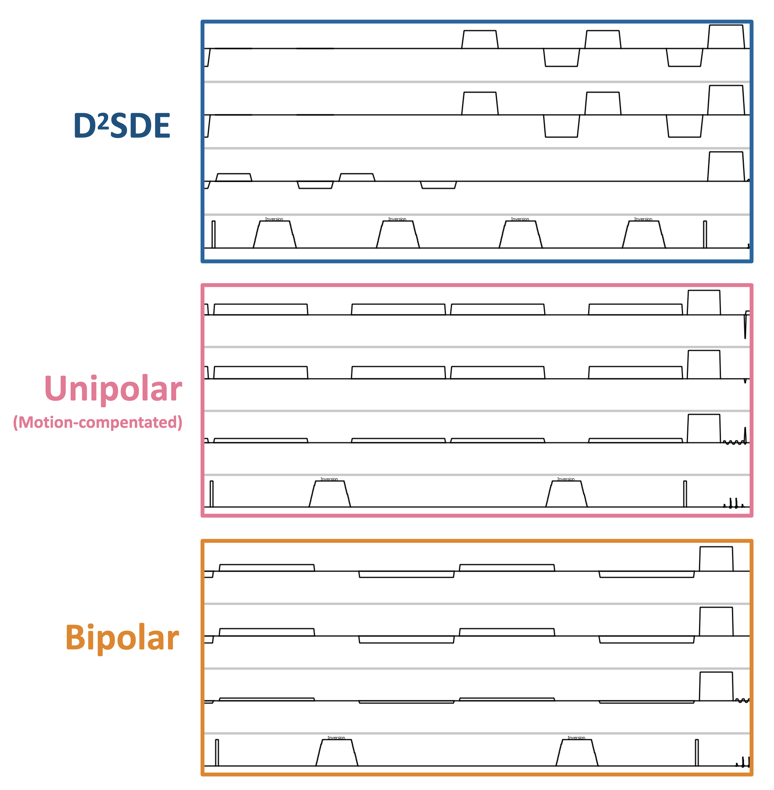
We compared the image quality of D2SDE prepared MR neurography with other types of MPG schemes (Fig.2) and SHINKEI.
A total of five volunteers were examined on a 3.0T system (Ingenia CX, Philips Healthcare). The study was approved by the local IRB, and written informed consent was obtained from all subjects.
Imaging parameters were; Axial, voxel size=2x2x3mm3, effective b-value=620s/mm2, D2SDE preparation time (prep-time)=90ms, gradient strength phase/freq/slice=30/30/10mT/m, shot interval=2800ms, flip angle=8°, turbo factor=30, ProSet 1331, C-SENSE=3.4, and total acquisition time=5m50s.
Results and Discussion
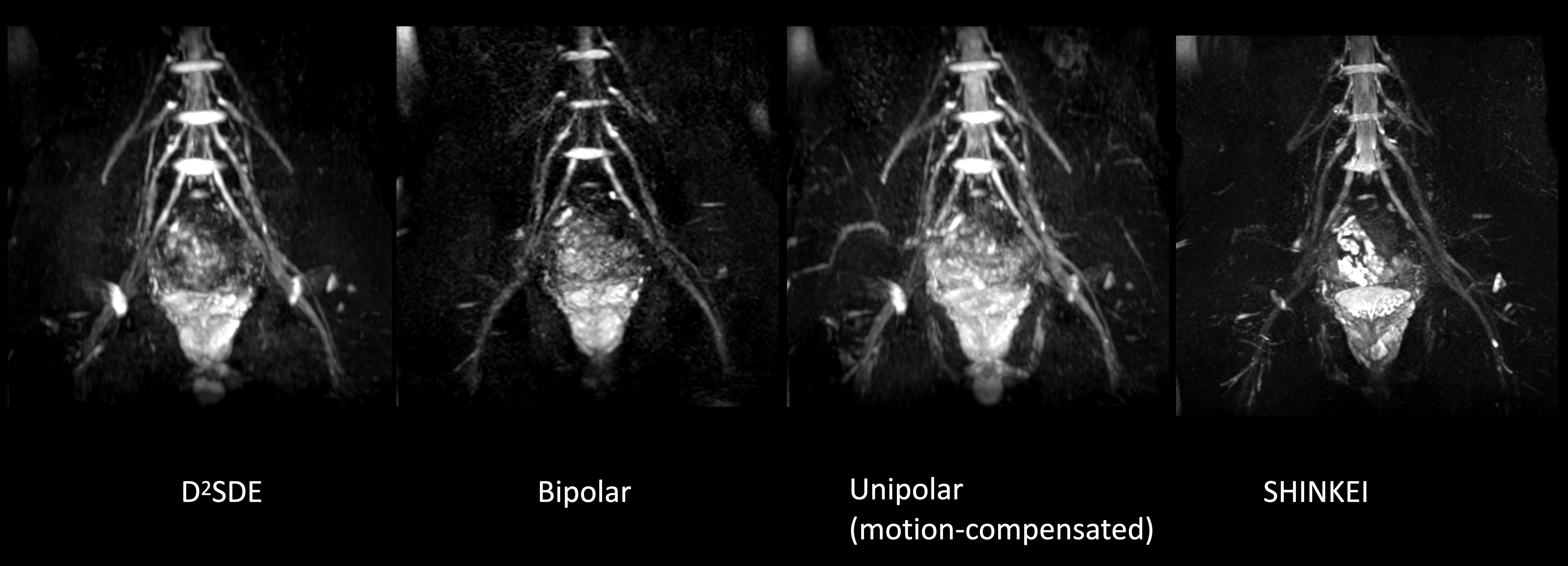
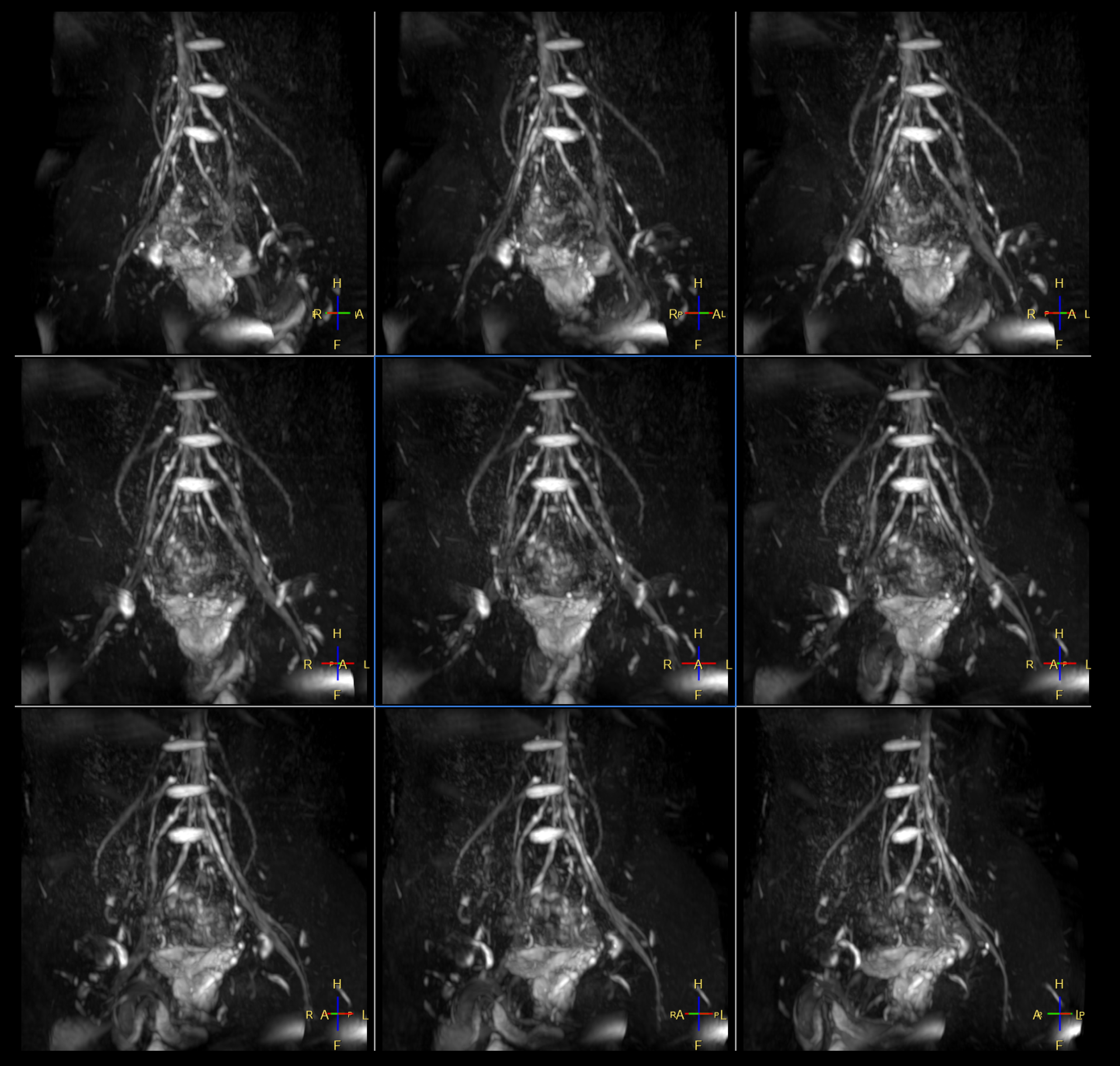
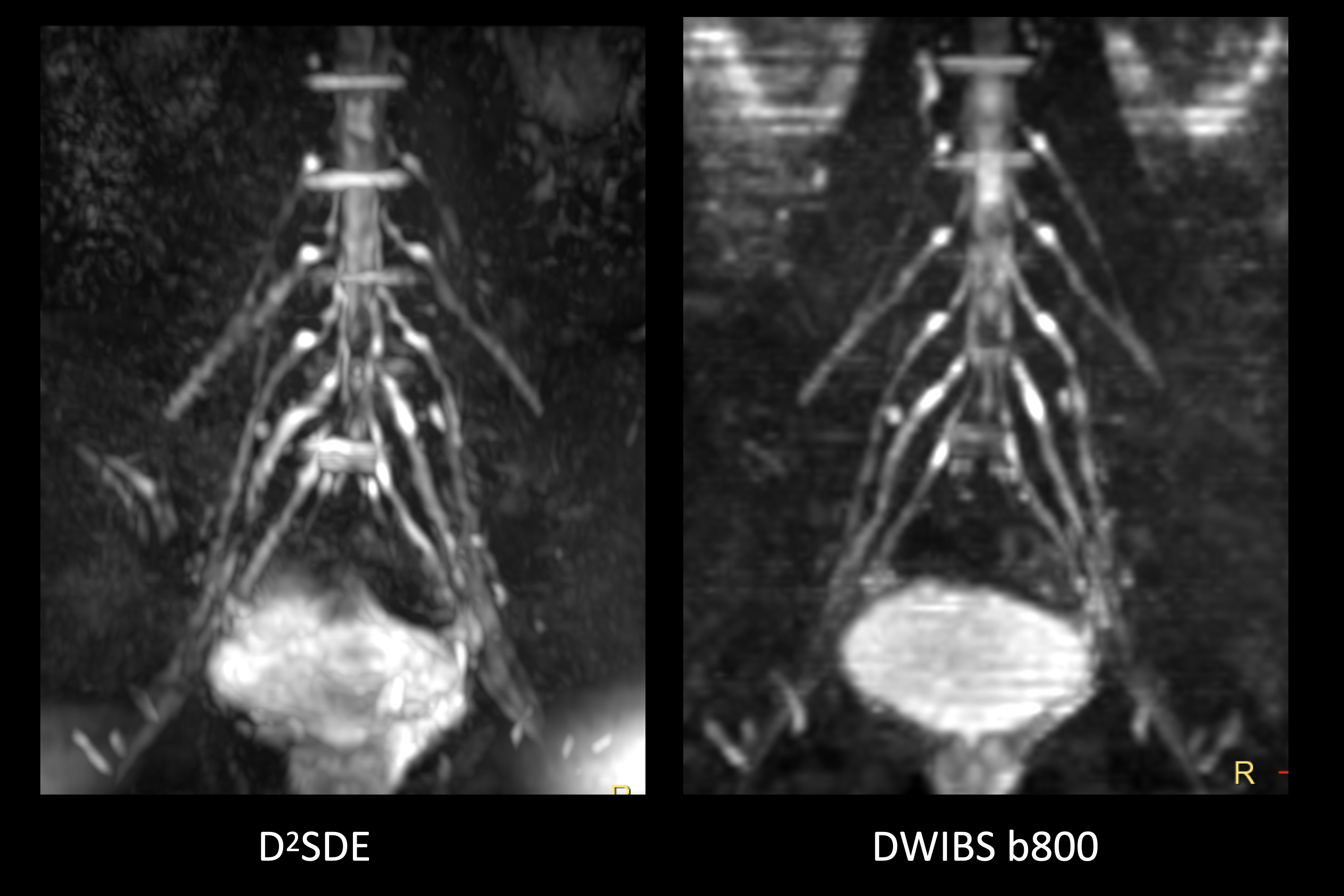
Figure 3 shows the representative MR neurography images obtained by the 3D phase-cycling TFE with D2SDE, conventional bipolar gradients, motion-compensated unipolar gradients and SHINKEI (MSDE-prepared 3D TSE) are shown. D2SDE provided best image quality, whereas bipolar gradients decreased the overall SNR, unipolar gradients maintained good SNR, but veins were not suppressed well, and SHINKEI did not depict peripheral nerves well. D2SDE has a good balance between SNR and venous suppression. Representative MIP images obtained by D2SDE are shown in Figure 4. Figure 5 shows the comparison of D2SDE and DWIBS using uni-directional MPG with b=800 s/mm2. Since D2SDE is a pure 3D acquisition, nerve conspicuity and continuity are better than DWIBS. Although further clinical evaluation is needed, D2SDE may be promising for improved visualization of the lumbosacral nerves.Conclusion
We developed a new prepulse scheme, D2SDE, inspired by filter-probe double diffusion scheme for improved visualization of MR Neurography in the lumbosacral and sciatic nerves. This technique may be useful for arriving at an accurate diagnosis of lumbosacral plexus pathology.Acknowledgements
No acknowledgement found.References
1. Chhabra A, et al. MR neurography: past, present, and future. AJR Am J Roentgenol. 2011 Sep;197(3):583-91. doi: 10.2214/AJR.10.6012.
2. Yoneyama M, et al. Rapid high resolution MR neurography with a diffusion-weighted pre-pulse. Magn Reson Med Sci. 2013;12:111–119.
3. Sollmann N, et al. Magnetic resonance neurography of the lumbosacral plexus at 3 Tesla - CSF-suppressed imaging with submillimeter resolution by a three-dimensional turbo spin echo sequence. Magn Reson Imaging. 2020 Sep;71:132-139. doi: 10.1016/j.mri.2020.06.009.
4. Tadenuma H, et al. Improved visualization of diffusion-prepared MR neurography (SHINKEI) in the lumbosacral plexus combining high-intensity reduction (HIRE) technique. Magn Reson Imaging. 2020 Jun;69:22-27. doi: 10.1016/j.mri.2020.02.006.
5. Yoneyama M, et al. High-resolution 3D Mr Neurography of the Wrist using Phase-Cycling Diffusion-Sensitized Driven-Equilibrium (pcDSDE). Proc. ISMRM 2015:0313.
6. Sakai T et al. Improvement of 3D diffusion-prepared MR neurography in the extremities using improved diffusion-sensitized driven-equilibrium (iDSDE) with phase-cycling turbo field echo sequence. Proc. ISMRM 2017:5027.
7. Cervantes B, et al. Orthogonally combined motion- and diffusion-sensitized driven equilibrium (OC-MDSDE) preparation for vessel signal suppression in 3D turbo spin echo imaging of peripheral nerves in the extremities. Magn Reson Med. 2018 Jan;79(1):407-415 doi: 10.1002/mrm.26660.
8. Yokota H, et al. Phase-cycling diffusion-sensitized driven-equilibrium (pcDSDE) for MR neurography of the crus. Proc. ISMRM 2020:2834.9. Yang G, et al. Double diffusion encoding MRI for the clinic. Magn Reson Med. 2018 Aug;80(2):507-520. doi: 10.1002/mrm.27043.
10. Budde MD, et al. Optimizing Filter-Probe Diffusion Weighting in the Rat Spinal Cord for Human Translation. Front Neurosci. 2017 Dec 19;11:706. doi: 10.3389/fnins.2017.00706.
11. Skinner NP, et al. Rapid in vivo detection of rat spinal cord injury with double-diffusion-encoded magnetic resonance spectroscopy. Magn Reson Med. 2017 Apr;77(4):1639-1649. doi: 10.1002/mrm.26243.
12. Skinner NP, et al. Filter-probe diffusion imaging improves spinal cord injury outcome prediction. Ann Neurol. 2018 Jul;84(1):37-50. doi: 10.1002/ana.25260.
13. Lee SY, et al. Diffusion-prepared fast spin echo for artifact-free spinal cord imaging. Magn Reson Med. 2021 Aug;86(2):984-994. doi: 10.1002/mrm.28751.
14. Kwee TC, et al. Diffusion-weighted whole-body imaging with background body signal suppression (DWIBS): features and potential applications in oncology. Eur Radiol. 2008; 18(9): 1937–1952. doi: 10.1007/s00330-008-0968-z.
15. Yoneyama M, et al. Cylindrical Inversion Pulse for the Reduction of Cardiac Motion Artifacts in Contrast-enhanced Breast MRI. Magn Reson Med Sci. 2018 Jan 10;17(1):80-85. doi: 10.2463/mrms.tn.2015-0150.
Figures