4519
Instability and hyper-connected pattern of dynamic functional network in MCI converters1Fudan University, Shanghai, China, 2Huashan Hospital, Shanghai, China
Synopsis
In this study, based on rs-fMRI, we analyzed the dynamic characteristics of 3 groups: MCI convert to AD within one-year follow-up (1Y_AD), stable in MCI within one-year follow-up (1Y_MCI) and revision from MCI within one-year follow-up (1Y_HC), including nodal entropy and measurements of dynamic brain functional network. We found that with the disease progression, the brain functional network was reconfigured into a highly-integrated/segregated pattern, and dwell time of highly-integrated pattern is associated with executive function in 1Y_AD. These findings provide the evidence for developing dynamic measurements as potential biomarkers for tracking mechanism of AD.
Introduction
Alzheimer’s disease (AD) , a common neurodegeneration disease, which is expected to affect 1 in 85 people worldwide by 2050 1, imposes heavy burden on society and patients’ families. Mild cognitive impairment (MCI) refers to the transition stage between normal aging and AD. It has been reported that more than half of MCI patients will progress to dementia within 5 years 2. Uncovering the pathophysiological mechanism of AD progress and developing effective biomarkers for identifying MCI converter is of important clinical significance. Resting-state functional magnetic resonance imaging (rs-fMRI), a non-invasive imaging technique, has been widely applied in AD-related pathological mechanism researches 3, 4. Recently, brain functional dynamics has attracted increasing interest in the fields of AD-related disease 5, 6 and found that both static and dynamic brain functional characteristics were affected in AD. However, there is limited studies focused on alterations of brain functional dynamics in MCI progression.Thus, in this study, we applied dynamic functional connectivity to explore the alteration of dynamical functional brain network in MCI progression, and evaluated the correlation between dynamic characteristics and neuropsychological scores.Methods
In this study, we enrolled 148 MCI patients, then divided them into 3 groups: 17 1Y_AD (MCI convert to AD within one-year follow-up), 106 1Y_MCI (stable in MCI within one-year follow-up) and 25 1Y_HC (revision from MCI within one-year follow-up). The rs-fMRI images were obtained using a single-shot gradient-recalled echo planar imaging (EPI) sequence with the following parameters: repetition time (TR) = 2000 ms, echo time (TE) = 35 ms, slice thickness = 4 mm, flip angle = 90°, slices = 33, field of view (FOV)= 256×256, matrix size = 64×64, number of volumes = 200. Shannon entropy was used to quantity functional flexibility, which characterizes inter-nodal heterogeneous connectivity over time. We calculated and compared the Shannon entropy of inter-nodal connectivity and the measurements of dynamical functional network among three groups and their correlation with the cognitive score were evaluated.Results
1Y_AD presented a significant increase in nodal entropy of left post cingulate compared with 1Y_MCI and 1Y_HC showed a significant increase in nodal entropy of right occipital and left cerebellum in comparison with 1Y_MCI. Group comparison of measurements of dynamic function network revealed that 1Y_AD showed significantly increased mean dwell time in state 4 (the brain states with strongly inter- and intra- network connectivity). Moreover, 1Y_AD presented the least and 1Y_HC presented the most number of transition among 3 groups. The average mean dwell time in state 4 was negatively correlated with the executive functional scores of 1Y_AD.Discussion
In the MCI progression, the configuration of different brain functional network state was disrupted, and the whole brain activity attends to the abnormal highly-integrated/segregated state. Task-fMRI studies have found that the motor execution depends on the intra-network integration and cognitive function depends on the inter-network connectome, which suggested that the effective functional brain network could keep the balance between integrated and segregated state. And the abnormal state configuration in MCI converters reveals a hallmark sign of MCI progression (increased dwell time in highly-integrated functional state). The reduction in transitioning between brain states could be interpreted as decreased functional dynamics and cognitive flexibility. With the disease deterioration, the lower rate of transitions may possibly be a measurement of reduced cognitive flexibility. In addition, the precuneus, the key region of default mode network, is associated with visual memory and processing. The increased functional flexibility of precuneus during conversion period may be related with the higher order function deficits in early stage of AD. We suggested that the MCI converters presented an increased instability and hyper-connected pattern in dynamic functional brain network, which would be the novel biomarkers for MCI conversion prediction.Conclusion
In this study, we analyzed the functional dynamics of MCI converters/non-converters and found that with MCI progression, the brain functional network was reconfigured into a highly integrated/segregated pattern and presented increased instability, which provides evidence for understanding of the pathophysiology and developing of novel biomarkers for MCI conversion.Acknowledgements
This work was supported by the National Natural Science Foundation of China (82102132, 82071200, 82001139), the Science and Technology Commission of Shanghai Municipality (20S31904300), the National Project of Chronic Disease of China (2016YFC1306402), the Clinical Research Plan of Shanghai Hospital Development Center (SHDC2020CR4007).References
1. Brookmeyer R, Johnson E, Ziegler-Graham K, et al. Forecasting the global burden of Alzheimer’s disease. Alzheimer's & Dementia. 2007;3(3): 186-191.
2. Busse A, Hensel A, Gühne U, et al. Mild cognitive impairment long-term course of four clinical subtypes. Neurology. 2006; 67(12): 2176-2185.
3. Biswal B, Yetkin F Z, Haughton V M, et al. Functional connectivity in the motor cortex of resting human brain using echo-planar MRI. MagnResonMed. 1995; 34(4): 537-541.
4.Khan W, Amad A, Giampietro V, et al. The heterogeneous functional architecture of the posteromedial cortex is associated with selective functional connectivity differences in Alzheimer's disease. Hum Brain Mapp. 2020; 41(6): 1557-1572.
5. Filippi M, Spinelli E G, Cividini C, et al. Resting State Dynamic Functional Connectivity in Neurodegenerative Conditions: A Review of Magnetic Resonance Imaging Findings. Front Neurosci. 2019; 13:657.
6. Hutchison R M, Womelsdorf T, Allen E A, et al. Dynamic functional connectivity: promise, issues, and interpretations. Neuroimage. 2013; 80: 360-378.
Figures
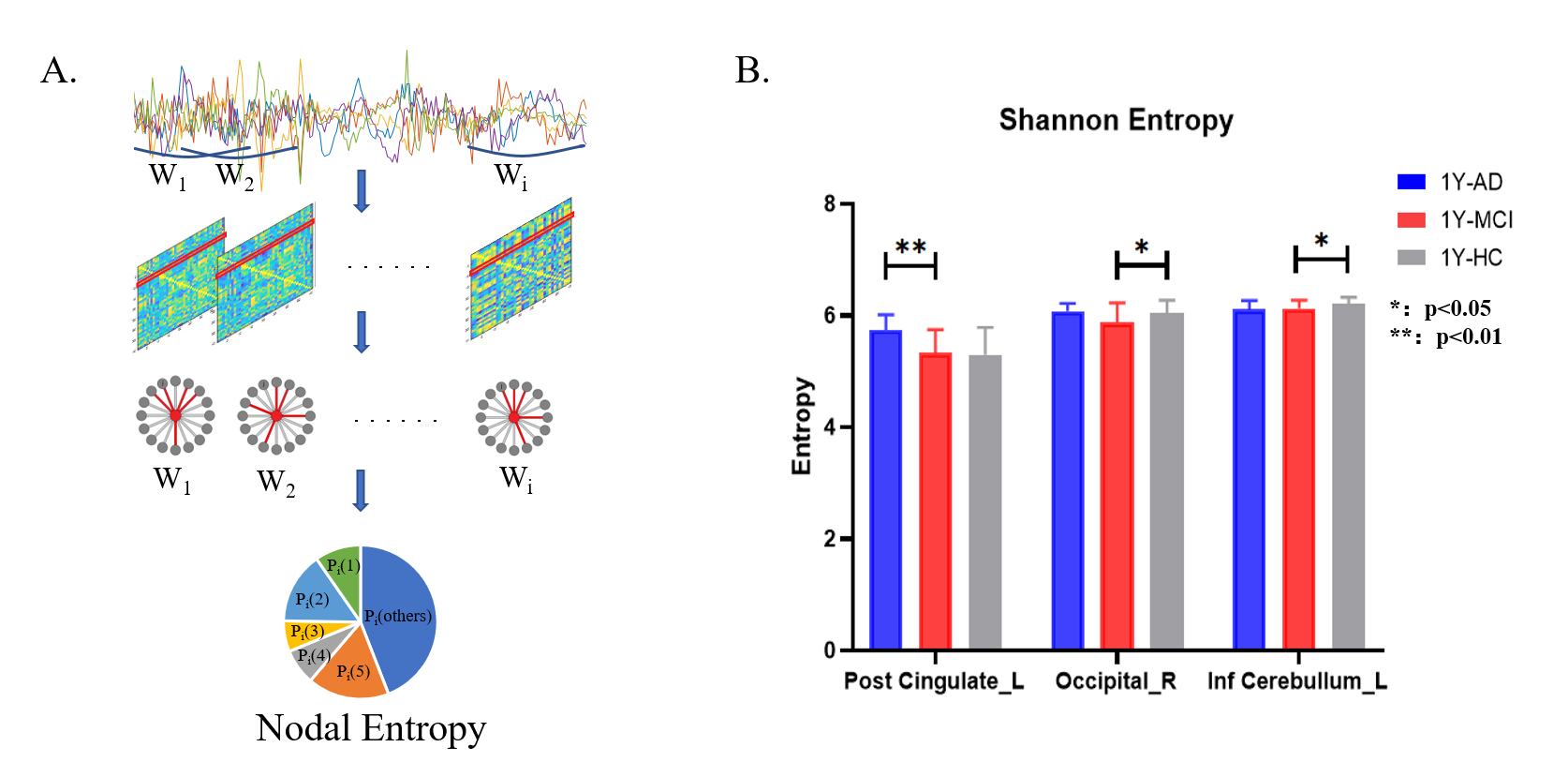
Fig 1 (A) the calculation diagram of nodal entropy (B) The significance difference of nodal entropy among 1Y-AD,1Y-MCI and 1Y-HC (FDR-corrected with post hoc test).
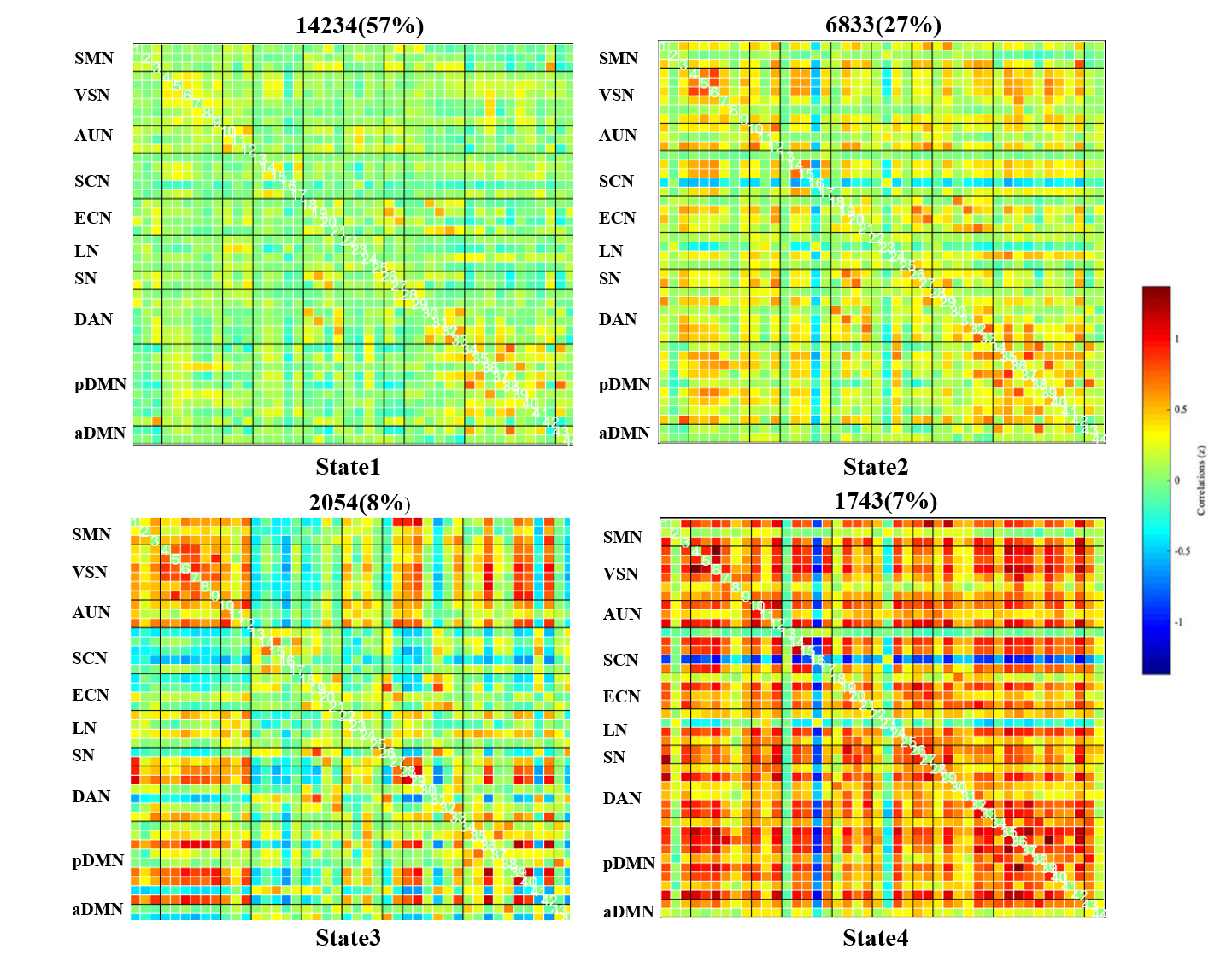
Fig 2 Functional network connectivity matrices for four states and their proportions across all sliding windows. The red color represents positive correlations, whereas the blue color indicates negative correlations.
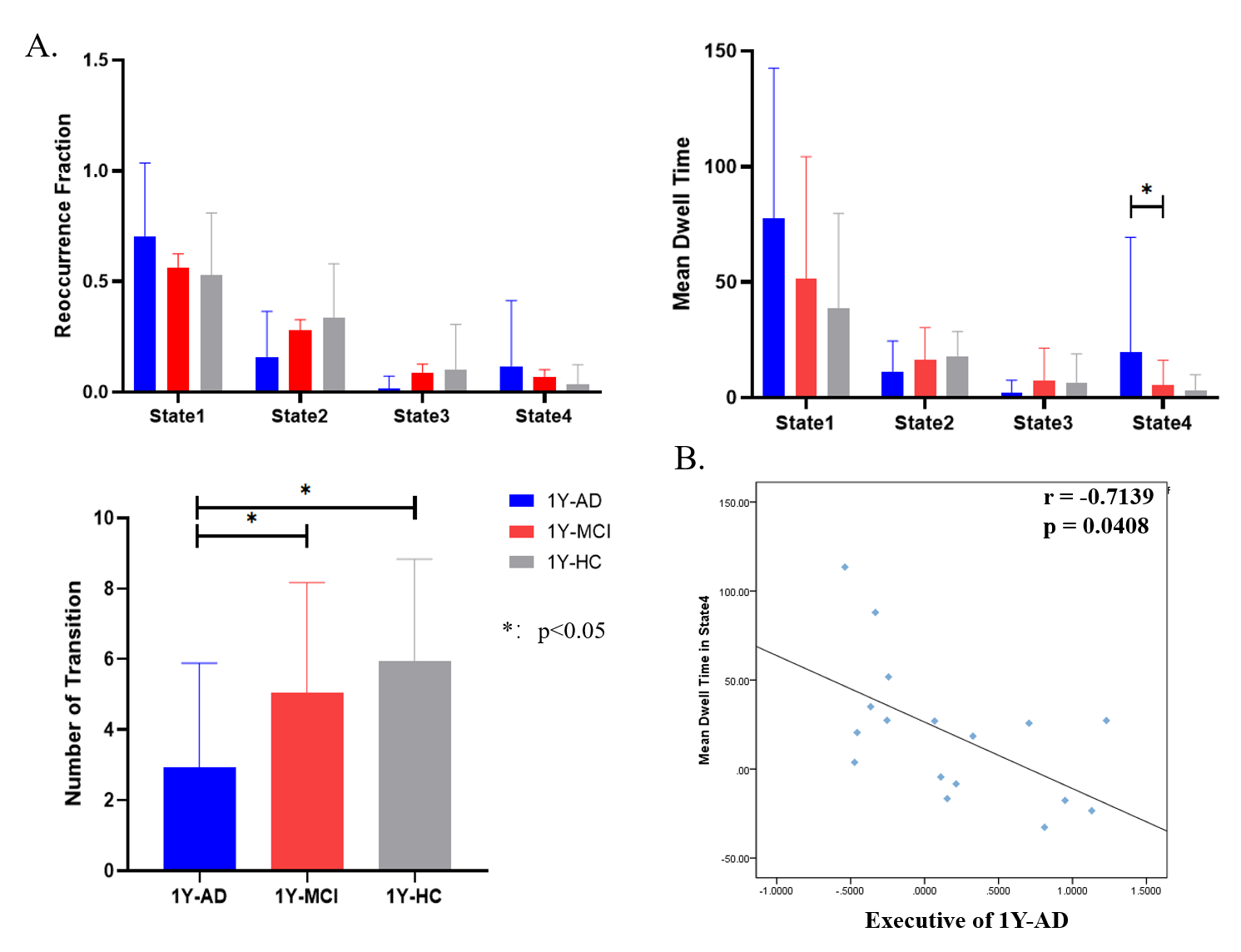
Fig 3 (A) Between-group differences in properties of dynamic functional connectivity states derived using the sliding window approach. (B) The Mean dwell Time in State 4 was negatively associated with Executive in 1Y-AD.