4514
Periventricular tissue damage in multiple sclerosis assessed by quantitative magnetization transfer1Department of Neurology, Division of Neurogeriatrics, Medical University of Graz, Graz, Austria, 2Medicinska fakulteta, Univerza v Ljubljani, Ljubljana, Slovenia, 3Department of Neurology, Division of General Neurology, Medical University of Graz, Graz, Austria, 4Neurology, Medical University of Graz, Graz, Austria, 5Department of Radiology, Division of Neuroradiology, Medical University of Graz, Graz, Austria
Synopsis
Periventricular tissue damage in multiple sclerosis has been suggested to be mediated by toxic soluble factors in the cerebrospinal-fluid. Magnetization transfer imaging has shown great potential to reveal microstructural tissue changes and has been used to study the association of periventricular normal-appearing white matter (NAWM) changes to the distance from the ventricle. We assessed high-resolution magnetization transfer saturation imaging (MTsat) in equidistant bands around the ventricle of MS patients and compared the resulting MTsat gradient of the thalamus and NAWM. While PV-MTsat values in NAWM are consistent to the literature, MTsat values in the outer thalamic bands were higher than controls, likely caused by increased iron levels.
Background
Cerebrospinal-fluid (CSF) mediated factors have been associated with periventricular (PV) tissue damage in multiple sclerosis (MS), which has been observed microstructurally using magnetization transfer ratio (MTR) in PV normal appearing white matter (NAMW),1-3 A gradient of tissue damage has also been observed in the thalamus using morphometry,4 R2*-mapping,5 T1/T2 ratio and diffusion imaging.6 However, thalamic tissue changes in relation to the CSF distance have not been investigated by magnetization transfer imaging so far, which could offer additional information regarding the underlying macromolecular tissue change.Objective
We here explored the CSF-related tissue changes in the thalamus and the PV-NAWM in MS patients, using high-resolution magnetization transfer saturation (MTsat) imaging.Methods
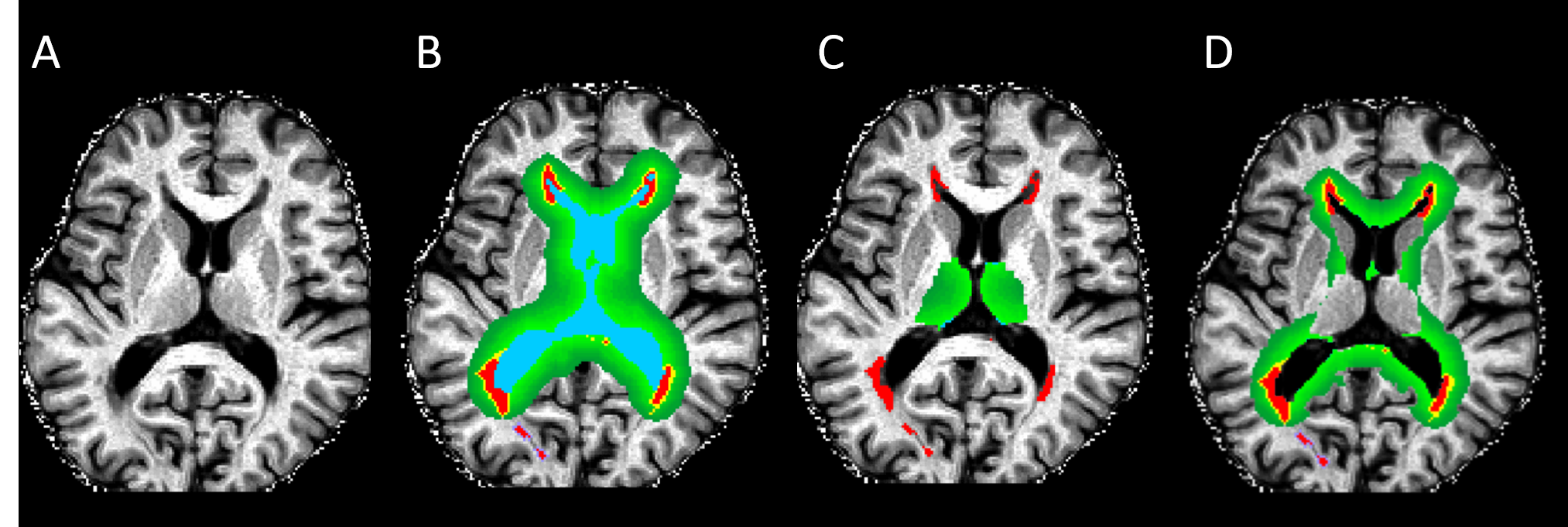
We performed MTsat imaging in 10 patients with clinically isolated syndrome (CIS) suggestive of MS (mean age 41 years) and 120 relapsing-remitting MS (RRMS) patients (mean age 37 years) as well as 9 healthy controls (HCs) (mean age 35 years). Band-wise MTsat values were assessed in the thalamus and PV-NAWM and were related to disability, total lesion load, normalized brain volume and cortical mean thickness (CMT). Using a 3.0T system with a 20-channel head coil (Siemens, Magnetom Prisma, VE11C) we acquired three multi-echo-gradient-echo sequences (MEGRE), a proton-density-weighted (TE=8.12ms, 13.19ms; TR=37ms; FA=6°) with and without MT-saturation pulse, and a T1-w sequence (TE=8.12ms, 13.19ms; TR=37ms; FA=35°), all with the same 1mm-isotropic resolution and acquisition matrix (224x168x192). Additionally, correction maps were acquired to account for transmit field inhomogeneities and the coil-sensitivity profile. A fluid attenuated inversion recovery (FLAIR) sequence (TE/TR/TI =95ms, 10000ms, 2500ms; resolution=0.9x0.9x3mm, matrix=256x192x44) was acquired to MS lesions, which were automatically segmented using the LST segmentation tool (freely available online https://www.applied-statistics.de/lst.html ). A synthetic T1 map (T1synth) was generated based on PD and apparent T1 maps using mri_synthesize (part of FreeSurfer, available online https://surfer.nmr.mgh.harvard.edu). MTsat-maps were processed according to Helms et.al.6 Brain volumetry, cortical mean thickness and Thalamic segmentation were calculated using FreeSurfer. To obtain MTsat values in equidistant bands around the ventricle, a CSF mask was generated using FSL-FAST (part of FSL https://fsl.fmrib.ox.ac.uk/), which was further dilated by one voxel in ten iterations.7 MTsat was bandwise evaluated in the FreeSurfer segmented thalamus mask and NAWM (WMH masks were subtracted from the global WM mask). All segmentations were visually inspected (figure 1). MTsat decrease of MS patients was computed relative to the average MTsat values of HCs in each band.Results
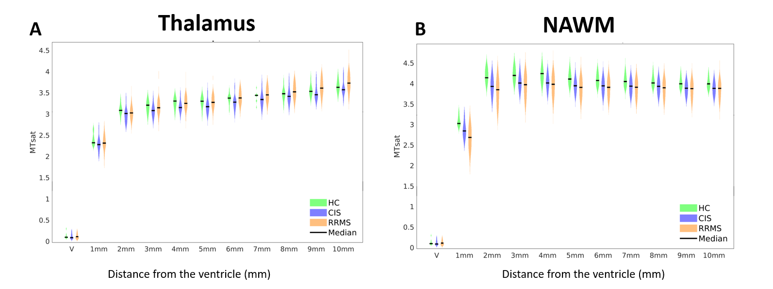
In the thalamus, MTsat values were reduced up to -4.2% in CIS and -2.0% in RRMS patients, relative to HCs (figure 2A). This relative decrease was more pronounced towards the lateral-ventricles with a slope of 0.47%/mm in CIS and 0.65%/mm in RRMS patients. However, in the outer bands towards thalamic WM (5mm-10mm), MTsat values were increased in RRMS patients, compared to HCs and CIS patients. In NAWM, MTsat values were decreased in all bands relative to HCs, the average decrease reached up to -11.7% in RRMS and -5.7% in CIS patients, relative to HCs (figure 2B). The slope over 10 bands towards the ventricle was 0.34%/mm in CIS and 0.58%/mm in RRMS patients.Discussion
For the first time, the PV tissue gradient was investigated using MTsat imaging in NAWM and the thalamus at a high resolution, with the benefit of reducing partial volume effects and increasing the sensitivity for macromolecular tissue changes in the ependymal zone. This area is considered as the interface between CSF and brain tissue and plays a major role within the circulatory system of the brain. A direct or indirect damage of the ependymal cells might therefore cause an accumulation of inflammatory factors solved in the CSF.8 In line with previous MTR studies conducted in PV-NAWM,1-3 also the MTsat values were decreased towards the ventricle. Further, RRMS patients were also stronger affected than CIS patients, confirming a more pronounced tissue damage with advancing disease. The comparison between the thalamic MTsat gradient and the gradient in NAWM highlights two important findings. First, a decrease relative to HCs could be observed towards the ventricle in both regions suggesting a common pathological process such as caused by an invasion of soluble factors from the CSF. Interestingly, in the outer bands of the thalamus (towards thalamic WM) we observed elevated MTsat values which were even higher for more advanced MS patients than for controls. This does not exclude demyelination and tissue destruction, but could be explained by a concomitant increase in iron concentration, since MTsat also scales with iron concentration due direct saturation effects of the MT saturation pulse.9Summary
In contrast to NAWM, where CIS and RRMS patients show a gradual decrease of MTsat values towards the CSF and are consistently lower than HCs, Thalamic MTsat values are increased in RRMS patients compared to HCs in the outer bands likely due to the accumulation of iron.Acknowledgements
No acknowledgement found.References
1. Liu Z, Pardini M, Yaldizli Ö, et al. Magnetization transfer ratio measures in normal-appearing white matter show periventricular gradient abnormalities in multiple sclerosis. Brain 2015; 138: 1239–1246.
2. Brown JWL, Pardini M, Brownlee WJ, et al. An abnormal periventricular magnetization transfer ratio gradient occurs early in multiple sclerosis. Brain 2017; 140: 387–398.
3. Poirion E, Tonietto M, Lejeune FX, et al. Structural and Clinical Correlates of a Periventricular Gradient of Neuroinflammation in Multiple Sclerosis. Neurology 2021; 96: e1865–e1875.
4. Fadda, G., Brown, R.A., Magliozzi, R., Aubert-Broche, B., O’Mahony, J., Shinohara, R.T., Banwell, B., Marrie, R.A., Yeh, E.A., Collins, D.L., Arnold, D.L., Bar-Or, A., 2019. A surface-in gradient of thalamic damage evolves in pediatric multiple sclerosis. Ann. Neurol. 85, 340–351. https://doi.org/10.1002/ana.25429
5. Louapre, C., Govindarajan, S.T., Giannì, C., Madigan, N., Sloane, J.A., Treaba, C.A., Herranz, E., Kinkel, R.P., Mainero, C., 2018. Heterogeneous pathological processes account for thalamic degeneration in multiple sclerosis: Insights from 7 T imaging. Mult. Scler. J. 24, 1433–1444. https://doi.org/10.1177/1352458517726382
6. Helms, G., Dathe, H., Dechent, P., 2008. Quantitative FLASH MRI at 3T using a rational approximation of the Ernst equation. Magn. Reson. Med. 59, 667–72. https://doi.org/10.1002/mrm.21542
7. Jehna, M., Pirpamer, L., Khalil, M., Fuchs, S., Ropele, S., Langkammer, C., Pichler, A., Stulnig, F., Deutschmann, H., Fazekas, F., Enzinger, C., 2015. Periventricular lesions correlate with cortical thinning in multiple sclerosis. Ann. Neurol. 78. https://doi.org/10.1002/ana.24461
8. Hatrock, D., Caporicci-Dinucci, N., Stratton, J., 2020. Ependymal cells and multiple sclerosis: Proposing a relationship. Neural Regen. Res. 15, 263–264. https://doi.org/10.4103/1673-5374.265551
9. Smith, S.A., Bulte, J.W.M., Van Zijl, P.C.M., 2009. Direct saturation MRI: Theory and application to imaging brain iron. Magn. Reson. Med. 62, 384–393. https://doi.org/10.1002/mrm.21980
Figures