4509
Improving Transmit and Receive Uniformity of 7T Brain MRI1Siemens Medical Solutions USA, Inc., Rochester, MN, United States, 2Siemens Medical Solutions USA, Inc., Urbana, IL, United States, 3Beckman Institute for Advanced Science and Technology, University of Illinois at Urbana-Champaign, Urbana, IL, United States, 4Bioengineering Department, University of Illinois at Urbana-Champaign, Urbana, IL, United States
Synopsis
Transmit and Receive inhomogeneity accentuate with increase in MR main magnetic field strength. Recently uniform combined reconstruction (UNICORN) was proposed to improve receive uniformity and applied for improving intensity uniformity of two-, three-, and N-dimensional 7T MRI data. In this work, we aim to improve both receive and transmit uniformity of 7T brain MRI by applying UNICORN in combination with transmit inhomogeneity correction methods. MP2RAGE images acquired with TrueForm, patient-specific and volume-selective B1 shimming methods were processed using the UNICORN algorithm. Significant improvement in uniformity was achieved using UNICORN compared to unnormalized images.
Introduction
7T MRI provides higher signal-to-noise ratio, contrast-to-noise ratio, and resolution compared to 3T/1.5 MRI. Increase in the field strength, however, results in higher transmit and receive non-uniformity. Reducing the impact of receive/transmit inhomogeneity would be useful to increase the clinical utility of ultra-high field MRI. Recently, uniform combined reconstruction (UNICORN) was prosed and applied to improve receive uniformity for two-, three-, and N-dimensional 7T MRI (1-2). In this work, we aim to improve both receive and transmit uniformity of 7T brain MRI by applying UNICORN in combination with transmit inhomogeneity correction methods.Methods
ImagingThree subjects were imaged on an investigational 7T pTX system (Siemens Healthcare, Erlangen, Germany) using a prototype 8-channel transmit 32-channel receive head coil (Nova Medical Inc., MA, USA). MP2RAGE (3) sequence, with typical imaging parameters, was used to acquire brain MRI volumes at inversion times of 840ms and 2370ms. Each subject was scanned using TrueForm (TF), volume-selective (VS), and patient-specific (PS) B1 shimming approaches. One of the three subjects (Subject 1) was scanned a second time with the MP2RAGE sequence using TF, VS, and PS B1 shimming options for reproducibility. 24 imaging volumes were acquired in total (4 × 2 inversions × 3 B1 shim options). The 24 imaging volumes were processed using the UNICORN algorithm.
UNICORN Algorithm
UNICORN ND algorithm proposed in (2) was used to improve receive uniformity. A prototype implementation of the UNICORN ND algorithm was used to combine the multichannel MP2RAGE data.
Uniformity Metric
The uniformity of the unnormalized and UNICORN normalized images was measured using difference between the mean intensities in two regions-of-interest (ROIs). One ROI was drawn in the frontal lobe. Another ROI was drawn in the cerebellum. Then, the uniformity (U) of the image was defined as shown in Equation 1.
$$ Uniformity \; (U) = 1-\left(\frac{A-B}{A+B}\right) \;\;\;\;\; [1] $$
Where A is the mean intensity within the ROI in the frontal lobe and B is the mean intensity within the ROI in the cerebellum.
Statistical Analysis
A two-sided paired two-sample (unnormalized and UNICORN) t-test was performed for the values of A, B, and the uniformity metric.
Results and Discussion
Figures 1 and 2 compare the uniformity of unnormalized and UNICORN normalized MP2RAGE MR images acquired using TF, VS, and PS B1 shimming methods in two subjects. It can be observed that UNICORN significantly improved the intensity in the inferior regions of the brain. A decrease in the hyper-intensity near the surface coil was also observed with UNICORN.Figure 3 shows the mean intensities within the two ROIs and the uniformity metric value for the twelve MP2RAGE volumes acquired with inversion time of 2370ms. Figure 4 shows the box plot of the mean intensities A and B within the ROIs and the measured uniformity. UNICORN normalization resulted in reduction in the hyperintensity in the frontal lobe, increase in the intensity in the cerebellum and improvement of uniformity. Figure 5 shows the subtraction between UNICORN normalized and unnormalized images demonstrating the reduction in hyper-intensity in superior regions and an increase in intensity in the inferior regions of the brain with UNICORN.
The p-value from the paired two-sample t-test for A was 0.009696, with mean difference of −110.9 between UNICORN and unnormalized images, and 95 percent confidence interval of [−189.08, −32.74] showing a significant decrease in hyper-intensity in frontal lobe with UNICORN. The p-value from the paired two-sample t-test for B was 0.02505, with mean difference of 39.9 between UNICORN and unnormalized images, and 95 percent confidence interval of [6.02, 73.81] showing a significant increase in intensity in the cerebellum with UNICORN. The p-value from the paired two-sample t-test for the uniformity metric was 7.477×10−06, with mean of difference 0.2125, and 95 percent confidence interval of [0.15, 0.27] demonstrating a significant improvement in measured uniformity with UNICORN.
It was observed that uniformity metric increased from TF to VS or PS B1 shimming methods for unnormalized images. The uniformity metric for TF, VS, and PS B1 shimming methods for UNICORN images was similar. One possible reason could be that in the regions of the ROIs analyzed in this work, the receive non-uniformity was more dominant than the transmit non-uniformity. Another reason could be the impact of coil combination method used by the B1 shimming methods during their optimization. We expect that using UNICORN for coil combination during B1 optimization could benefit image uniformity further.
One of the limitations of this work is that B1 shimming methods do not completely compensate for transmit inhomogeneities. Usage of fast online-customized (FOCUS) pulses for pTx has been proposed recently to efficiently correct transmit inhomogeneity (4). Using pTx approaches such as FOCUS was beyond the scope of this work.
Conclusion
Significant improvement in uniformity was achieved using UNICORN compared to unnormalized images. Future work would explore the use of UNICORN with FOCUS and for coil combination during B1 shim optimization.Acknowledgements
No acknowledgement found.References
1. Chebrolu VV, Kollasch PD, Deshpande V, et al. Uniform combined reconstruction of multichannel 7T knee MRI receive coil data without the use of a reference scan. J Magn Reson Imaging 2019; 50:1534–1544.
2. Chebrolu VV, Zhong X, Liebig P, Heidemann R. Uniform Combined Reconstruction for Improving Receive Intensity Homogeneity of N-dimensional 7T MRI. Proceedings ISMRM & SMRT Annual Meeting & Exhibition. 2021: 2609.
3. Marques JP, Kober T, Krueger G, van der Zwaag W, Van de Moortele PF, Gruetter R: MP2RAGE, a self bias- eld corrected sequence for improvedsegmentation and T1-mapping at high eld. Neuroimage 2010.
4. Herrler J, Liebig P, Gumbrecht R, et al. Fast online-customized (FOCUS) parallel transmission pulses: A combination of universal pulses and individual optimization
Figures
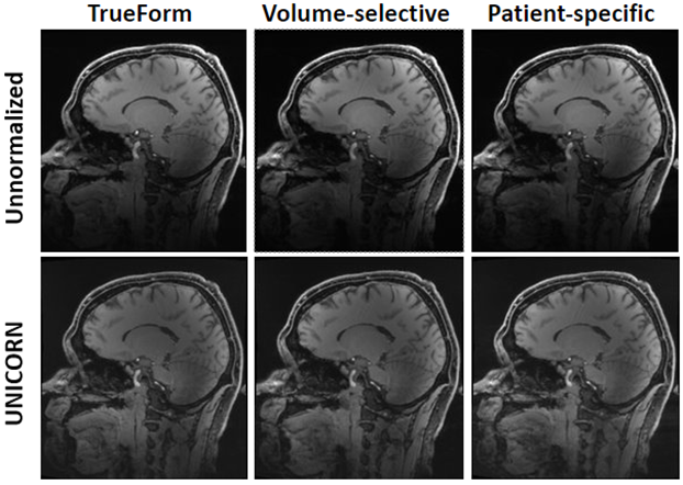
Figure 1: Comparison of uniformity between 7T brain MP2RAGE MR images acquired using TrueForm (TF), volume-selective (VS), and patient-specific (PS) B1 shimming methods in Subject 1. Top row: Images with no normalization or receive uniformity correction. Bottom row: Top row images processed using UNICORN normalization. All the images shown are from MP2RAGE volumes acquired with inversion time of 2370ms. The same window-width (WW) and window-level (WL) was used for all images. Significant improvement in conspicuity of the inferior regions of the brain can observed with UNICORN.
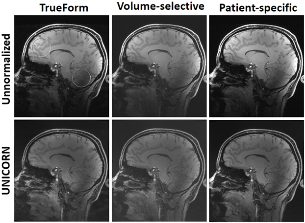
Figure 2: Comparison of uniformity between 7T brain MP2RAGE MR images acquired using TrueForm (TF), volume-selective (VS), and patient-specific (PS) B1 shimming methods in Subject 3. Top row: Images with no normalization or receive uniformity correction. Bottom row: Top row images processed using UNICORN normalization. All the images shown are from MP2RAGE volumes acquired with inversion time of 2370ms. The same window-width (WW) and window-level (WL) was used for all images. ROIs used for measuring the uniformity metric are shown on the unnormalized TrueFrom image.
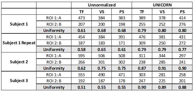
Figure 3: Mean intensity within two ROIs and the uniformity metric value for the different data sets analyzed in this work. TF: TrueForm B1 shimming; PS: patient-specific B1 shimming; and VS: volume-selective B1 shimming. ROI 1 was placed within the frontal lobe and ROI 2 was placed within the cerebellum. A is the mean intensity in ROI 1 and B is the mean intensity in ROI 2. ROI analysis was performed on MP2RAGE volumes acquired with inversion time of 2370ms. Improvement in intensity homogeneity and the uniformity metric was observed with UNICORN.
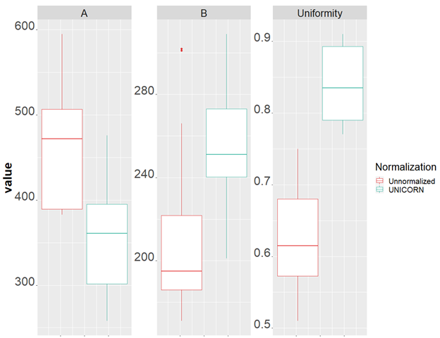
Figure 4: Box plots showing the spread of the mean intensity in ROI 1 (A) and the mean intensity in ROI 2 (B), and the uniformity metric in unnormalized and UNICORN normalized images. Reduction in the hyperintensity in the frontal lobe, increase in the intensity in the cerebellum and improvement of uniformity can be observed with UNICORN.
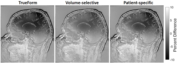
Figure 5: Percent difference between UNICORN and unnormalized images of the 7T brain MP2RAGE MRI acquired using TrueForm (TF), volume-selective (VS), and patient-specific (PS) B1 shimming methods in Subject 1. The percentage difference was computed at each pixel as (UNICORN intensity – Unnormalized intensity)/M, where M is the average of the maximum values within UNICORN and unnormalized image slices shown in the figure. The images demonstrate a reduction in hyperintensity in the superior regions and increase in the intensity in the inferior regions of the brain with UNICORN.