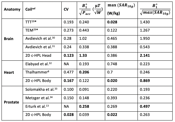
4502
In-silico shimming performance of a bore-sized 2-D UHF volume coil for different anatomies at 7T1Radiology, Weill Cornell Medicine, New York, NY, United States, 2College of Human Ecology, Cornell University, Ithaca, NY, United States, 3General Electric Healthcare, Aurora, OH, United States
Synopsis
We propose a two-dimensional cylindrical high-pass ladder (2D c-HPL) coil concept and investigate the contribution of the longitudinal dimension to utilize a bore-mounted general-purpose clinical volume coil for ultra-high field MRI. Theory and in-silico results show that this architecture exhibits improved B1 field uniformity for specific anatomies and regions such as brain (a coefficient of variation of 12.3%), cerebellum (4.9%), heart (16.7%), prostate (2.8%), and whole chest (36.8%). This could prove particularly useful for whole-body clinical imaging and could open new pathways for clinical diagnostics in UHF MRI.
Introduction
Ultra-high-field (UHF) magnetic resonance imaging (MRI) (7 T and above) promises improved signal-to-noise-ratio (SNR), hence improved spatial/temporal resolution over 3 T MRI.1-3 At UHF, the wavelength related to the intrinsic Larmor frequency shortens (~12 cm in tissue for 7 T)4, and typical body-sized birdcage designs fail due to wavelength effects. As a result, current UHF imaging most commonly employs localized surface array solutions driven by parallel transmission (pTx) benefiting from the space diversity of decoupled elements to create uniform B1+ for specific anatomies, often at the cost of higher specific absorption rate (SAR).5-15Although these array-based solutions can offer good SNR and B1+ uniformity, especially for smaller field of views (FOV), their use requires delicate placement of elements and sophisticated algorithms impeding clinical use as discussed before.16 A general-purpose bore-mounted body coil could facilitate more universal clinical use and hence clinical acceptance of UHF technology.
The goal of this work is to research the feasibility of a general-purpose, birdcage-like, whole-body coil at 7T the way it is routinely used at 3T. The first bore-size experimental body array at 7 T was presented more than a decade ago,17 with recent follow-up studies using microstrip elements18-20. Recent simulation studies on 7T volume coils show that bore-mounted circumferential coils can exhibit similar B1+ and SAR performance to local coils21-23.
In this work, we use an inductively coupled planar ladder structure, which inherently exhibits high pass characteristics and thus lends itself to a birdcage-like volume coil (Figure 1). It has been shown that these ladders can be used as MRI coils.22-26 We propose to use a 2D high-pass ladder (HPL) architecture in cylindrical form (2D c-HPL) and analyzed its in-silico shimming capabilities for various anatomies.
Methods
The 2D c-HPL coils designed before23, 24 are used as volumetric body coils for head and body imaging. We briefly compare body-sized design to 24-leg birdcage and transverse electromagnetic (TEM) designs of the same dimensions for homogeneous phantom loaded (spherical phantom, 34 cm diameter, εr=55, σ=0.6 S/m) and realistic body model loaded (Ella27) cases. TEM and birdcage coils were driven by using four active ports evenly distributed in their circumference direction to excite a circular polarization (CP) mode with 1 W of accepted power. For homogeneity evaluation, the coefficient of variation (CV) (ratio of B1+ standard deviation to B1+ mean (σ/μ)) is calculated.B1+ homogeneity for the whole brain (shimming volume of 20×20×13 cm3) and the cerebellum (shimming volume of 10×10×8 cm3) is presented for the head-sized coil designed earlier24 by using Ella. B1+ uniformity is also evaluated for the heart by using Ella (shimming volume of 10×10×10 cm3), for the chest by using Ella (shimming volume of 30×20×30 cm3) and for the prostate by using Duke27 (shimming volume of 4×5×4 cm3), with all anatomies located at the isocenter.
Here, we use a static phase-only B1+ shimming approach28 based on the Nelder-Mead optimization method, using initial phase values of 0, π/2, π, and 3π/2 for active elements in each row (i.e., CP excitation), and the CV as the cost function. B1+ shimming results and SAR efficiency (B1+/sqrt(SAR10g)) metrics are compared with the existing literature.
Results
Comparison of bore-sized architectures in CP mode is given in Figure 2. Here, the 2D c-HPL architecture offers the best SAR efficiency (B1+/√SAR-10g, 0.78 𝜇T/√(W/kg)).RF Shimming results of 2D c-HPL coils are provided in Figure 3. A CV of 12.3% for the whole head volume, a CV of 4.9% for the cerebellum, a CV of 16.7% for the heart, and a CV of 2.8% for the prostate is achieved.
Large FOV shimming results are given in Figure 4. A CV of 36.8% for the whole chest volume with an in-plane CV of 11.3% for the central transverse slice is achieved.
A comparison of B1+ efficiency, CV, SAR, and SAR efficiency between the 2D c-HPL architectures and existing designs from literature is provided in Table1. The 2D c-HPL architecture offers better homogeneity (i.e., lower CV) and better SAR efficiency for most anatomies (i.e., brain, cerebellum, and heart). However, in the case of heart and prostate imaging, the B1+ efficiency of the 2D c-HPL is decreased compared to localized surface coil solutions, which is expected due to the increased distance of a bore-wall volume coil design.
Conclusion
A cylindrical coil architecture that extends the tried-and-tested 1D birdcage topology to 2D, suitable for UHF MRI applications, is proposed and demonstrated in-silico. Simulation results of bore-sized coils with CP mode transmission demonstrated that the proposed 2D c-HPL coil performs better than the similar TEM coil and the birdcage designed in this study, both in terms of B1+ uniformity and SAR efficiency. B1+ shimming optimization yielded a CV of 12.3% for the head, 4.9% for the cerebellum, 16.7% for the heart, 2.8% for the prostate, and 36.8% for the whole chest anatomies. An improvement of ≥20% in B1+ homogeneity and improved SAR efficiency for most of the anatomies is observed compared to the literature. We believe that this 2D c-HPL architecture is a promising candidate to serve as a new generation clinical volume body coil for UHF MRI applications.Acknowledgements
This work was supported in part by the National Institute of Health (NIH) through R00EB024341. The authors are also grateful to GE Healthcare for the equipment support.
References
1. Pohmann, R, Speck, O, Scheffler, K. Signal-to-noise ratio and MR tissue parameters in human brain imaging at 3, 7, and 9.4 tesla using current receive coil arrays. Magnetic Resonance in Medicine 2016;75(2):801-809.
2. Trattnig, S, Springer, E, Bogner, W, et al. Key clinical benefits of neuroimaging at 7T. NeuroImage 2018;168:477-489.
3. Hendriks, AD, D'Agata, F, Raimondo, L, et al. Pushing functional MRI spatial and temporal resolution further: High-density receive arrays combined with shot-selective 2D CAIPIRINHA for 3D echo-planar imaging at 7 T. NMR in Biomedicine 2020;33(5):e4281.
4. Elabyad, IA, Terekhov, M, Lohr, D, et al. A Novel Mono-surface Antisymmetric 8Tx/16Rx Coil Array for Parallel Transmit Cardiac MRI in Pigs at 7T. Scientific Reports 2020;10(1).
5. Ipek, Ö. Radio-frequency coils for ultra-high field magnetic resonance. Analytical biochemistry 2017;529:10-16.
6. Rosenkrantz, AB, Zhang, B, Ben-Eliezer, N, et al. T2-weighted prostate MRI at 7 tesla using a simplified external transmit-receive coil array: Correlation with radical prostatectomy findings in two prostate cancer patients. Journal of Magnetic Resonance Imaging 2015;41(1):226-232.
7. Arteaga De Castro, CS, Van Den Bergen, B, Luijten, PR, et al. Improving SNR and B1 transmit field for an endorectal coil in 7 T MRI and MRS of prostate cancer. Magnetic Resonance in Medicine 2012;68(1):311-318.
8. Thalhammer, C, Renz, W, Winter, L, et al. Two-Dimensional sixteen channel transmit/receive coil array for cardiac MRI at 7.0 T: Design, evaluation, and application. Journal of Magnetic Resonance Imaging 2012;36(4):847-857.
9. Rietsch, SHG, Quick, HH, Orzada, S. Impact of different meander sizes on the RF transmit performance and coupling of microstrip line elements at 7 T. Medical Physics 2015;42(8):4542-4552.
10. Metzger, GJ, Snyder, C, Akgun, C, et al. LocalB1+ shimming for prostate imaging with transceiver arrays at 7T based on subject-dependent transmit phase measurements. Magnetic Resonance in Medicine 2008;59(2):396-409.
11. Bing, W, Chunsheng, W, Kelley, DAC, et al. Shielded Microstrip Array for 7T Human MR Imaging. IEEE Transactions on Medical Imaging 2010;29(1):179-184.
12. Steensma, BR, Voogt, IJ, Leiner, T, et al. An 8-channel Tx/Rx dipole array combined with 16 Rx loops for high-resolution functional cardiac imaging at 7 T. Magnetic Resonance Materials in Physics, Biology and Medicine 2018;31(1):7-18.
13. Erturk, MA, Raaijmakers, AJ, Adriany, G, et al. A 16-channel combined loop-dipole transceiver array for 7 Tesla body MRI. Magn Reson Med 2017;77(2):884-894.
14. Clément, J, Gruetter, R, Ipek, Ö. A combined 32‐channel receive‐loops/8‐channel transmit‐dipoles coil array for whole‐brain MR imaging at 7T. Magnetic Resonance in Medicine 2019.
15. Oezerdem, C, Winter, L, Graessl, A, et al. 16-channel bow tie antenna transceiver array for cardiac MR at 7.0 tesla. Magnetic Resonance in Medicine 2016;75(6):2553-2565.
16. Paška, J, Cloos, MA, Wiggins, GC. A rigid, stand-off hybrid dipole, and birdcage coil array for 7 T body imaging. Magnetic Resonance in Medicine 2018;80(2):822-832.
17. Vaughan, JT, Snyder, CJ, DelaBarre, LJ, et al. Whole-body imaging at 7T: Preliminary results. Magnetic Resonance in Medicine 2009;61(1):244-248.
18. Orzada, S, Bitz, AK, Johst, S, et al. Analysis of an Integrated 8-Channel Tx/Rx Body Array for Use as a Body Coil in 7-Tesla MRI. Frontiers in Physics 2017;5.
19. Orzada, S, Solbach, K, Gratz, M, et al. A 32-channel parallel transmit system add-on for 7T MRI. PLOS ONE 2019;14(9):e0222452.
20. Hernandez, D. Three-Line Microstrip Array for Whole-Body MRI System at 7 T. Applied Sciences 2020;11(1):73.
21. Fiedler, TM, Orzada, S, Flöser, M, et al. Performance analysis of integrated RF microstrip transmit antenna arrays with high channel count for body imaging at 7 T. NMR in Biomedicine 2021;34(7).
22. Gokyar, S, Voss, HU, Robb, F, et al. An Electrically Long Ultra-High Field MRI Volume Body Coil Design. International Conference on Electromagnetics in Advanced Applications (ICEAA). Honolulu, HI, USA: IEEE; 2021.
23. Gokyar, S, Voss, HU, Taracila, V, et al. A Comparative Study Highlighting a Novel UHF MRI Volume Body Coil Design. Proc. Intl. Soc. Mag. Reson. Med. 29 (2021). 1580. ISMRM; 2021:p.1580.
24. Gokyar, S, Voss, HU, Taracila, V, et al. A 2D High-Pass Ladder RF Coil Architecture for UHF MRI. Proc. Intl. Soc. Mag. Reson. Med. 29 (2021). 1589. ISMRM; 2021:p.1589.
25. Voss, HU, Ballon, DJ. High-pass two-dimensional ladder network resonators for magnetic resonance imaging. IEEE Trans Biomed Eng 2006;53(12 Pt 2):2590-2593.
26. Ballon, D, Meyer, K. Two-dimensional ladder network resonators. Journal of Magnetic Resonance, Series A 1994;111(1):23-28.
27. IT’IS foundation, Z, Switzerland.
28. Padormo, F, Beqiri, A, Hajnal, JV, et al. Parallel transmission for ultrahigh-field imaging. NMR in Biomedicine 2016;29(9):1145-1161.
29. Krishnamurthy, N, Santini, T, Wood, S, et al. Computational and experimental evaluation of the Tic-Tac-Toe RF coil for 7 Tesla MRI. PLOS ONE 2019;14(1):e0209663.
30. Avdievich, NI, Solomakha, G, Ruhm, L, et al. Bent folded‐end dipole head array for ultrahigh‐field MRI turns “dielectric resonance” from an enemy to a friend. Magnetic Resonance in Medicine 2020;84(6):3453-3467.
31. Avdievich, NI, Solomakha, G, Ruhm, L, et al. Folded-end dipole transceiver array for human whole-brain imaging at 7 T. NMR in Biomedicine 2021;34(8):e4541.
32. Elabyad, IA, Terekhov, M, Schreiber, LM. Development and RF Shimming of an 8Tx/16Rx Antisymmetric Transceiver Coil Array for Parallel Transmit Cardiac MRI in Humans at 7T. Proc Intl. Soc. Mag. Reson. Med. (ISMRM). 2020.
33. Solomakha, G, Svejda, JT, Van Leeuwen, C, et al. A self-matched leaky-wave antenna for ultrahigh-field magnetic resonance imaging with low specific absorption rate. Nature Communications 2021;12(1).
34. Metzger, GJ, Van De Moortele, P-F, Akgun, C, et al. Performance of external and internal coil configurations for prostate investigations at 7 T. Magnetic Resonance in Medicine 2010;64(6):1625-1639.
Figures
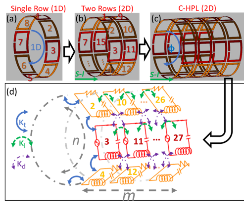
Figure 1. Schematics of the proposed 2D cylindrical high-pass ladder (2D c-HPL) architecture (not on scale). (a) Single row coil (1D architecture), (b) a sample two-row coil cascaded in superior to inferior (S-I) direction, (c) a 4×16 2-D c-HPL coil, and (d) an equivalent circuit model with transverse, longitudinal, and diagonal coupling coefficients as reported before [24].
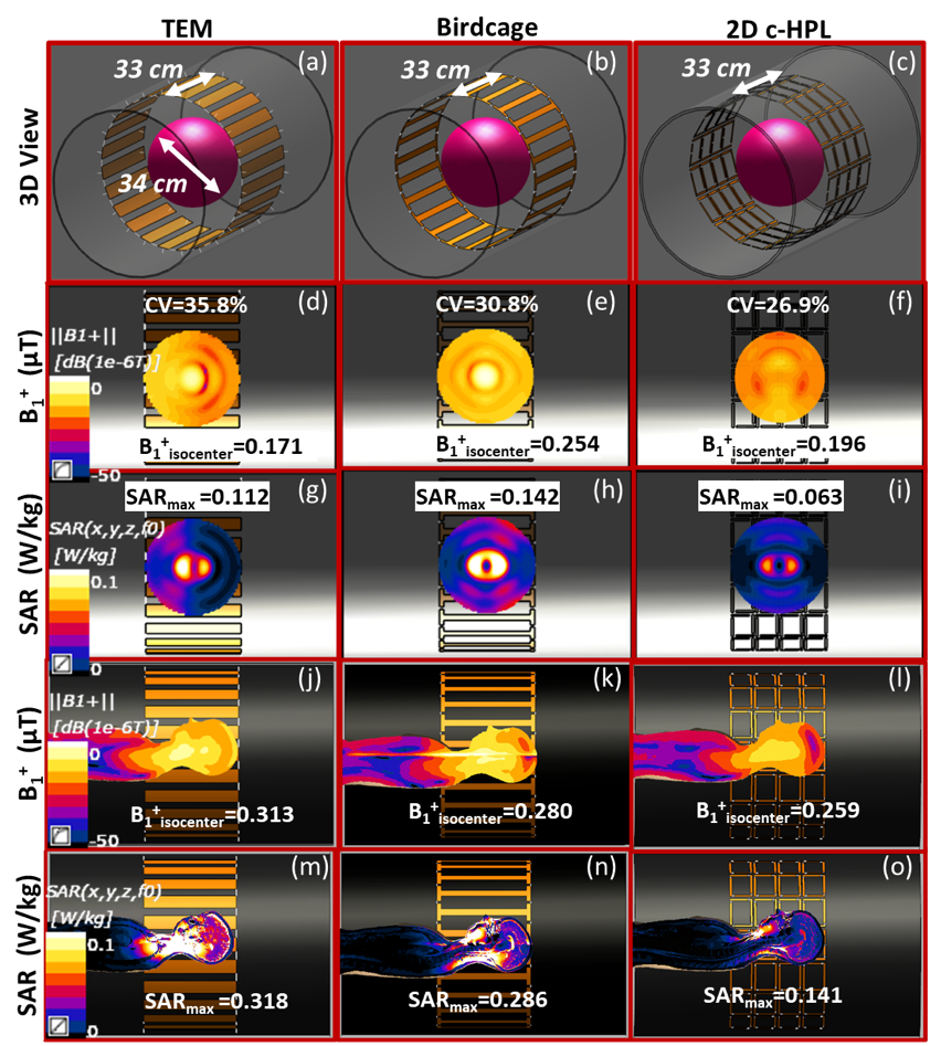
Figure 2. Comparison of standard body-sized coils in circular polarization (CP) mode, (a) transverse electromagnetic (TEM), (b) birdcage, and (c) 2D c-HPL. B1+ maps with a spherical phantom positioned at the isocenter are shown in (d-f). SAR maps with a spherical phantom are shown in (g-i). Here, the 2D c-HPL exhibits the highest SAR efficiency (0.78 𝜇T/√(W/kg)). B1+ maps with a realistic body model, Ella, are shown in (j-l) and SAR maps in (m-o). Here, the 2D c-HPL design exhibits the highest SAR efficiency (0.69 𝜇T/√(W/kg)) compared to TEM and birdcage.
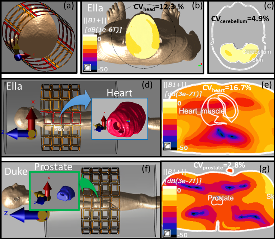
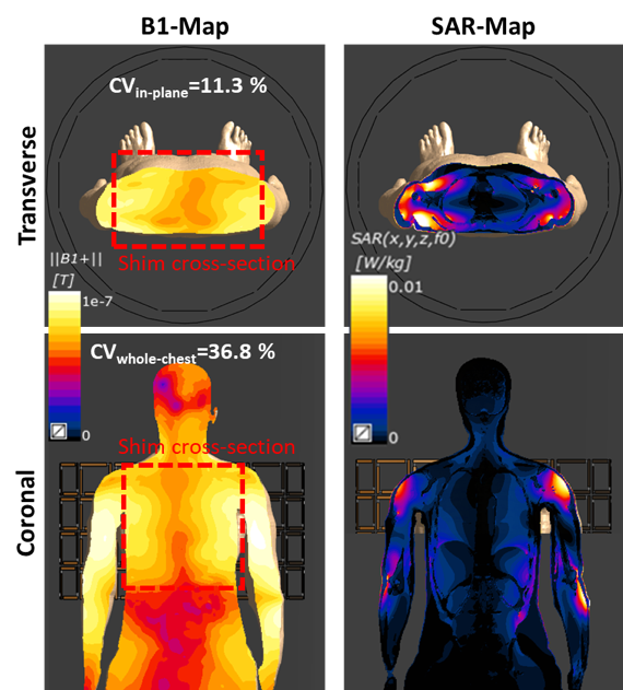
Figure 4. B1+ shimming performance of the 2-D c-HPL architecture for larger shimming regions at 7T. When a transverse slice (30 × 25 cm2) containing the whole torso is selected for shimming, a CV of 11.3% is achieved with a maximum SAR-1g of 0.44 W/kg for 1W of input power. For a volumetric region of whole chest (30 × 25 × 30 cm3 in x- y- and z- directions, respectively), a CV of 36.8% is observed with a maximum SAR-1g (outside the region of interest) of 0.48 W/kg for 1 W accepted RF power. These inhomogeneities are comparable to 3T studies.