4434
Deblurring 3D FSE Images via Regularized Iterative Demodulation of T2 Decay1GE Healthcare, New York, NY, United States, 2Department of Radiology and Imaging, Hospital for Special Surgery, New York, NY, United States
Synopsis
The 3D FSE sequence is widely used in clinical MRI for its capability to collect many phase encoding lines in each RF excitation. However, the signals during the long echo train readout are modulated by the T2 decay, resulting in blurring. An algorithm is presented to retrospectively demodulate estimated T2 decay from the actual signal. In the preliminary data, the proposed algorithm demonstrated success in deblurring the images.
INTRODUCTION
The 3D FSE sequence is widely used in clinical MRI for its capability to collect many phase encoding lines in each RF excitation. However, the signals during the long echo train readout are modulated by the T2 decay, resulting in the blurring of the images. Here, we present an algorithm to retrospectively demodulate such T2 decay from the actual signal, and we hypothesize that this will improve the image sharpness.METHODS
The effect of T2 decay on the FSE signal is determined using FSE Bloch equations for a targeted tissue relaxation characteristic and the specific view-order from a given scan. The estimated FSE signal was used to increase the signal of kspace that were acquired in later echoes. The direct upscaling of kspace often lead to the introduction of artifacts such as noise amplification and ringing. Therefore, the upscaled kspace was instead solved iteratively while applying a weighted generalized Tikhonov regularization:$$X^{'}=argmin_{X}||EFX-S||^{2}_{2}+\lambda||W(F^{-1}S-X)||^{2}_{2}$$
Where $$$E$$$ is the kspace profile of the FSE signal with T2 decay that was determined using FSE Bloch equations for an assumed tissue relaxation characteristic and the specific view-order for a given scan, $$$F$$$ is Fourier transformation, $$$S$$$ is the kspace of the original image, $$$\lambda$$$ is the regularization parameter for the generalized Tikhonov regularization, and $$$W$$$ is the weighting. The weighting was estimated as a function of the signal intensity to minimize the various artifacts mentioned above.
Under an IRB-approved study, the 3D Cartesian T2w-FSE brachial plexus images of 5 patients were acquired on a 3T clinical GE Signa Premier scanner (1x1x1mm, TR/TE=3300/60ms, time=6.5 minutes). The kspace was extracted from the DICOM images of the product reconstruction, and the proposed algorithm was applied while targeting the relaxivity values of nerve (T1=1410ms, T2=180ms). For comparison, the product images were also sharpened using unsharp masking (1).
RESULTS
An example of kspace in ky/kz plane before and after applying the proposed algorithm is shown in Figure 1. As shown in Figure 2, while the unsharp masking method does improve the sharpness of the nerves, it did not recover the caps between the nerve that are present in the results of the proposed algorithm.DISCUSSION
The preliminary results demonstrated the feasibility of retrospective demodulation of the effects of T2 decay, and that one of the benefits of such demodulation is the deblurring of the images. A limitation of the current implementation is that the whole kspace is demodulated according to the relaxivity values of one targeted tissue type, this could contribute to artifacts in other tissues with vastly different relaxivity values. It is possible that the regularization term helped suppressing such artifacts, but it is more appropriate to implement the algorithm to target multiple relaxivity values.CONCLUSION
In the preliminary data, the proposed algorithm demonstrated success in deblurring the images. Further evaluations of the proposed algorithm in more applications are warranted, and the algorithm may be further improved to perform demodulation on multiple relaxivity values.Acknowledgements
No acknowledgement found.References
1. M. A. Badamchizadeh and A. Aghagolzadeh, "Comparative study of unsharp masking methods for image enhancement," Third International Conference on Image and Graphics (ICIG'04), 2004, pp. 27-30, doi: 10.1109/ICIG.2004.50.
Figures
Figure 1. In a representative case, the kspace before and after demodulation shown in ky/kz plane.
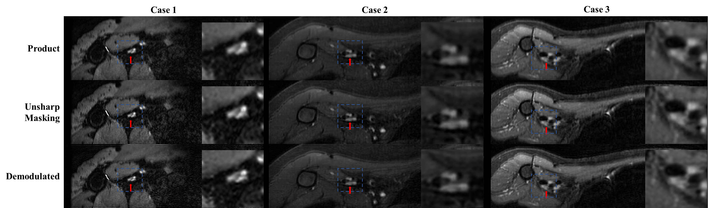
Figure 2. Representative cases of 3D T2w-FSE brachial plexus scan reformatted to the Y/Z plane. Red arrow indicates the sharpened of nerve in the results from the proposed algorithm.